. 2021 Jul 14;6(1):269.
doi: 10.1038/s41392-021-00669-2.

A novel dephosphorylation targeting chimera selectively promoting tau removal in tauopathies

Affiliations

A novel dephosphorylation targeting chimera selectively promoting tau removal in tauopathies

Jie Zheng et al. Signal Transduct Target Ther. .

Abstract

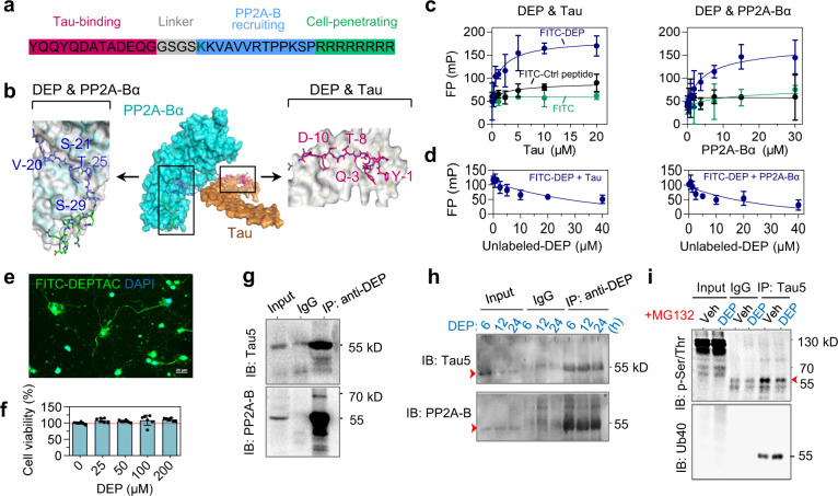
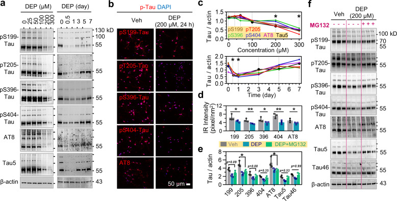
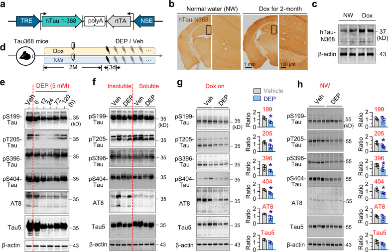
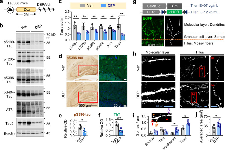
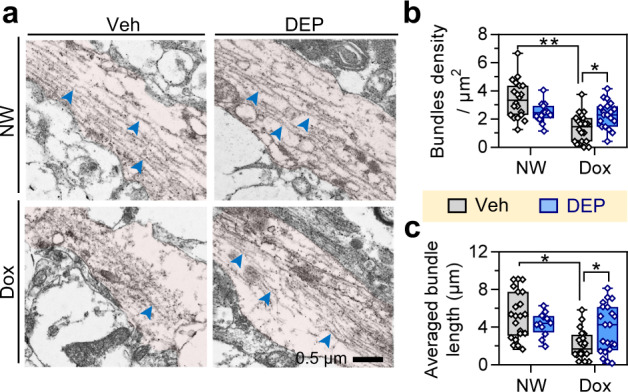
Intraneuronal accumulation of hyperphosphorylated tau is a hallmark pathology shown in over twenty neurodegenerative disorders, collectively termed as tauopathies, including the most common Alzheimer's disease (AD). Therefore, selectively removing or reducing hyperphosphorylated tau is promising for therapies of AD and other tauopathies. Here, we designed and synthesized a novel DEPhosphorylation TArgeting Chimera (DEPTAC) to specifically facilitate the binding of tau to Bα-subunit-containing protein phosphatase 2A (PP2A-Bα), the most active tau phosphatase in the brain. The DEPTAC exhibited high efficiency in dephosphorylating tau at multiple AD-associated sites and preventing tau accumulation both in vitro and in vivo. Further studies revealed that DEPTAC significantly improved microtubule assembly, neurite plasticity, and hippocampus-dependent learning and memory in transgenic mice with inducible overexpression of truncated and neurotoxic human tau N368. Our data provide a strategy for selective removal of the hyperphosphorylated tau, which sheds new light for the targeted therapy of AD and related-tauopathies.


Conflict of interest statement

J-Z.W. and J.Z. have filed for a patent related to this work (CN109824787A). The remaining authors declare no competing interests.

Figures

Fig. 1
Fig. 1
DEPTAC binds to tau and simultaneously recruits PP2A-Bα. a Functional motifs and amino acid sequence of DEPTAC (DEP). b The estimated triadic interaction of DEPTAC with tau (khaki) and PP2A-Bα (cyan) based on their 3D-structures in ZDOCK Server. Key binding residues of DEPTAC were as labeled. c, d Direct interactions of FITC-conjugated DEPTAC to tau and PP2A-Bα measured by fluorescence polarization binding (c) and the competing (d) assays. e High cell penetrability of DEPTAC in cultured primary rat neurons. Scale bar, 20 μm. f No cytotoxicity of the DEPTAC (to 200 μM) detected by CCK8 assay. Data were normalized by the mean value of the 0-μM group. One-way ANOVA followed by Tukey’s multiple comparisons tests. F(4, 25) = 1.47, P = 0.24. g The triadic interaction of DEPTAC with tau and PP2A-B was confirmed by immunoprecipitation using DEPTAC antibody in primary cultured rat neurons (12 h). h Co-immunoprecipitation of DEPTAC with tau and PP2A-B can be detected 6–24 h of post-DEPTAC treatment, with a gradual decrease of total tau. i DEPTAC decreased tau phosphorylation at Ser/Thr residues with upregulated tau ubiquitination in the presence of MG132
Fig. 2
Fig. 2
DEPTAC facilitates tau dephosphorylation and degradation in primary neurons with hTau-overexpression. ad DEPTAC promoted tau dephosphorylation at the AD-related pS199, pT205, pS396, pS404, and AT8 epitope with reduced total tau probed by Tau5 antibody, in a dose- and time-dependent manner in cultured primary rat neurons with lentivirus-mediated hTau-overexpression (a, c). Data were normalized to the level of β-actin. One-way ANOVA followed by Tukey’s multiple comparisons tests, *p < 0.01 compared with baseline, n = 3 cell wells for each group. By immunofluorescence staining, DEPTAC (200 μM for 24 h) also reduced the averaged immunoreactive (IR) intensity of phospho-tau (b, d). Scale bar, 50 μm. Two-way ANOVA followed by Tukey’s multiple comparisons tests. *p < 0.05, **p < 0.01, n = 3 cell wells in each group. e, f DEPTAC promoted tau dephosphorylation with reduced efficiency in the presence of MG132. Two-way ANOVA followed by Tukey’s multiple comparisons tests, *p < 0.05, n = 3 cell wells in each group
Fig. 3
Fig. 3
DEPTAC reduces phosphorylated and total tau in the hippocampus of Tau368 mice. a The expression of hTau-N368 was controlled by the NSE promoter and a tet-on system. b, c Prominent hTau-N368 expression was detected by immunohistochemistry (b) and Western blotting (c) in the dorsal hippocampus of the dox-, but not normal water (NW)-drinking group. d Experimental procedures for drugs administration. DEPTAC or vehicle was administrated for once or a consecutive month during dox on. e DEPTAC for once reduced phosphorylated and total tau prominently at 5 mM and 12–72 h of post intra-cerebroventricular injection in the hippocampus of Tau368 mice. f DEPTAC decreased both soluble and insoluble tau in the hippocampus of Tau368 mice. g, h DEPTAC for a consecutive significantly reduced hippocampal phospho-tau and total tau (/ 10 μg total protein) after dox administration for 4 months (g), while the efficiency of DEPTAC on mouse tau was relatively limited in NW-administrated Tau368 mice (/ 30 μg total protein) (h). Unpaired t-tests, *p < 0.05, n = 3–4 mice in each group
Fig. 4
Fig. 4
DEPTAC promotes tau dephosphorylation and improves neurite plasticity in dox-treated Tau368 mice. a Experimental procedure showing that DEPTAC or vehicle was administrated for a consecutive month after stopping the dox. b, c DEPTAC downregulated phospho-tau and total tau levels in the hippocampi of Tau368 mice. Two-way ANOVA followed by Tukey’s multiple comparisons tests. **p < 0.01, n = 3 mice in each group. df DEPTAC ameliorated the level of pS396-tau immunostaining (e) and thioflavin T (ThT)-stained tau aggregation (f) in the DG subset (red boxes). Unpaired t-tests, **p < 0.01, n = 4–5 mice in each group. (g) Sparse labeling of dentate granular cells by a mixture of AAVs. hj DEPTAC increased the densities of overall and the mushroom-like dendritic spines (i) and averaged area of mossy fiber puncta (j) of granular cells. The insert (i) illustrated the morphology of different types of dendrite spines: T thin, M mushroom-like, S stubby. Two-way ANOVA followed by Tukey’s multiple comparisons tests or unpaired t-tests, *p < 0.05, n = 5 mice in each group
Fig. 5
Fig. 5
DEPTAC improves microtubule assembly in dox-treated Tau368 mice. a Representative electron microscopy images. Pink areas indicated the neurites of hippocampal neurons, and the blue arrowheads indicate the representative polymerized microtubules. Scale bar, 500 nm. b, c Dox for 2 months decreased the density (b) and length (c) of microtubule bundles in Tau368 mice, while both of which were significantly reversed by 1-month DEPTAC treatment after stopping the dox. Two-way ANOVA followed by Tukey’s multiple comparisons tests, *p < 0.05, n = 5 mice and 12–23 view fields (circles) in each group. Data were presented as Min-Median-Max
Fig. 6
Fig. 6
DEPTAC improves hippocampus-dependent learning and memory in dox-treated Tau368 mice. af 1-month DEPTAC treatment after stopping the dox rescued the hTau-induced object-place (ac) and novel-object (df) recognition impairments in Tau368 mice compared with the vehicle controls. Cartoons (a, d) show paradigms of the behavioral tests. Representative heatmaps (b, e) show the time-location of mice traveled in test chambers. Two-way ANOVA followed by Tukey’s multiple comparisons tests, *p < 0.05, n = 10–15 mice in each group. g DEPTAC significantly alleviated the hTau-induced spatial learning deficits in Morris water maze test. Repeated measures ANOVA followed by Tukey’s multiple comparisons tests, **p < 0.01, *p < 0.05. h, i DEPTAC attenuated spatial memory deficits in dox-treated Tau368 mice shown by the increased time staying in h and number of entries into (i) the target quadrant in water maze test detected at day 6 after removed the platform. Two-way ANOVA followed by Tukey’s multiple comparisons, *p < 0.05. j Representative traces of the mice traveled in the water maze at day 6. Green areas indicate the target quadrants. Data were presented as Min-Median-Max

Similar articles

Cited by

References

    1. Li C, Gotz J. Tau-based therapies in neurodegeneration: opportunities and challenges. Nat. Rev. Drug Discov. 2017;16:863–883. doi: 10.1038/nrd.2017.155. - DOI - PubMed
    1. Grundke-Iqbal I, et al. Abnormal phosphorylation of the microtubule-associated protein tau (tau) in Alzheimer cytoskeletal pathology. Proc. Natl Acad. Sci. USA. 1986;83:4913–4917. doi: 10.1073/pnas.83.13.4913. - DOI - PMC - PubMed
    1. DeVos SL, et al. Tau reduction prevents neuronal loss and reverses pathological tau deposition and seeding in mice with tauopathy. Sci. Transl. Med. 2017;9:g481. doi: 10.1126/scitranslmed.aag0481. - DOI - PMC - PubMed
    1. Boxer AL, et al. Safety of the tau-directed monoclonal antibody BIIB092 in progressive supranuclear palsy: a randomised, placebo-controlled, multiple ascending dose phase 1b trial. Lancet Neurol. 2019;18:549–558. doi: 10.1016/S1474-4422(19)30139-5. - DOI - PubMed
    1. Lu M, et al. Discovery of a Keap1-dependent peptide PROTAC to knockdown Tau by ubiquitination-proteasome degradation pathway. Eur. J. Med. Chem. 2018;146:251–259. doi: 10.1016/j.ejmech.2018.01.063. - DOI - PubMed

Publication types

MeSH terms