. 2021 Jul 15;12(7):705.
doi: 10.1038/s41419-021-03996-y.

DHA exhibits synergistic therapeutic efficacy with cisplatin to induce ferroptosis in pancreatic ductal adenocarcinoma via modulation of iron metabolism

Affiliations

DHA exhibits synergistic therapeutic efficacy with cisplatin to induce ferroptosis in pancreatic ductal adenocarcinoma via modulation of iron metabolism

Jing Du et al. Cell Death Dis. .

Abstract

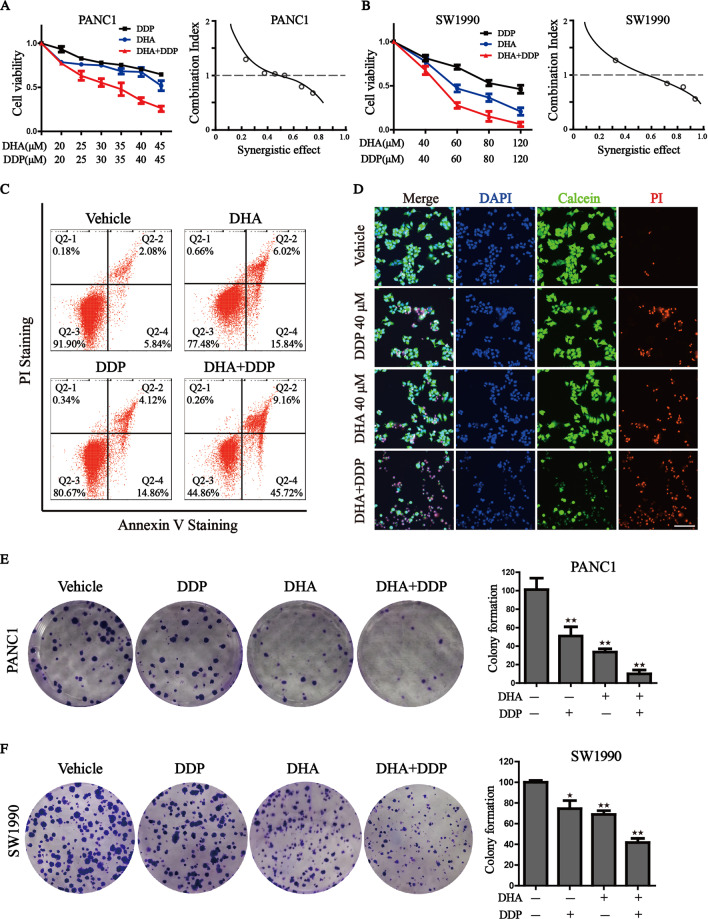
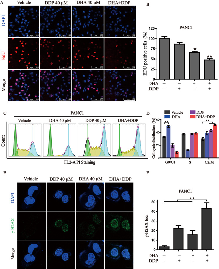
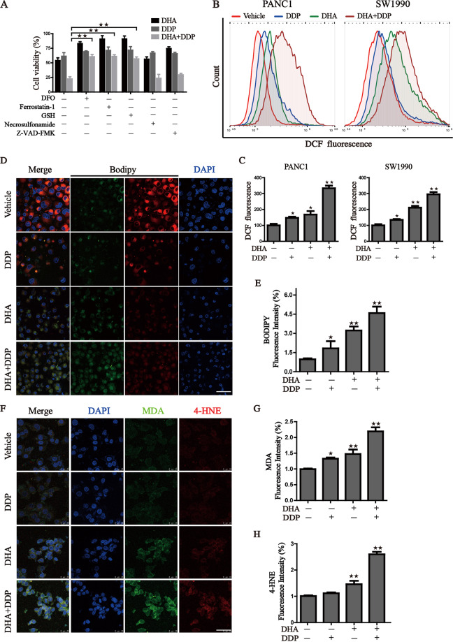
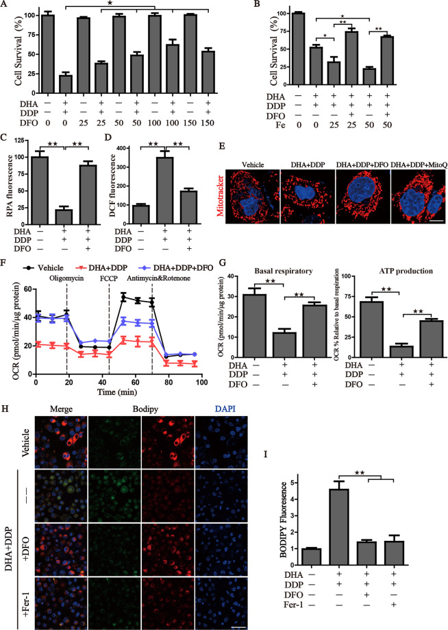
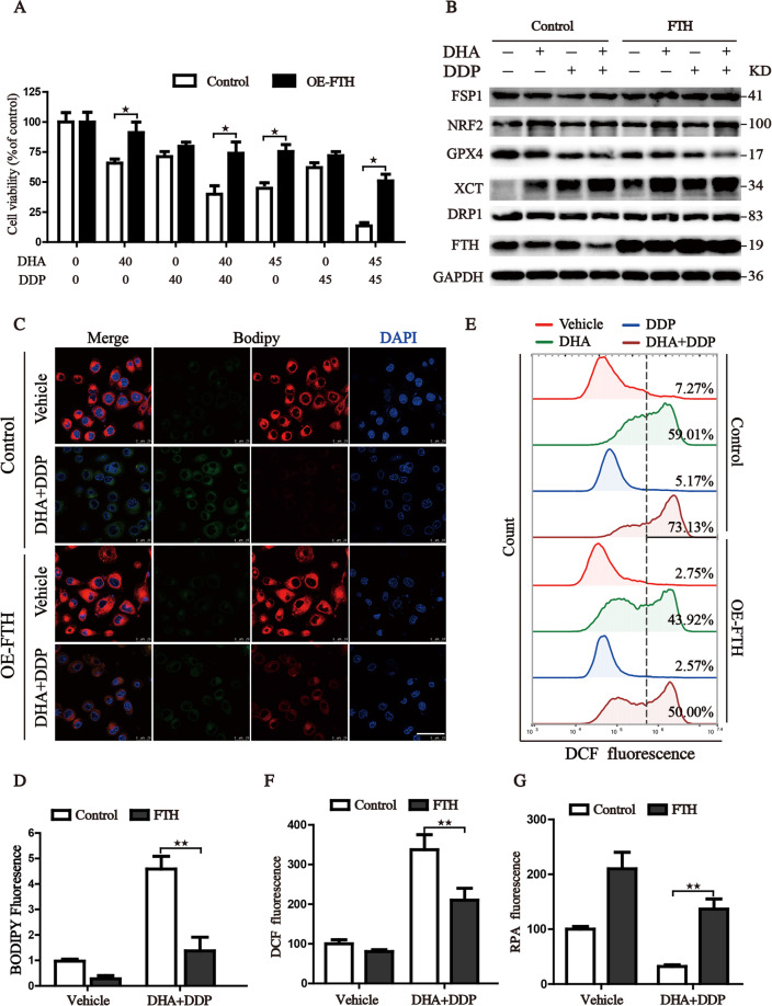
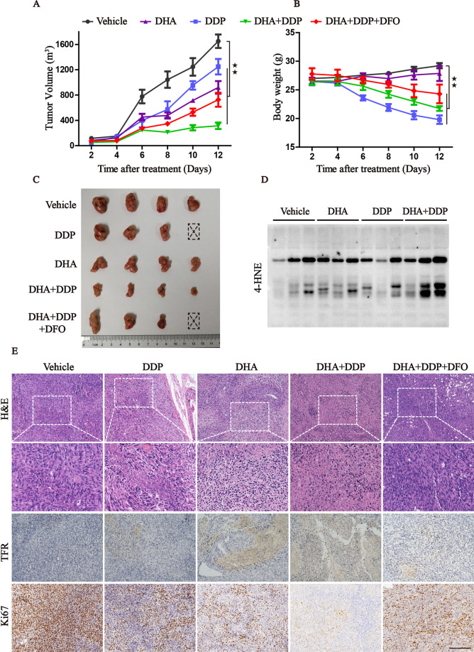
Pancreatic ductal adenocarcinoma (PDAC) is an extremely lethal cancer with limited treatment options. Cisplatin (DDP) is used as a mainstay of chemotherapeutic agents in combination with other drugs or radiotherapy for PDAC therapy. However, DDP exhibits severe side-effects that can lead to discontinuation of therapy, and the acquired drug resistance of tumor cells presents serious clinical obstacles. Therefore, it is imperative to develop a more effective and less toxic therapeutic strategy. We and others have previously discovered that dihydroartemisinin (DHA) represents a safe and promising therapeutic agent to preferentially induce cancer cell ferroptosis. In the present study, we find that DHA could intensively strengthen the cytotoxicity of DDP and significantly reduce its effective concentrations both in vitro and in vivo. Combination of DHA and DDP synergistically inhibits the proliferation and induces DNA damage of PDAC cells. Mechanically, the combinative treatment impairs mitochondrial homeostasis, characterized by destroyed mitochondrial morphology, decreased respiratory capacity, reduced ATP production, and accumulated mitochondria-derived ROS. Further studies show that ferroptosis contributes to the cytotoxic effects in PDAC cells under the challenge of DHA and DDP, together with catastrophic accumulation of free iron and unrestricted lipid peroxidation. Moreover, pharmacologic depleting of the free iron reservoir or reconstituted expression of FTH contributes to the tolerance of DHA/DDP-induced ferroptosis, while iron addition accelerates the ferroptotic cell death. In summary, these results provide experimental evidence that DHA acts synergistically with DDP and renders PDAC cells vulnerable to ferroptosis, which may act as a promising therapeutic strategy.


Conflict of interest statement

The authors declare no competing interests.

Figures

Fig. 1
Fig. 1. Synergistic antitumor effect of DHA in combination with DDP.
A, B Pancreatic cancer cells were subjected to different concentrations of DHA and DDP treatment for 24 h and cell viability was detected by CCK-8 assay. Combination index (CI) values were calculated by Compusyn software. C PANC1 cells were subjected to 40 µM DHA or/and DDP treatment for 24 h and followed by Annexin V-FITC/PI assay. D PANC1 cells were pretreated with 40 µM DHA or/and DDP for 24 h and subjected to the Calcein-AM/PI staining (Calcein AM: live cells, PI: dead cells). Scale bar: 100 µm. E, F The colony formation assay of PANC1 and SW1990 cells were performed under mono or combination treatment of DHA (5 µM for PANC1 or 10 µM for SW1990) and DDP (5 µM for PANC1 or 10 µM for SW1990) for 7 days. The representative images and the corresponding quantitative histograms from three independent experiments are shown (values represented mean ± SD. P < 0.05, ★★P < 0.01 versus control).
Fig. 2
Fig. 2. Combinative treatment of DHA and DDP inhibits proliferation and induces DNA damage in pancreatic cancer cells.
A, B Cell proliferation of PANC1 was detected by 5-ethynyl-2′-deoxyuridine (EdU) incorporation assay under mono or combination treatment of DHA and DDP for 12 h. Representative images and statistical histograms are shown. Scale bar: 100 µm. C, D PANC1 cells were treated with mono or combination of DHA and DDP for 12 h, cell cycle distribution was determined by flow cytometry. E, F PANC1 cells were treated with mono or combination of DHA and DDP for 12 h and stained with γ-H2AX antibody. DAPI was used for nucleus staining. Images were acquired with confocal laser scanning microscopy. Quantification of γ-H2AX immunofluorescence was shown on the right. Scale bar: 10 µm. P < 0.05, ★★P < 0.01 versus control.
Fig. 3
Fig. 3. DHA co-treatment with DDP synergistically impairs mitochondrial homeostasis.
A, B Observation of the changes in mitochondrial morphology. The treated PANC1 cells were stained with MitoTracker probe (100 nM) and DAPI (10 μg/mL) and then photographed by confocal laser microscope. Scale bars: 10 μm. C, D Mitochondrial oxygen consumption rate (OCR) was carried out with a Seahorse analyzer after the sequential addition of oligomycin, FCCP, and Antimycin A/ Rotenone. The OCR values of maximal respiration, ATP and non-mitochondrial oxygen consumption were normalized to the basal respiration. E, F Flow cytometry was performed to measure mitochondrial ROS by mitoSOX probe (3 μM) (values represented mean ± SD. P < 0.05, ★★P < 0.01 versus control).
Fig. 4
Fig. 4. Ferroptosis contributed to the cytotoxic effects in PDAC cells under the treatment with DHA and DDP.
A Cell viability was detected by CCK8 assay under treatment of DHA and DDP (45 μM) in the presence or absence of several cell death inhibitors. B, C Flow cytometry was performed to measure cellular ROS, and quantitation of fluorescence intensities was shown. D, E Representative images of BODIPY staining were photographed by confocal laser microscope with the designed treatment. Scale bars: 50 μm. Statistical results of the fluorescence intensities were shown on the right. FH Immunofluorescence staining of MDA and 4-HNE were captured by confocal laser microscope with the designed treatment. Scale bars: 50 μm. Statistical results of the fluorescence intensities were shown on the right (values represented mean ± SD. P < 0.05, ★★P < 0.01 versus control).
Fig. 5
Fig. 5. Iron accumulation in pancreatic cancer cells induced by the combination treatment of DHA and DDP.
A, B Intracellular Fe2+ was measured by the staining of RPA and photographed by the confocal microscope after indicated treatment. Scale bars: 50 μm. C, D Detection of cell surface TFR levels by immunofluorescence staining of PANC1 cells after indicated treatment. E The expression of iron metabolism proteins in DHA- and DDP-treated cells, which was determined by western blot assay (values represented mean ± SD. P < 0.05, ★★P < 0.01 versus control).
Fig. 6
Fig. 6. Pharmacologic depleting of the free iron reservoir attenuates the DHA/DDP-induced ferroptosis.
A PANC1 cells were treated with DHA/DDP (45 μM) in the presence or absence of DFO. B PANC1 cells were treated with DHA/DDP (30 μM) in the presence or absence of DFO and Fe2+, cell survival was detected by CCK8. C, D After indicated treatment, PANC1 cells were loaded with RPA or DCF-DA probe for 30 min, and fluorescence intensities were detected by microplate spectrophotometer and normalized to the corresponding cell number. E MitoTracker Red labeled pancreatic cancer cells were subjected to the confocal microscope for observing the changes of mitochondrial morphology after the indicated treatment. Scale bars: 10 μm. F, G OCR of PANC1 cells was carried out with a Seahorse analyzer after the addition of oligomycin, FCCP, and Antimycin A/Rotenone. The basal respiration and ATP production were calculated on the right. H, I To assess lipid ROS production, pancreatic cancer cells were treated with DHA and DDP with or without DFO, fer-1. Treated cells were loaded with BODIPY C11 probe for 30 min followed by confocal laser microscope. Scale bars: 50 μm. Statistical results of the fluorescence intensities were shown on the right (values represented mean ± SD. P < 0.05, ★★P < 0.01 versus control).
Fig. 7
Fig. 7. Reconstituted expression of FTH contributes to the tolerance of DHA/DDP-induced ferroptosis.
A PANC1 cells were treated with various concentrations of DHA and DDP for 24 h, followed by CCK8 assay. B Proteins isolated from the treated cells were assayed by western blot to detect the expression of ferroptosis-related proteins. C, D Indicated cells were stained with BODIPY C11 probe for the assessment of lipid ROS production and observed under the confocal laser microscope. Scale bars: 50 μm. Cells with indicated treatment were stained with RPA and DCF-DA probe for 30 min and followed by flow cytometry to assess intracellular Fe2+ and ROS level (EG) (values represented mean ± SD. P < 0.05, ★★P < 0.01 versus control).
Fig. 8
Fig. 8. The combination of DHA and DDP inhibits the growth of xenograft in vivo.
A, B Xenografts were established in mice and treated with vehicle, DHA, DDP, DHA/DDP, and DHA/DDP plus DFO, respectively. The tumor volume and body weight were measured every 2 days. C The dissected xenografts were photographed at the end of the experiment. D Western blots analysis for 4-HNE on lysates isolated from xenografts in different groups. E H&E staining and IHC analysis for TFR, Ki67 in indicated tumor specimens. Scale bars: 200 μm (values represented as mean ± SD. ★★P < 0.01 versus control).

References

    1. Hingorani SR, Petricoin EF, Maitra A, Rajapakse V, King C, Jacobetz MA, et al. Preinvasive and invasive ductal pancreatic cancer and its early detection in the mouse. Cancer Cell. 2003;4:437–50. doi: 10.1016/S1535-6108(03)00309-X. - DOI - PubMed
    1. Bryant KL, Mancias JD, Kimmelman AC, Der CJ. KRAS: feeding pancreatic cancer proliferation. Trends Biochem Sci. 2014;39:91–100. doi: 10.1016/j.tibs.2013.12.004. - DOI - PMC - PubMed
    1. Buscail L, Bournet B, Cordelier P. Role of oncogenic KRAS in the diagnosis, prognosis and treatment of pancreatic cancer. Nat Rev Gastroenterol Hepatol. 2020;17:153–68. doi: 10.1038/s41575-019-0245-4. - DOI - PubMed
    1. Tsai LH, Hsu KW, Chiang CM, Yang HJ, Liu YH, Yang SF, et al. Targeting interleukin-17 receptor B enhances gemcitabine sensitivity through downregulation of mucins in pancreatic cancer. Sci Rep. 2020;10:17817. doi: 10.1038/s41598-020-73659-z. - DOI - PMC - PubMed
    1. Siegel RL, Miller KD, Jemal A. Cancer statistics, 2019. CA Cancer J Clin. 2019;69:7–34. doi: 10.3322/caac.21551. - DOI - PubMed

Publication types

MeSH terms