. 2021 Jul 14;12(1):4308.
doi: 10.1038/s41467-021-24631-6.

The HIF target MAFF promotes tumor invasion and metastasis through IL11 and STAT3 signaling

Affiliations

The HIF target MAFF promotes tumor invasion and metastasis through IL11 and STAT3 signaling

Eui Jung Moon et al. Nat Commun. .

Abstract

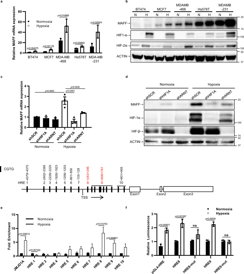
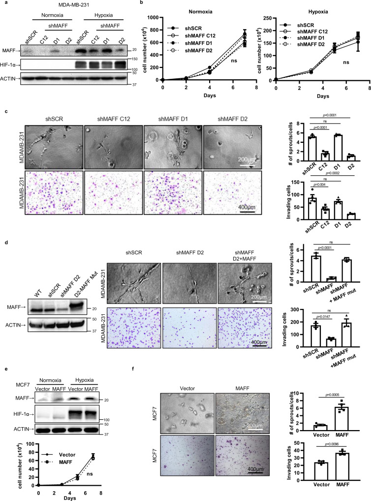
Hypoxia plays a critical role in tumor progression including invasion and metastasis. To determine critical genes regulated by hypoxia that promote invasion and metastasis, we screen fifty hypoxia inducible genes for their effects on invasion. In this study, we identify v-maf musculoaponeurotic fibrosarcoma oncogene homolog F (MAFF) as a potent regulator of tumor invasion without affecting cell viability. MAFF expression is elevated in metastatic breast cancer patients and is specifically correlated with hypoxic tumors. Combined ChIP- and RNA-sequencing identifies IL11 as a direct transcriptional target of the heterodimer between MAFF and BACH1, which leads to activation of STAT3 signaling. Inhibition of IL11 results in similar levels of metastatic suppression as inhibition of MAFF. This study demonstrates the oncogenic role of MAFF as an activator of the IL11/STAT3 pathways in breast cancer.


Conflict of interest statement

The authors declare no competing interests.

Figures

Fig. 1
Fig. 1. Hypoxia-induced MAFF regulates tumor invasion and acts as a prognostic indicator.
a 50 hypoxia-inducible genes were identified from RCC4-VHL, a clear-cell renal cell carcinoma line with reconstituted VHL, using microarray analysis. TCGA data from cbioportal showed that these genes were altered in most of tumor cases, specifically in breast cancer. b Dataset from GEO showed that combined expression of 50 hypoxia genes were significantly correlated with overall survival in breast cancer patients (Log-rank test, p = 0.0039, GSE19783, n = 56 with low expression, n = 56 with high expression). c DAVID gene functional classification tool, classified 50 hypoxia-inducible genes into functional groups, which are correlated with “Glucose Metabolism”, “Regulation of apoptosis”, and “Positive regulation of cell migration”. d RNAi screening was performed to determine genes regulating cell invasion using endoribonuclease prepared siRNA (esiRNA) libraries (MilliporeSigma). Collagen assay with tdTomato labeled MDA-MB-231 cells showed that MAFF regulates tumor cell invasion the most among 50 hypoxia-inducible genes (bar = 500μm) n = 3 biological replicates. One-Way ANOVA with multiple comparisons. p < 0.0001. e MAFF expression on breast cancer patient tissues was evaluated by staining tissue microarrays. When compared to primary tumor tissues using ImageJ, lymph node (LN) metastatic tissues showed higher intensity of MAFF expression (n = 46 for primary tissues, n = 36 for LN metastatic tissues). One-Way ANOVA with multiple comparisons. p < 0.0001. f While MAFF expression in breast cancer patients significantly correlated with hypoxia signatures (Log-rank test, p < 0.0001, R2 = 0.314), g it was also a prognostic indicator for overall survival (OS) and distant metastasis-free survival (DMFS) in all or basal-like breast cancer patients (OS: GSE42568, n = 52 with low MAFF expression, n = 52 with high MAFF expression, p = 0.0029, DMFS: KM Plotter, n = 117 with low MAFF expression, n = 115 with high MAFF expression, p = 0.0190). Log-rank test. Graphs represent the mean per group and error bars represent the SEM.
Fig. 2
Fig. 2. MAFF induction under hypoxia is regulated by HIF-1.
a MAFF mRNA expression was elevated under hypoxia in breast cancer cell lines. n = 3 biological replicates. Unpaired t-test. b While basal expression of MAFF was different, hypoxia increased MAFF expression in all breast cancer cell lines. c, d Knocking down HIF1A or ARNT (HIFβ) prevented MAFF mRNA and protein induction under hypoxia. One-Way ANOVA with multiple comparisons. e ChIP assay was performed to identify a HIF-1 binding site on the MAFF promoter (HRE8 and 9) (TSS: transcription start site). n = 4 biological replicates. Unpaired t-test. f Luciferase reporter assays showed that transcription activity of HRE8 or HRE9 was increased under hypoxia compared to mutated HRE8 or HRE9. pGL4-HRE, which contains 4x HRE regions, was used as a positive control. n = 3 biological replicates. Unpaired t-test. Graphs represent the mean per group and error bars represent the SEM. ns: not significant.
Fig. 3
Fig. 3. MAFF regulates tumor cell invasion.
a MAFF was knocked down using three different clones of shRNA against MAFF. Western blot data showed that while two of clones (C12 and D2) decreased MAFF expression significantly, one clone had little effect on MAFF expression (D1), which we decided to use as a negative control. b MAFF inhibition using shRNA did not affect tumor cell growth in MDA-MB-231 cells both under normoxia and hypoxia. n = 3 biological replicates. Unpaired t-test. c MAFF knockdown in MDA-MB-231 cells decreased tumor cell invasion through collagen (upper panels, n = 3 biological replicates) or Matrigel-coated transwell membranes (lower panels, n = 4 biological replicates). Quantification is included in graphs. One-Way ANOVA with multiple comparisons. d MAFF knockdown was rescued by overexpressing MAFF cDNA with mutations in the shRNA targeting region. Decreased tumor cell invasion by MAFF inhibition was reactivated when MAFF was re-expressed (mut: mutant). Tumor cell invasion was determined using collagen assay (upper panels) or Matrigel-coated transwell membrane assay (lower panels). n = 3 biological replicates. One-Way ANOVA with multiple comparisons. e MAFF overexpression in MCF7 cells did not show any significant difference in cell growth rates (ns: not significant). n = 3 biological replicates. Unpaired t-test. f Increased invasive phenotypes were observed in MAFF overexpressing MCF7 using collagen assay (upper panels, n = 3 biological replicates) and transwell invasion assay (lower panels, n = 4 biological replicates). Unpaired t-test.(Graphs represent the mean per group and error bars represent the SEM. ns: not significant).
Fig. 4
Fig. 4. MAFF regulates in vivo tumor metastasis.
MDA-MB-231 cells were orthotopically injected into mammary fat pads of nude mice (106 cells/50 μl/mouse) to determine the role of MAFF in tumor growth. a MAFF inhibition did not affect tumor growth in vivo (ns: not significant). n = 9 shSCR, n = 7 shMAFF. Two-Way ANOVA. b While cell proliferation (Ki67 positive staining) was not changed, microvessel density (MECA32 positive staining) significantly decreased when MAFF was inhibited (n = 4 shSCR, n = 5 shMAFF). Unpaired t-test. c Lung colonization of tumor cells was evaluated either by H&E staining (n = 4) or qPCR to measure human GAPDH expression (n = 9 shSCR, n = 7 shMAFF). Unlike primary tumor growth, MAFF knockdown reduced lung metastasis. Unpaired t-test. d Intraperitoneal injection of OVCAR8 cells also showed that inhibition of MAFF expression was significantly correlated with reduced tumor metastasis. Arrow indicates tumor nodules formed in intraperitoneal cavity. n = 6 shSCR, n = 8 shMAFF. Unpaired t-test. Graphs represent the mean per group and error bars represent the SEM. ns: not significant.
Fig. 5
Fig. 5. MAFF target gene identification by genome-wide analysis.
MDA-MB-231 cells were treated under normoxia or hypoxia with or without siMAFF for 24 h. MAFF target genes were determined by analyzing RNA-sequencing and ChIP-sequencing data. a When MAFF was knocked down, 415 or 240 genes were significantly altered under normoxia or hypoxia, respectively and among them 106 genes were affected both under normoxia and hypoxia. b Volcano plot showed that 192 genes were downregulated and 223 genes were upregulated under normoxia. Under hypoxia, 68 genes were downregulated and 172 genes were upregulated. c DAVID, analysis determined that genes involved in pathways of “Blood vessel development”, “TGFβ signaling”, and “Regulation of cell adhesion/morphogenesis” were significantly altered in the absence of MAFF expression. d GSEA analysis revealed that genes involved in metastasis (normoxia) or migration (hypoxia) were regulated by MAFF expression. e The genomic location of MAFF binding sites was identified by cis-regulatory element annotation system (CEAS). f A MAFF binding motif based on MARE was identified using the motif discovery algorithm, Multiple Expectation Maximizations for Motif Elicitation (MEME). g When we combined RNA-sequencing and ChIP-sequencing, 292 genes were regulated under normoxia and 70 genes under hypoxia. 66 genes were regulated both under normoxia and hypoxia. h ChIP-sequencing data showed enriched DNA sequence tags, which were highlighted in red using the UCSC genome browser. While HMOX1 showed two peaks from previously known MAFF binding sites, MAFF binding peak was found in IL11 promoter, which included the MARE/ARE sequence (GCGAGCTCA).
Fig. 6
Fig. 6. MAFF-mediated IL11 regulates tumor cell invasion.
a Knocking down IL11 significantly decreased invasive phenotypes of MDA-MB-231 through collagen (upper panels) and Matrigel-coated transwell membranes (lower panels). n = 3 biological replicates, Unpaired t-test. b The rescue effect of IL11 expression on decreased tumor cell invasion was evaluated in MDA-MB-231 cells with or without MAFF knockdown. n = 3 (collagen assay, upper panels) or 4 (transwell membrane assay, lower panels) biological replicates. One-Way ANOVA with multiple comparisons. c STAT3 activation was evaluated in MDA-MB-231 cells treated under normoxia or hypoxia by Western blotting. Hypoxia increased phospho-STAT3 (Tyr705), while MAFF knockdown inhibited its induction both under normoxia and hypoxia. d, e The downstream effect of IL11 on STAT3 activation was determined after IL11 was knocked down (d) or overexpressed with or without MAFF inhibition (e). Phospho-STAT3 expression was investigated by Western blotting. f, g Endothelial tube formation assay was performed using GFP-labeled HUVEC cells. Cell culture media were transferred from MDA-MB-231 cells infected with IL11 Cas9-CRISPR (f) or MAFF shRNA with or without IL11 overexpression (g). n = 3 biological replicates, Unpaired t-test (f) or One-Way ANOVA with multiple comparisons (g). Graphs represent the mean per group and error bars represent the SEM.
Fig. 7
Fig. 7. MAFF binds to BACH1 to activate IL11-STAT3 pathways.
a MAFF binding proteins were immunoprecipitated using MAFF antibody and analyzed by mass spectrometry. In MDA-MB-231 cells, BACH1 was a main binding partner of MAFF. The graph indicates the unique peptide number identifying MAFF binding compared to IgG control. n = 3 biological replicates. Unpaired t-test. b Co-immunoprecipitation confirmed the binding of MAFF and BACH1, which was further increased under hypoxia. c When BACH1 was knocked down, mRNA expression of IL11 was decreased. n = 3 biological replicates. Unpaired t-test. d The downstream effect of BACH1 on the STAT3 pathway was determined by western blotting after BACH1 was knocked down in MDA-MB-231 both under normoxia and hypoxia. e ChIP-qPCR confirmed that MAFF and BACH1 bindings on the MARE sequence of IL11 promoter, which was identified from ChIP-sequencing. Bindings were further increased under hypoxia. Unpaired t-test. f Luciferase assay was performed using pGL4-luciferease vector, which was subcloned with the identified MARE sequence on IL11 promoter. While hypoxia increased the luciferase activity, BACH1 or MAFF knockdown decreased the transcriptional activity of the identified MARE. n = 3 biological replicates. Unpaired t-test. Knocking down BACH1 or both MAFF and BACH1 decreased tumor cell invasion through Matrigel coated transwells (g) and tube formation of HUVEC representing angiogenic features (h) (CM: cell media). However, IL11 overexpression rescued the reduction in cell invasion and tube formation. n = 3 biological replicates. One-Way ANOVA with multiple comparisons. Graphs represent the mean per group and error bars represent the SEM.
Fig. 8
Fig. 8. MAFF enhances tumor metastasis through IL11/STAT3 pathways in vivo and their combined expression predicts patient survival.
Spontaneous lung metastasis model of MDA-MB-231 was studied by injecting 2 × 106 cells into the mammary fat pad of NSG mice (2 × 106 cells/75 μl/mouse). a In MAFF knockdown group, spontaneous lung metastasis was significantly decreased while IL11 overexpression rescued the reduction. Lung weight was measured right after sacrificing mice. Also, lung tumor area was determined using ImageJ after H&E staining (n = 10 for shSCR and shMAFF, n = 9 for shMAFF+IL11). One-Way ANOVA with multiple comparisons. b IL11 and IL6 levels in mouse serum were determined using ELISA. While IL11 was decreased with MAFF knockdown, elevated IL11 levels were confirmed in shMAFF with IL11 overexpression group. However, IL6 levels remained unchanged in every group (n = 10 for shSCR, shMAFF, n = 7 or 6 for shMAFF+IL11). Unpaired t-test. c, d Expression of phospho-STAT3 and MECA32 were evaluated from primary tumor tissues and we observed consistent changes with in vitro studies suggesting that MAFF and IL11 pathways regulated phospho-STAT3 and angiogenesis (n = 10 for shSCR, shMAFF, n = 9 for shMAFF+IL11). Arrows indicate positive MECA-32 staining for vessels. One-Way ANOVA with multiple comparisons. e Distant metastasis-free survival of breast cancer patients was evaluated using KM plotter. Combined expression of MAFF and IL11, MAFF, and BACH1, or MAFF, IL11, and BACH1 showed the correlation with patient survival, indicating prognostic significance of these factors. Log-rank. Graphs represent the mean per group and error bars represent the SEM. ns: not significant.
Fig. 9
Fig. 9. Summary of hypoxia-induced MAFF and its role in tumor invasion and metastasis through IL11/STAT3 pathways.
Hypoxia-induced MAFF binds to BACH1 to transcriptionally activate IL11. Increased IL11 leads to elevated phosphorylation of STAT3, which results in enhanced tumor invasion, angiogenesis, and metastasis.

References

    1. Brown JM, Giaccia AJ. The unique physiology of solid tumors: opportunities (and problems) for cancer therapy. Cancer Res. 1998;58:1408–1416. - PubMed
    1. Moon EJ, Brizel DM, Chi JT, Dewhirst MW. The potential role of intrinsic hypoxia markers as prognostic variables in cancer. Antioxid. Redox Signal. 2007;9:1237–1294. doi: 10.1089/ars.2007.1623. - DOI - PubMed
    1. Wilson WR, Hay MP. Targeting hypoxia in cancer therapy. Nat. Rev. Cancer. 2011;11:393–410. doi: 10.1038/nrc3064. - DOI - PubMed
    1. Semenza GL. Molecular mechanisms mediating metastasis of hypoxic breast cancer cells. Trends Mol. Med. 2012;18:534–543. doi: 10.1016/j.molmed.2012.08.001. - DOI - PMC - PubMed
    1. LaGory EL, Giaccia AJ. The ever-expanding role of HIF in tumour and stromal biology. Nat. Cell Biol. 2016;18:356–365. doi: 10.1038/ncb3330. - DOI - PMC - PubMed

Publication types

MeSH terms