. 2021 Jun 21:20:101482.
doi: 10.1016/j.jcot.2021.101482. eCollection 2021 Sep.

Comprehensive review of surgical approaches to the elbow

Affiliations

Comprehensive review of surgical approaches to the elbow

Saurabh Aggarwal et al. J Clin Orthop Trauma. .

Erratum in

  • Erratum regarding previously published articles.
    [No authors listed] [No authors listed] J Clin Orthop Trauma. 2021 Jul 30;20:101539. doi: 10.1016/j.jcot.2021.101539. eCollection 2021 Sep. J Clin Orthop Trauma. 2021. PMID: 34405084 Free PMC article.

Abstract

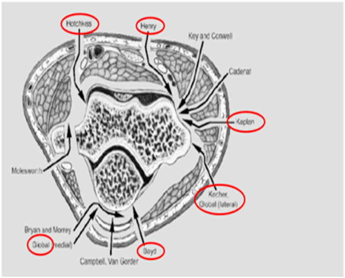
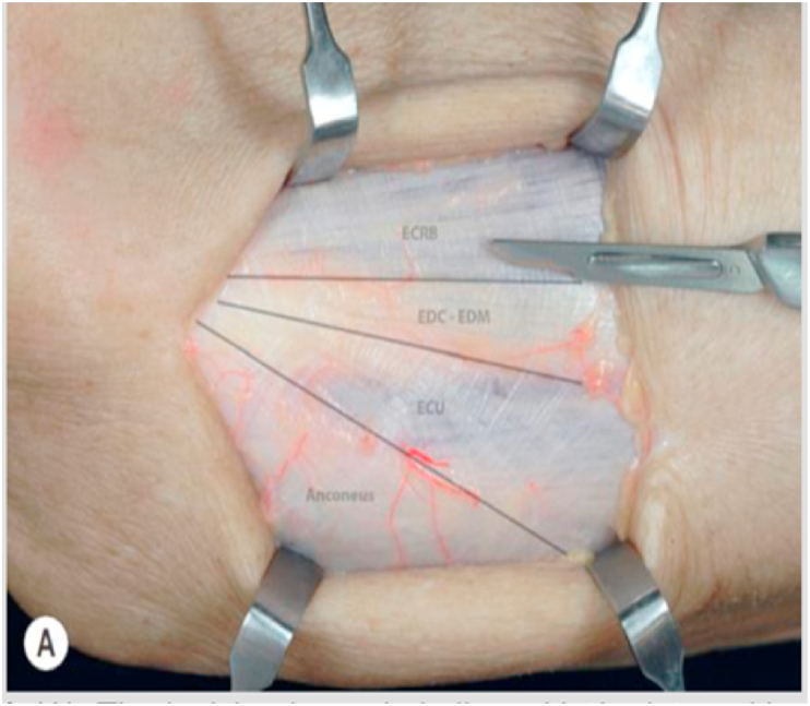
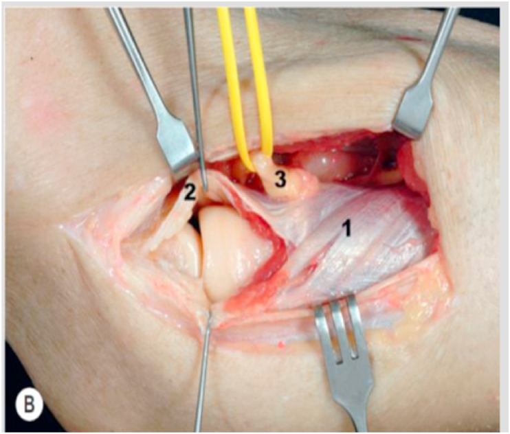
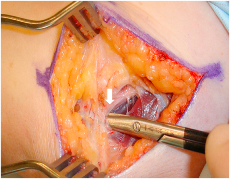
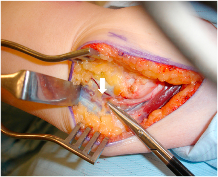
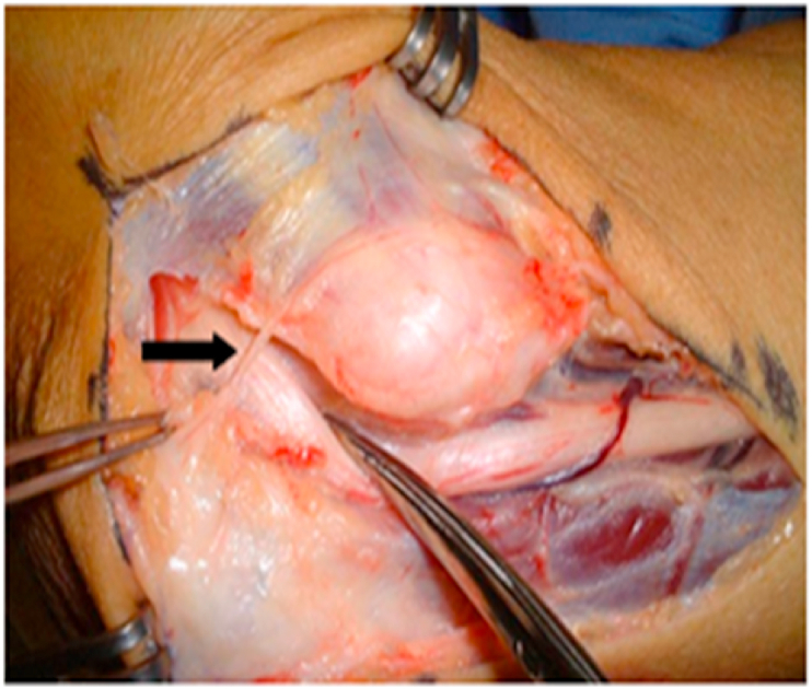
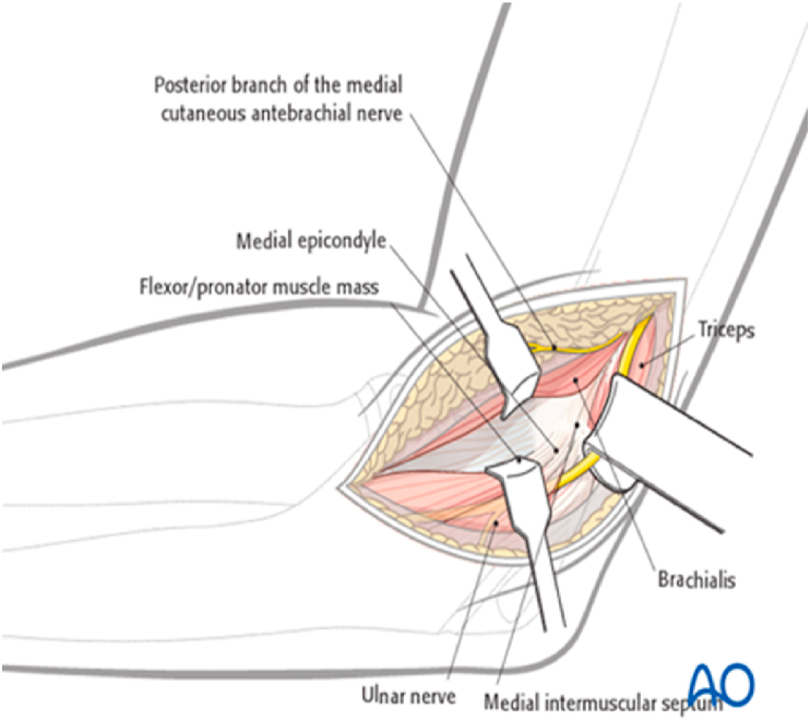
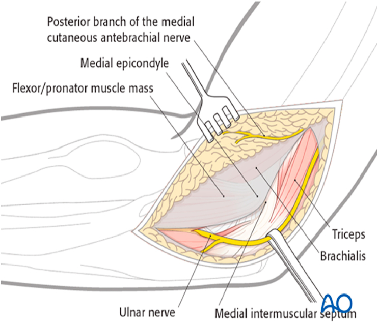
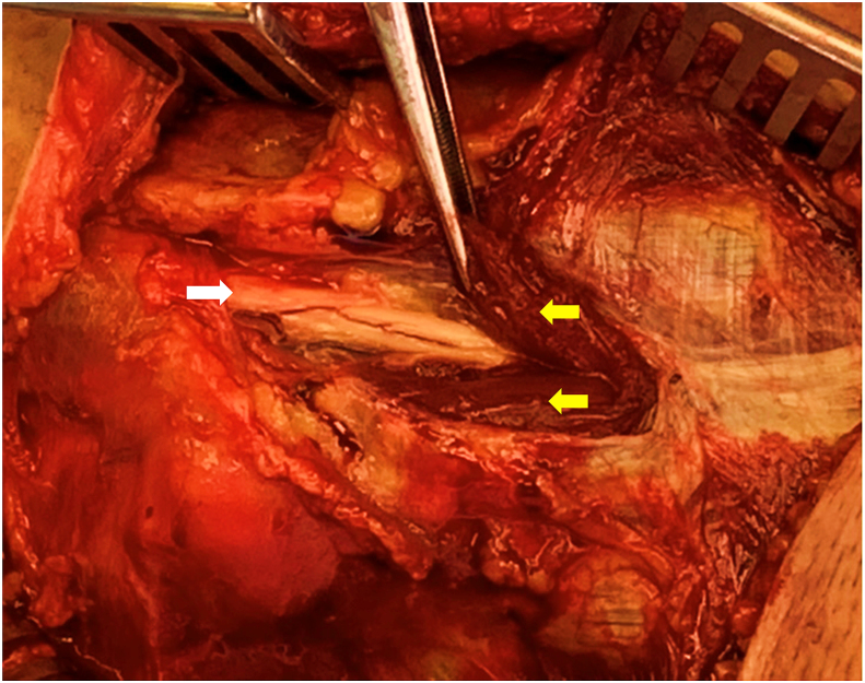
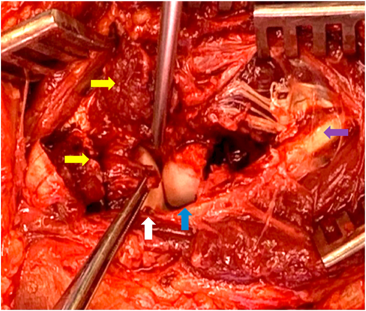
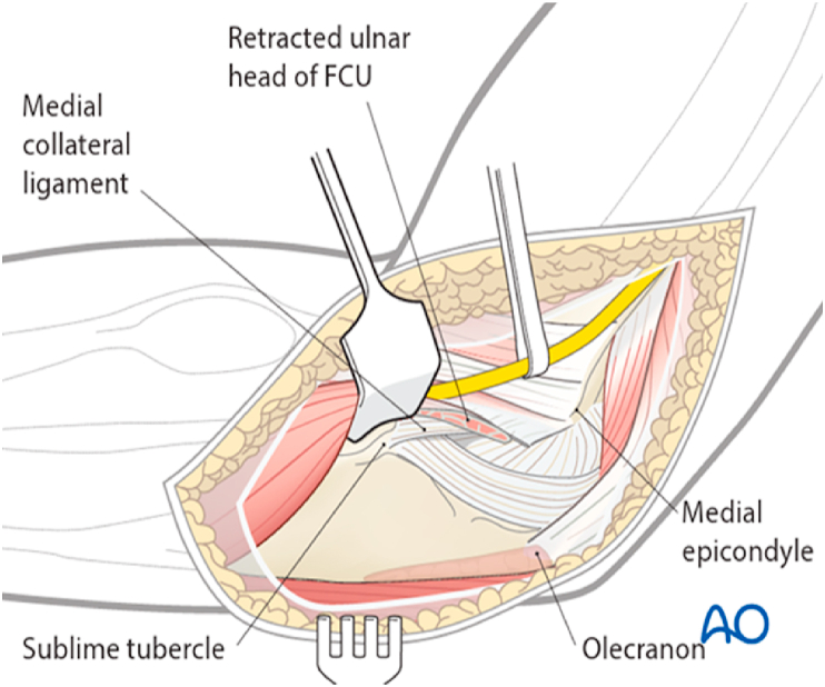
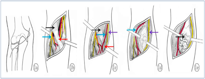
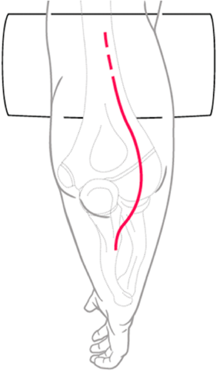
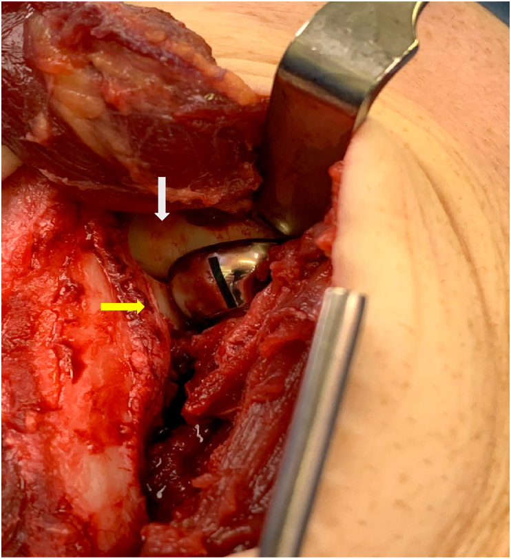
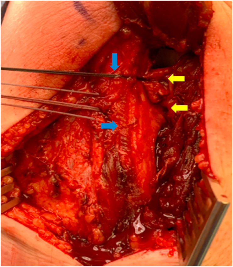
The choice of the most suitable surgical approach to the elbow forms the foundation of any successful elbow surgery. The surgical approach is based on the injury or pathology to be addressed and therefore specific anatomical details need to be considered. The surgeon must be comfortable with the bony, ligamentous and neurovascular anatomy of the elbow to consider and execute the best approach for each problem. This is an imperative to avoid iatrogenic injury. This article provides a detailed analysis, valuable technical tips, advantages and disadvantages of the most common approaches to the elbow. The lateral approaches include the Kocher, Kaplan and Extensor Digitorum Communis (EDC) Split approaches, the medial approaches include the Hotchkiss, Flexor carpi ulnaris (FCU) splitting approach, the Taylor and Scham approach. The anterior approach includes the anterior neurovascular interval approach and the posterior approaches include the Olecranon osteotomy, triceps sparing, triceps reflecting approach and finally the Boyd interval approach. The text and illustrations will provide a structured overview for the practicing surgeon.

Keywords: Anterior approach; Elbow approaches; Kaplan approach; Kocher approach; Olecranon osteotomy approach; Triceps on and triceps off approaches.


Figures

Fig. 1
Fig. 1
Overview of the surgical approaches of the elbow.
Fig. 2
Fig. 2
Modified kocher's approach where lateral elbow capsule is identified and incised anteriorly to the lateral ulna collateral ligament.
Fig. 3
Fig. 3
Black lines show the Kocher and EDC Split approach. The interval between the anconeus and extensor carpi ulnaris (ECU) can be identified by a thin strip of fat distally.
Fig. 4
Fig. 4
Shows that with a Kocher approach we can see the posterior half of the radial head where as with the EDC split approach we can see the anterior half of the radial head. C = capitellum, R = radial head, B = ECU + half of EDC, white arrow represents retracted anconeus, black arrow represents the split EDC.
Fig. 5
Fig. 5
Incision is made in line with the interval between the extensor carpi radialis brevis (ECRB) muscle and the extensor digitorum communis (EDC).
Fig. 6
Fig. 6
The underlying capsule shown with white arrow should be longitudinally incised, allowing access to the most anterior part of the radial head. Yellow arrow represents EDC, Red arrow represents ECRB.
Fig. 7
Fig. 7
Shows division of Osborne ligament shown by white arrow.
Fig. 8
Fig. 8
Shows 2 heads of FCU shown by white arrow.
Fig. 9
Fig. 9
Shows black arrow towards medial antebrachial cutaneous nerve.
Fig. 10
Fig. 10
Hotchkiss approach. Shows the top retractor retracting pronator teres and flexor carpi radialis. Bottom retractor is retracting palmaris longus and FCU.
Fig. 11
Fig. 11
FCU split approach.
Fig. 12
Fig. 12
White arrow shows ulna nerve and 2 yellow arrows show two heads of FCU.
Fig. 13
Fig. 13
White arrow shows anterior band of MCL avulsed from the sublime tubercle. 2 yellow arrows show cut surface of flexor muscle origin, purple arrow shows ulna nerve with preserved vascularity decompressed in situ through the two heads of FCU, blue arrow shows displaced anteromedial facet fracture.
Fig. 14
Fig. 14
Taylor and Scham approach - Shows ulna nerve in sling. Retractor is retracting the entire flexor pronator mass.
Fig. 15
Fig. 15
a shows S type incision across the anterior elbow crease. b red arrow shows bicipital aponeurosis covering brachial artery and median nerve. Black arrow shows biceps muscle, blue arrow shows radial nerve dividing into superficial radial nerve and posterior interosseous nerve. 15 c Blue arrow shows brachial artery, violet arrow shows median nerve, black arrow shows biceps, red arrow shows brachial artery dividing into radial and ulna artery. 15 d Shows split brachialis muscle. Blue arrow shows the retracted radial half of the brachialis muscle and brachial artery radially. Violet arrow shows median nerve and the medial half of the brachialis muscle retracted medially. 15 e shows exposure to the coronoid process and the trochlea after splitting the capsule. Black arrow points to the coronal shear fracture of trochlea which can be addressed using this approach.
Fig. 16
Fig. 16
Shows patient positioned laterally with a curved incision, curving laterally avoiding the ulna nerve.
Fig. 17
Fig. 17
Shows a chevron shaped osteotomy with complete retraction of the triceps along with the olecranon tip bringing a wide exposure to the distal posterior humerus. Bi column plate fixation seen. White arrow -chevron olecranon osteotomy. Black arrow - Ulna Nerve. Yellow arrow Radial nerve pointed by the forceps.
Fig. 18
Fig. 18
Shows plate fixation for olecranon osteotomy pointed by a yellow arrow. Forceps holding Anconeus muscle part of the triceps anconeus pedicle flap (TRAP flap) shown by white arrow.
Fig. 19
Fig. 19
Triceps split and triceps reflection both require a complete removal of triceps from its reflection both require a complete removal of triceps from its attachment from olecranon. For an olecranon osteotomy or triceps sparing approach the triceps attachment is left intact. Although with the osteotomy, the olecranon is completely removed from the ulna.
Fig. 20
Fig. 20
After isolation of ulnar nerve (1), the triceps (2) is subperiosteally detached from the posterior aspect of the humerus (3).
Fig. 21
Fig. 21
The triceps is dissected from its attachment on the olecranon (4) and is reflected laterally together with the anconeus (5).
Fig. 22
Fig. 22
Showing- Ulnar nerve (1), Triceps (2), humerus (3).
Fig. 23
Fig. 23
Patient is positioned in a lateral position. Incision is around 16–20 cm centred over the olecranon. After developing skin flaps, ulna nerve is identified and protected (1). Then triceps (2) is subperiosteally detached from the posterior aspect of the humerus (3) on both sides, preserving its insertion on the olecranon. Expose the elbow joint through the medial and lateral windows on each side of the triceps.
Fig. 24
Fig. 24
Shows reflected Anconeus muscle pointed by a white arrow, black arrow shows the supinator crest. Forceps is holding the annular ligament, capsule and the LUCL complex, red arrow points to radial head, violet arrow points to supinator muscle which is partially reflected.
Fig. 25
Fig. 25
Shows a radial head replacement in situ, yellow arrow points to lesser sigmoid notch. White arrow points to capitellum.
Fig. 26
Fig. 26
Shows transosseous suture repair pointed by yellow arrows to LUCL, annular ligament and capsule complex. White arrow shows green sutures representing Mitek anchor for annular ligament repair. Repair done over supinator crest as shown by a blue arrow.

References

    1. William D., Hakim L., Steven R., Whitcomb P J. Objective analysis of lateral elbow exposure with the extensor digitorum communis split compared with the kocher interval. J Bone Joint Surg Am. 2014;96:387–393. - PubMed
    1. Van Leeuwen D.H., Guitton T.H., Lambers K., Ring D. Quantitative measurement of radial head fracture location. J Shoulder Elbow Surg. 2012. Aug;21(8):1023. 7Epub 2011 Nov 09. - PubMed
    1. Schimizzi A., MacLennanA, Meier K.M., Chia B., Lw3rd Catalano, Gilckel S.Z. Defining a safe zone of dissecting during the extensor digitorum communis splitting approach to the proximal radius and forearm : an anatomical study. J Hand Surg Am. 2009 Sep;34(7):125. 5. - PubMed
    1. https://surgeryreference.aofoundation.org/orthopedic-trauma/adult-trauma...
    1. https://surgeryreference.aofoundation.org/orthopedic-trauma/adult-trauma...

LinkOut - more resources