

Review
doi: 10.1016/j.jcot.2021.101485.
eCollection 2021 Sep.
Clinical anatomy and biomechanics of the elbow
Affiliations
- PMID: 34262850
- PMCID: PMC8258984
- DOI: 10.1016/j.jcot.2021.101485
Item in Clipboard
Review
Clinical anatomy and biomechanics of the elbow
J Clin Orthop Trauma.
.
Erratum in
-
Erratum regarding previously published articles.J Clin Orthop Trauma. 2021 Jul 30;20:101539. doi: 10.1016/j.jcot.2021.101539. eCollection 2021 Sep. J Clin Orthop Trauma. 2021. PMID: 34405084 Free PMC article.
Abstract
The anatomy of the elbow joint had been studied extensively over the last 2 decades. The increased understanding of the anatomy and contribution of the anatomical structures to the elbow biomechanics had enabled surgeons to improve the results of surgical reconstruction and fracture fixation. This review articles intend to summarise the salient functional and clinical anatomical and relevant biomechanical data that had been published recently.
Keywords: Anatomy; Biomechanics; Elbow.
© 2021 Delhi Orthopedic Association. All rights reserved.
Figures
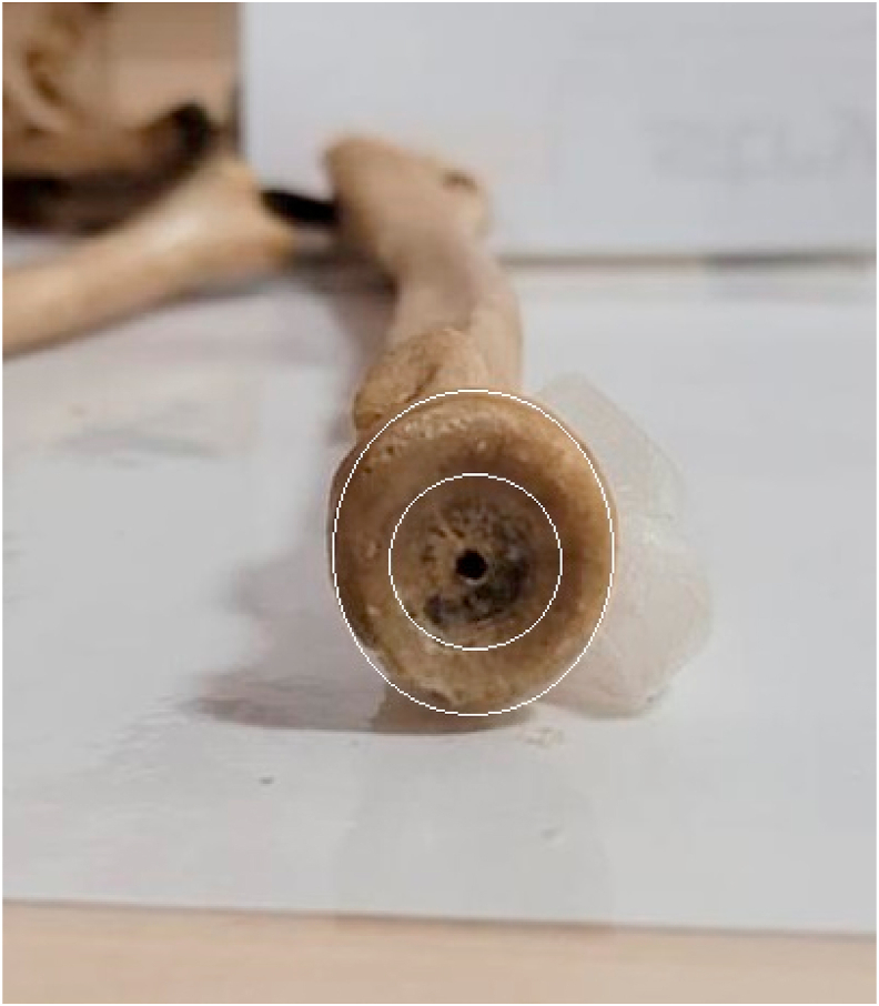
Showing apex posterior, anterior angulated distal humerus on sagittal plane with the capitellum flexed anteriorly by approximately 30–40°. Centre drill hole on the capitellum is the site of origin of the lateral collateral ligament complex.
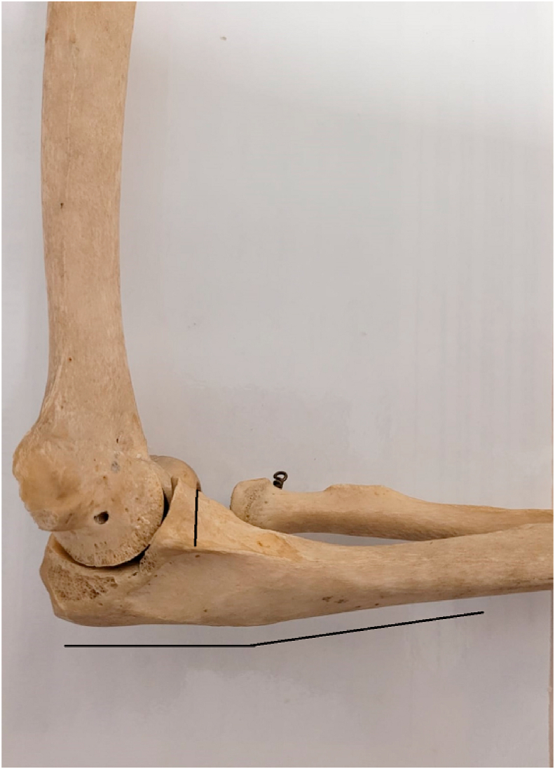
Showing the valgus angulation of articular segment relative to the shaft and the trochlear notch angle (angulated line), of average 142°.
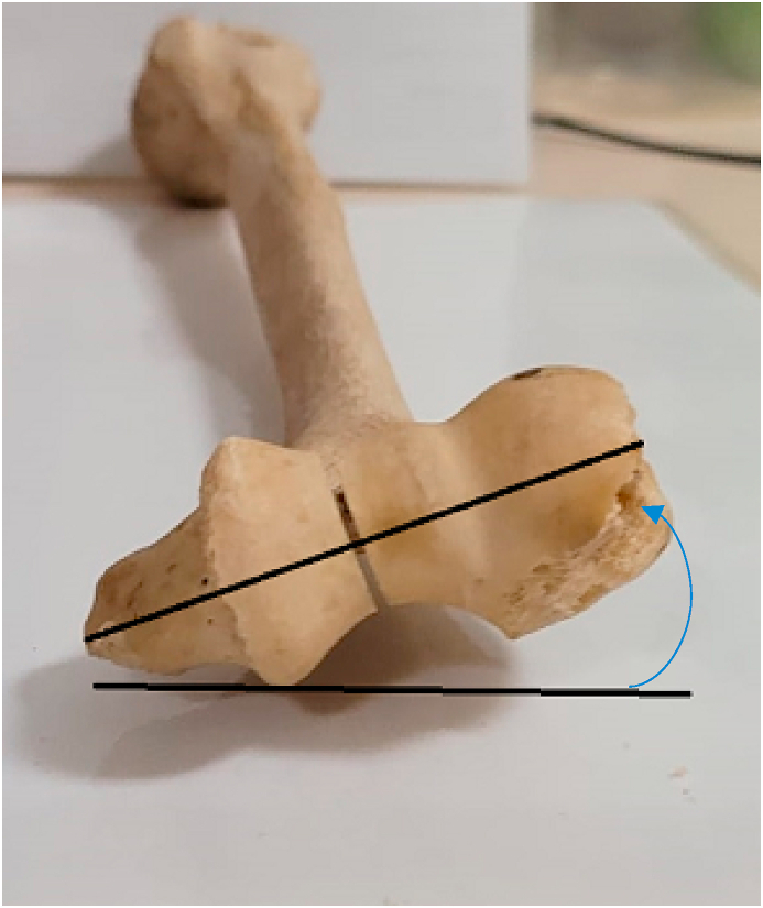
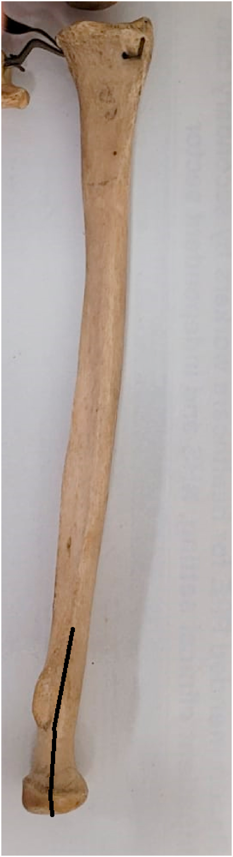
The distal humeral articular segment and the trans-epicondylar line (oblique line) are internally rotated (arrow) relative to the posterior flat surface of the humeral shaft (horizontal line).
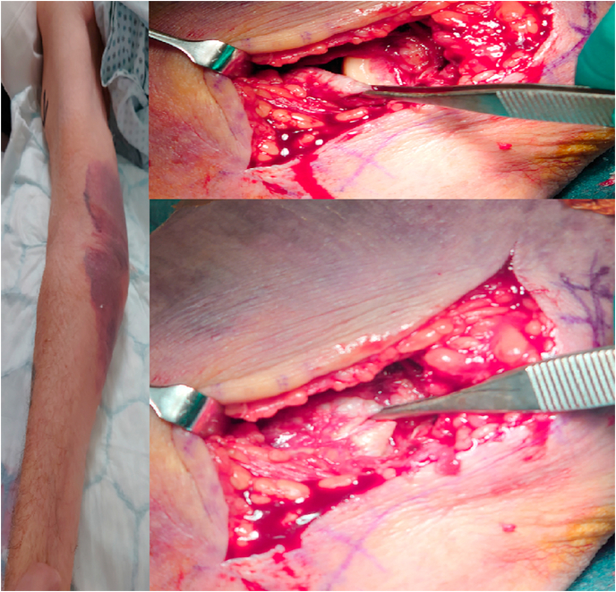
Showing MCL rupture from its attachment at the inferior medial epicondyle in a patient with posteromedial elbow dislocation. The forceps is holding the ruptured ligament exposing the origin in top right photo and opposition of the ligament back to the origin in the bottom right photo.
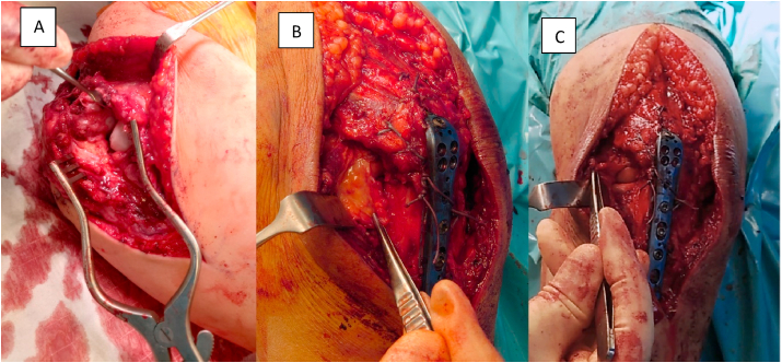
Showing rare rupture of the LUCL from its attachment to the supinator crest(A). forceps holding the ruptured ligament(B), reflected ligament revealing the radial head and capitellum (C).
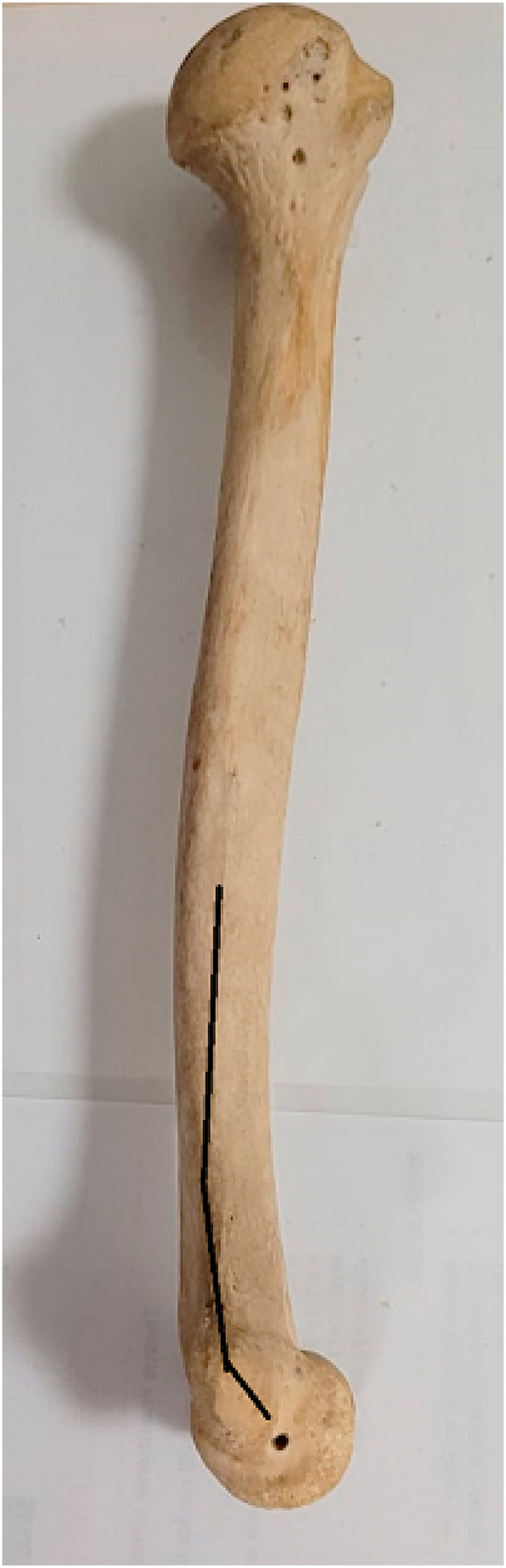
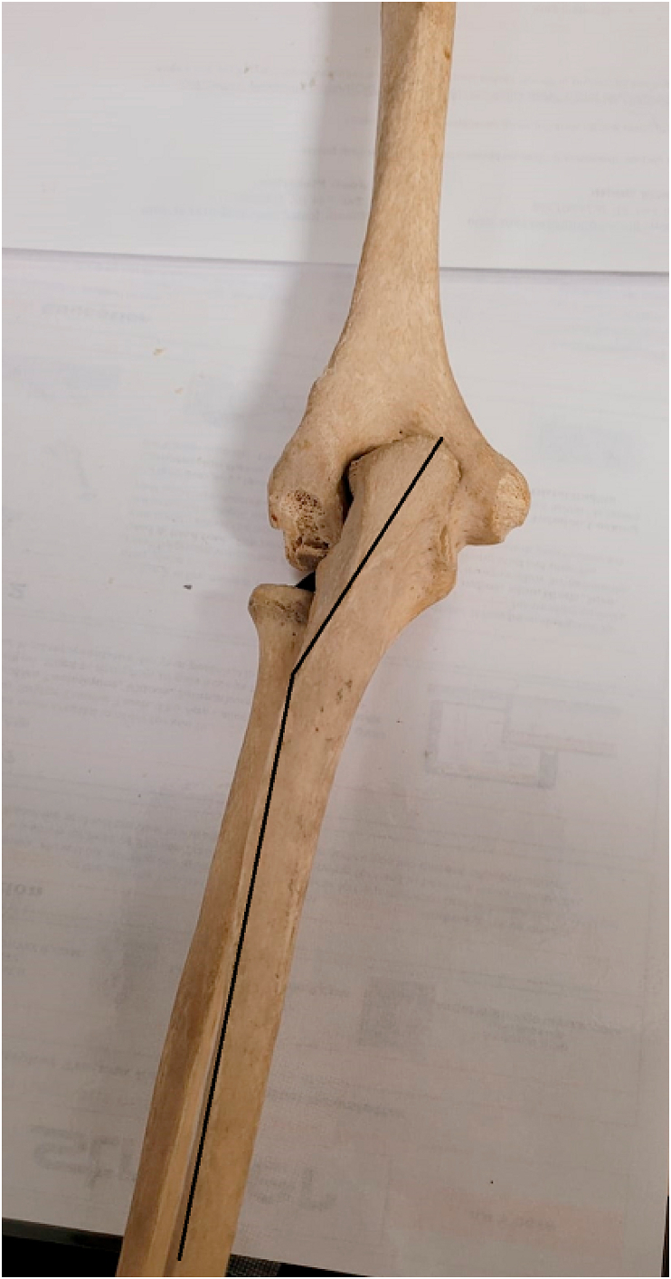
Showing the PUDA (horizontal lines); vertical line showing the coronoid height with the sublime tubercle at the bottom of the line.
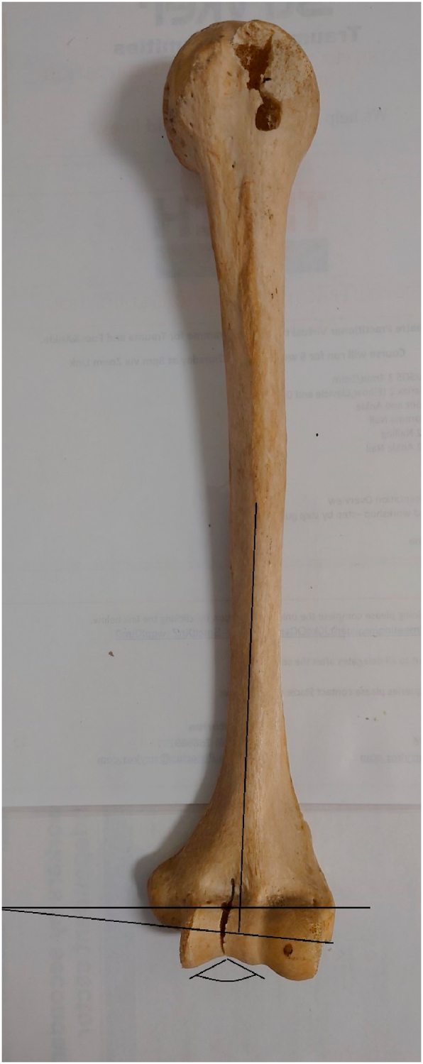
Showing varus angulation of the proximal ulna.
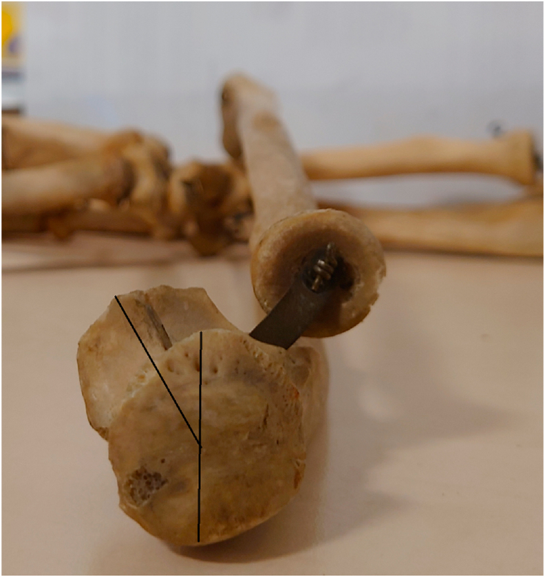
Showing torsional angle of the olecranon relative to the coronoid.
Showing maximum and minimum diameter of the elliptical radial head.
Showing the neck-shaft and neck-head angles.
References
-
- Brownhill J.R., King G.J., Johnson J.A. Morphologic analysis of the distal humerus with special interest in elbow implant sizing and alignment. J Shoulder Elbow Surg. 2007;16(3 Suppl):S126–S132. - PubMed
-
- Goldberg S.H., Omid R., Nassr A.N., Beck R., Cohen M.S. Osseous anatomy of the distal humerus and proximal ulna: implications for total elbow arthroplasty. J Shoulder Elbow Surg. 2007;16(3 Suppl):S39–S46. - PubMed
-
- Miyasaka K.C. Anatomy of the elbow. Orthop Clin N Am. 1999;30(1):1–13. - PubMed
-
- Sabo M.T., McDonald C.P., Ng J., Ferreira L.M., Johnson J.A., King G.J. A morphological analysis of the humeral capitellum with an interest in prosthesis design. J Shoulder Elbow Surg. 2011;20(6):880–884. - PubMed
Publication types
LinkOut - more resources
Full Text Sources