

doi: 10.1016/bs.ant.2020.12.001.
Epub 2021 Feb 11.
Molecular mechanisms of aluminum neurotoxicity: Update on adverse effects and therapeutic strategies
Affiliations
- PMID: 34263089
- PMCID: PMC8276946
- DOI: 10.1016/bs.ant.2020.12.001
Item in Clipboard
Molecular mechanisms of aluminum neurotoxicity: Update on adverse effects and therapeutic strategies
Adv Neurotoxicol.
2021.
No abstract available
Figures
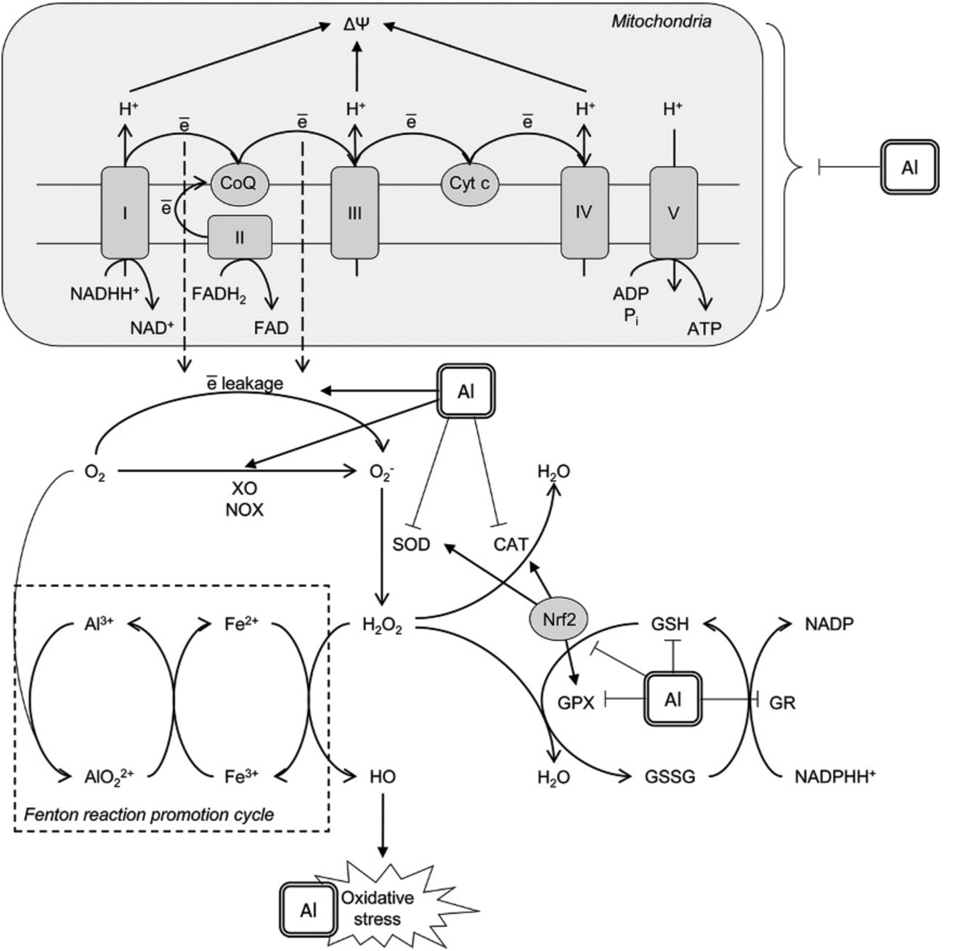
Mechanisms underlying prooxidant effect of aluminium (Al3+). Al3+ was shown to affect mitochondrial electron transport chain thus increasing electron leakage from Complex I and III with subsequent formation of superoxide anion radical . Another mechanism contributing to superoxide production involves Al-dependent increase in xanthine oxidase (XO) and NADPH-oxidase (NOX) activity. Al3+ cation is directly involved in the formation of highly reactive Al superoxide semi-reduced radical ion that was shown to promote prooxidant activity of Fe2+ in Fenton reaction with generation of hydroxyl radical (HO•). Prooxidant activity of Al is also aggravated by its inhibitory effect on enzymatic antioxidants including superoxide dismutase (SOD), catalase (CAT), glutathione peroxidase (GPX), and glutathione reductase (GR). The latter results in reduced glutathione (GSH) depletion. Moreover, Al was shown to down-regulate nuclear factor erythroid 2–related factor 2 (Nrf2), being the key regulator of the antioxidant system. Taken together, these mechanisms result in development of oxidative stress with increased oxidative modification of lipids, proteins and nucleic acids observed in brain/neuronal cell lines under Al exposure.
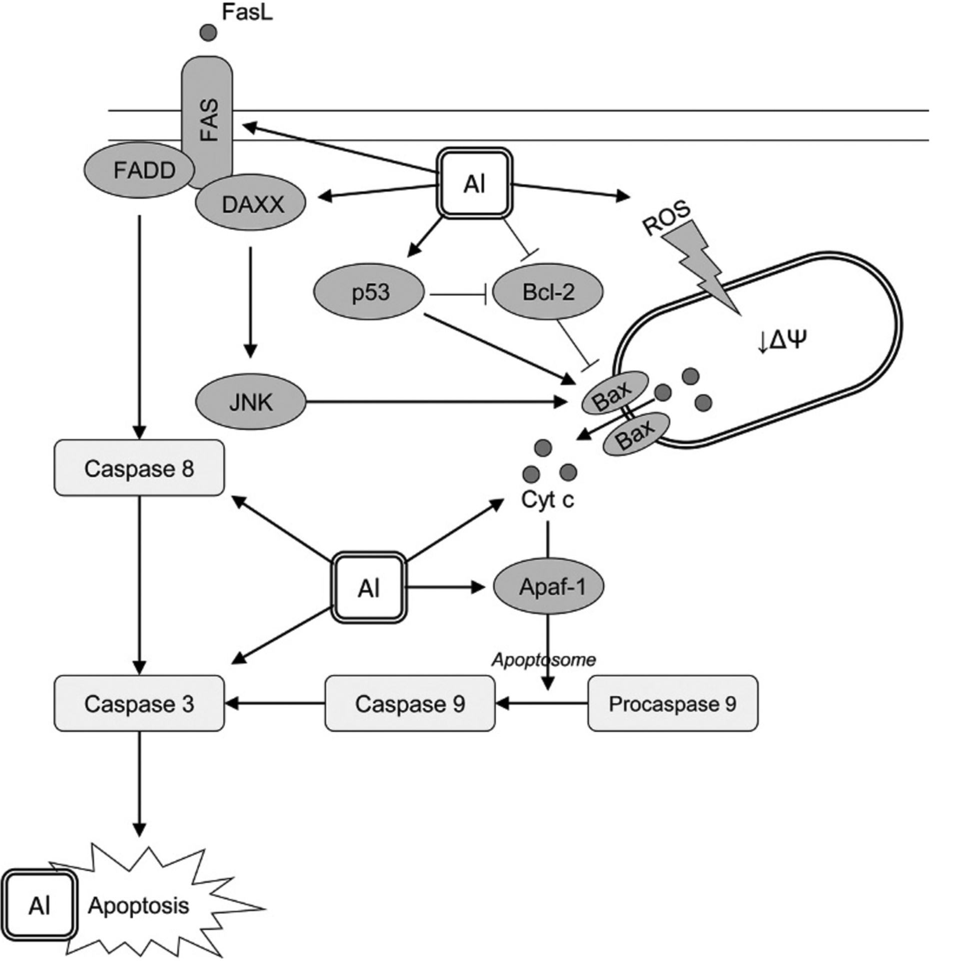
The proposed model of aluminium-induced neuronal apoptosis. Al was shown to induce apoptosis in neuronal cells through distinct pathways. Mitochondrial pathway is directly related to Al-induced mitochondrial dysfunction and subsequent Bax-mediated cytochrome c release. The latter is also stimulated due to positive and negative regulation of p53 and Bcl-2 expression, respectively. Al was shown to increase the rate of cytochrome c binding to Apaf-1 with subsequent formation of apoptosome and caspase 9 activation with subsequent caspase 3 cleavage and activation promoting apoptosis. Another pathway of Al proapoptotic effect involves Fas/FasL signaling with activation of caspase 3 following caspase 8 stimulation. In addition, Al3+-induced up-regulation of DAXX results in JNK signaling that also possesses a stimulatory effect on Bax. Prooxidant activity of Al3+ is also expected to underlie proapoptotic effect of the metal through increased oxidative modification of biomolecules and particularly nucleic acids.
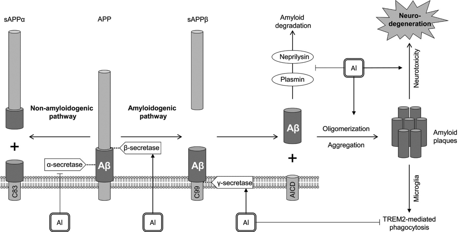
The role of Al in amyloid β generation. Al exposure promotes amyloidogenic pathway through activation of β- (BACE1) and γ-secretase (presenilin-1), as well as down-regulating non-amyloidogenic pathway due to inhibition of α-secretase. Along with promotion of amyloid oligomerization and aggregation, Al3+ also inhibits neprilysin and plasmin that are known to be involved in amyloid degradation. In addition, Al exposure was shown to impair TREM2-mediated phagocytosis of amyloid proteins by microglia. Taken together, these effects of Al exposure result in accumulation of amyloid plaques and Alzheimer disease-like neurodegeneration.
References
-
- Ahmed GA, Khalil SK, Abbas L, Sherif HH, Abdel-Rahman EA, Saber SH, Ali SS, 2020a. ATR-IR and EPR spectroscopy for detecting the alterations in cortical synaptosomes induced by aluminium stress. Spectrochim. Acta A Mol. Biomol. Spectrosc 228, 117535. - PubMed
-
- Ahmed GAR, Khalil SK, El Hotaby W, Abbas L, Farrag ARH, Aal WEA, Hassan MH, 2020b. ATR-IR and EPR spectroscopy for following the membrane restoration of isolated cortical synaptosomes in aluminium-induced Alzheimer’s disease–Like rat model. Chem. Phys. Lipids 231, 104931. - PubMed
-
- Akinrinade ID, Memudu AE, Ogundele OM, 2015a. Fluoride and aluminium disturb neuronal morphology, transport functions, cholinesterase, lysosomal and cell cycle activities. Pathophysiology. 22 (2), 105–115. - PubMed
-
- Akinrinade ID, Memudu AE, Ogundele OM, Ajetunmobi OI, 2015b. Interplay of glia activation and oxidative stress formation in fluoride and aluminium exposure. Pathophysiology. 22 (1), 39–48. - PubMed
Grants and funding
LinkOut - more resources
Full Text Sources