

Advances in the diagnosis and treatment of keratoconus
- PMID: 34263132
- PMCID: PMC8246497
- DOI: 10.1177/25158414211012796
Advances in the diagnosis and treatment of keratoconus
Abstract
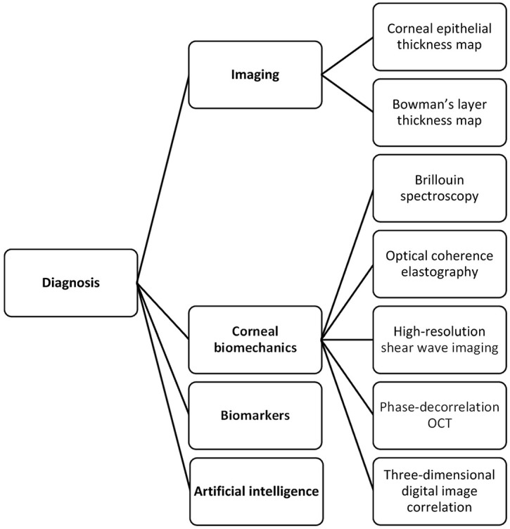
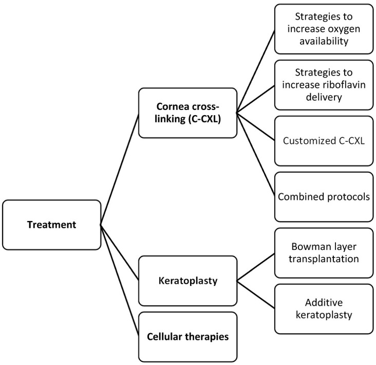
Keratoconus had traditionally been considered a rare disease at a time when the imaging technology was inept in detecting subtle manifestations, resulting in more severe disease at presentation. The increased demand for refractive surgery in recent years also made it essential to more effectively detect keratoconus before attempting any ablative procedure. Consequently, the armamentarium of tools that can be used to diagnose and treat keratoconus has significantly expanded. The advances in imaging technology have allowed clinicians and researchers alike to visualize the cornea layer by layer looking for any early changes that might be indicative of keratoconus. In addition to the conventional geometrical evaluation, efforts are also underway to enable spatially resolved corneal biomechanical evaluation. Artificial intelligence has been exploited in a multitude of ways to enhance diagnostic efficiency and to guide treatment. As for treatment, corneal cross-linking treatment remains the mainstay preventive approach, yet the current main focus of research is on increasing oxygen availability and developing new strategies to improve riboflavin permeability during the procedure. Some new combined protocols are being proposed to simultaneously halt keratoconus progression and correct refractive error. Bowman layer transplantation and additive keratoplasty are newly emerging alternatives to conventional keratoplasty techniques that are used in keratoconus surgery. Advances in tissue engineering and regenerative therapy might bring new perspectives for treatment at the cellular level and hence obviate the need for invasive surgeries. In this review, we describe the advances in the diagnosis and treatment of keratoconus primarily focusing on newly emerging approaches and strategies.
Keywords: advances; diagnosis; keratoconus; treatment.
© The Author(s), 2021.
Conflict of interest statement
Conflict of interest statement: The authors declared no potential conflicts of interest with respect to the research, authorship, and/or publication of this article.
Figures

References
-
- Kennedy RH, Bourne WM, Dyer JA. A 48-year clinical and epidemiologic study of keratoconus. Am J Ophthalmol 1986; 101: 267–273. - PubMed
-
- Ma JX, Wang L, Weikert MP, et al. Evaluation of the repeatability and reproducibility of corneal epithelial thickness mapping for a 9-mm zone using optical coherence tomography. Cornea 2019; 38: 67–73. - PubMed
-
- Pircher N, Beer F, Holzer S, et al. Large field of view corneal epithelium and bowman’s layer thickness maps in keratoconic and healthy eyes. Am J Ophthalmol 2020; 209: 168–177. - PubMed
-
- Matalia H, Francis M, Gangil T, et al. Noncontact quantification of topography of anterior corneal surface and bowman’s layer with high-speed OCT. J Refract Surg 2017; 33: 330–336. - PubMed
-
- Chandapura R, Salomão MQ, Ambrósio R, Jr, et al. Bowman’s topography for improved detection of early ectasia. J Biophotonic 2019; 12: e201900126. - PubMed
Publication types
LinkOut - more resources
Full Text Sources
Research Materials
