. 2021 May 22;3(13):3799-3815.
doi: 10.1039/d0na01086c. eCollection 2021 Jun 30.

Biocompatibility, uptake and subcellular localization of bacterial magnetosomes in mammalian cells

Affiliations

Biocompatibility, uptake and subcellular localization of bacterial magnetosomes in mammalian cells

Frank Mickoleit et al. Nanoscale Adv. .

Abstract

Magnetosomes represent biogenic, magnetic nanoparticles biosynthesized by magnetotactic bacteria. Subtle biological control on each step of biomineralization generates core-shell nanoparticles of high crystallinity, strong magnetization and uniform shape and size. These features make magnetosomes a promising alternative to chemically synthesized nanoparticles for many applications in the biotechnological and biomedical field, such as their usage as biosensors in medical diagnostics, as drug-delivery agents, or as contrast agents for magnetic imaging techniques. Thereby, the particles are directly applied to mammalian cells or even injected into the body. In the present work, we provide a comprehensive characterization of isolated magnetosomes as potential cytotoxic effects and particle uptake have not been well studied so far. Different cell lines including cancer cells and primary cells are incubated with increasing particle amounts, and effects on cell viability are investigated. Obtained data suggest a concentration-dependent biocompatibility of isolated magnetosomes for all tested cell lines. Furthermore, magnetosome accumulation in endolysosomal structures around the nuclei is observed. Proliferation rates are affected in the presence of increasing particle amounts; however, viability is not affected and doubling times can be restored by reducing the magnetosome concentration. In addition, we evidence magnetosome-cell interactions that are strong enough to allow for magnetic cell sorting. Overall, our study not only assesses the biocompatibility of isolated magnetosomes, but also evaluates effects on cell proliferation and the fate of internalized magnetosomes, thereby providing prerequisites for their future in vivo application as biomedical agents.


Conflict of interest statement

There are no conflicts to declare.

Figures

Fig. 1
Fig. 1. (A) Transmission electron microscopy (TEM) micrograph of a representative cell of the wildtype strain of M. gryphiswaldense. The latter biosynthesizes ∼40 magnetosomes per cell arranged in a chain-like manner ad midcell. (B) TEM micrograph of a suspension of isolated magnetosomes (negatively stained) containing well-dispersed particles of ∼35 nm in diameter. Magnetosomes consist of a cuboctahedral, monocrystalline core of pure magnetite (Fe3O4) that is surrounded by an electron-light organic shell (indicated by blue arrows), representing the magnetosome membrane. (C) Schematic illustration of a single magnetosome nanoparticle. The magnetite core is enveloped by a phospholipid bilayer (magnetosome membrane) that harbours a set of magnetosome specific proteins (Mam proteins).
Fig. 2
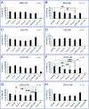
Fig. 2. Cell viability of BeWo (A/B), FaDu (C/D), HCC78 (E/F) and hPC-PL (G/H) cell lines incubated with different concentrations of isolated magnetosomes. PrestoBlue assays were performed to assess potential cytotoxic effects of the indicated amounts of isolated magnetosomes when added to the respective cell line (incubation for 24 or 48 h). Viability values are given as percentage of the value obtained for untreated cells (negative control). Cells incubated with 0.02% Triton X-100 served as positive control. As illustrated by the bar charts, for 24 h incubation time magnetosome concentrations up to 50 μg cm−2 (194.4 μg mL−1) were considered to be biocompatible, with viability values ≥69% (classified as moderate viability; EN ISO 10993-5:2009). For 48 h of incubation, trends towards decreasing viability rates were observed, however, sensitivity to magnetosome treatment differed depending on the tested cell line, with FaDu exhibiting the highest viability rates (>90% for 100 μg Fe cm−2). Number of biological replicates (n) as indicated, statistically significant differences are denoted as follows: p < 0.05 (*), p < 0.01 (**), p < 0.001 (***). Statistical analysis and p-values are provided in Table S2. Please note, that statistically significant differences to Triton X-100 (which applies to the majority of samples tested) are not included in the figure, but listed in Table S2.
Fig. 3
Fig. 3. Viability rates of magnetosome-treated BeWo, FaDu, HCC78 and hPC-PL cells determined by SYTOX™ staining assay. 500 000 cells of the respective cell lines were incubated with isolated magnetosomes (100 μg cm−2) for 24 or 48 h (“w M”). Afterwards, cell death rates were determined using the SYTOX™ assay followed by flow cytometry analysis. Untreated cells (“w/o M”) and cells treated with 0.1% Triton X-100 served as controls. The obtained values (provided in Table S3†) were subsequently taken to calculate cell viability rates. Incubation with Triton X-100 resulted in viability rates ≤10%. Whereas for the magnetosome-treated FaDu and HCC78 cells viability was comparable to the corresponding untreated fractions, the more sensitive BeWo and hPC-PL exhibited a slight but partially significant viability decrease, which is in agreement with the results from PrestoBlue assays (Fig. 2). Data are presented as mean ± standard deviation, n ≥ 2. Differences considered as statistically significant are specified as follows: p < 0.05 (*), p < 0.01 (**), or p < 0.001 (***). Statistical analysis is provided in Table S4.
Fig. 4
Fig. 4. Internalization of magnetosomes by FaDu cells. Confocal laser scanning microscopy (cLSM) analysis of FaDu cells incubated with fluorescent, DyLight488-labelled WT magnetosomes (10 or 25 μg Fe cm−2) for 24 h. cLSM indicated that the particles are taken up by the cells and localize in endolysosomes around the nucleus (white arrows). Fluorescent magnetosomes, green; actin cytoskeleton (phalloidin-AF633), red; nuclei (hoechst33258), blue.
Fig. 5
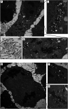
Fig. 5. Magnetosome uptake by FaDu cells and subcellular localization. FaDu cells were grown on coverslips and incubated with WT magnetosomes. After fixation, dehydration and flat embedding, ultrathin serial sections were cut and screened for particle internalization using transmission electron microscopy. (A) TEM micrographs of ultrathin sections demonstrate magnetosome uptake, which is indicated by high amounts of electron-dense particles being internalized in vesicle-like structures (iii), partially located in close proximity to the nucleus (i). In addition to magnetosomes taken up by the cells, well-dispersed particles can be found in the area surrounding the cells (ii), forming the typical chain-like arrangements (inset). (B) TEM image of a representative dividing FaDu cell, showing internalized magnetosomes. Even for dividing cells, vesicle-like structures could be found, filled with high amounts of electron-dense nanoparticles (shown in more detail in the magnifications (i) and (ii)).
Fig. 6
Fig. 6. Real-time analysis of the proliferation behavior of magnetosome-treated FaDu-NR cells. In order to investigate potential effects on cellular growth, transduced FaDu-NR cells were incubated with the indicated magnetosome concentrations, ranging from 5 to 100 μg cm−2. The IncuCyte ZOOM system was used to determine the number of red-fluorescent nuclei (denominated as “red object count”) as a measure for the number of vital cells. Untreated FaDu-NR cells and cells incubated with HEPES buffer served as positive control, cells treated with 0.1% Triton X-100 or PEI-coated nanoparticles (50 μg cm−2) were used as negative control, as incubation with the FaDu-NR cells led to cell death. For the magnetosome-treated fractions, a concentration-dependent decrease of the fluorescence signal was observed, indicating impaired proliferation. Number of biological replicates: n = 3, each measured in quadruplicates.
Fig. 7
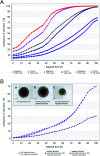
Fig. 7. Cellular growth and spheroid formation of FaDu-NR cells after magnetosome loading. (A) FaDu-NR cells were incubated with 25 μg cm−2 magnetosomes for 24 h. After magnetic enrichment by subjecting the mixture to MACS MS columns, the indicated, defined cell numbers were seeded into fresh medium to allow for proliferation. The time-dependent cellular growth was monitored for 102 h (“Magnetos.”) and compared to the untreated cell fractions (“Control”). For both, the untreated and the corresponding positive fractions, nearly identical growth patterns were observed, and proliferation rates gradually increased with the number of FaDu-NR cells seeded. Proliferation is given as “confluency of cell layer [%]”, directly correlating with the cellular growth. (B) 150 000 FaDu-NR cells were subjected to magnetic separation after incubation with 25 μg cm−2 magnetosomes. The majority of the magnetosome-treated FaDu-NR cells was retained in the matrix and only eluted after removal of the magnetic field (“positive fraction”). Afterwards, the cellular growth was analyzed, which was comparable to untreated FaDu-NR cells (“w/o magnetosomes, w/o magnetic separation”). Only a low number of magnetosome-treated FaDu-NR cells was directly eluted from the column (“negative fraction”). Therefore, due to the reduced number of cells in the proliferation assay, decreased growth rates (compared to those of the “positive fraction”) were observed. As a further proof of adequate cell behavior, the formation of spheroids was analyzed. For that purpose, FaDu-NR cells were seeded in u-bottom shaped 96-well plates and monitored in the IncuCyte system for 96 h. Inset: (a) FaDu-NR cells without magnetosome-incubation, without magnetic separation (5000 cells seeded); (b) FaDu-NR cells with magnetosome-incubation, magnetic separation – positive fraction (5000 cells seeded); (c) FaDu-NR cells with magnetosome-incubation, magnetic separation – negative fraction (2000 cells seeded).
Fig. 8
Fig. 8. Interaction of magnetosomes with FaDu-NR cells and magnetic separation of particle-loaded cells. (A) 1 × 10⁶ FaDu-NR cells or 2.5 × 10⁶ leukocytes were incubated with magnetosomes (10–100 μg mL−1) for 8 min. The suspensions were subsequently separated using magnetic columns, and cells of the flow-through (negative fraction) and cells retained in the magnetic field (positive fraction) were counted. Whereas for the leukocytes only weak interactions were detected (most cells were found in the flow-through), up to 60% of the FaDu-NR cells could be retained in the columns, indicating a sufficient particle-labelling for magnetic separation. Cell loss: 2–20%, n = 2. (B) 125 000 FaDu-NR cells were mixed with 2.5 × 10⁶ leukocytes and subsequently treated with magnetosomes (25 μg Fe mL−1) for different incubation times (up to 20 min). As described in (A), the suspensions were magnetically separated and cells of the positive and negative fraction were counted. Flow cytometry was used to determine the portion of FaDu-NR cells. With increasing incubation times, a significant increase of the portion of particle-labelled FaDu-NR cells was observed in the positive fraction, whereas the total cell number stayed constant in the range from 2–5%. These finding argue for increased interaction of magnetosomes with the tumor cell line (compared to leukocytes). Cell loss <15%, n = 4. Statistically significant differences are denoted as follows: p < 0.05 (*), p < 0.01 (**), p < 0.001 (***).

References

    1. Haun J. B. Yoon T. J. Lee H. Weissleder R. Wiley Interdiscip. Rev.: Nanomed. Nanobiotechnol. 2010;2:291–304. - PubMed
    1. Hepel M. Magnetochemistry. 2020;6:3. doi: 10.3390/magnetochemistry6010003. - DOI - PubMed
    1. Sun C. Lee J. S. Zhang M. Adv. Drug Delivery Rev. 2008;60:1252–1265. doi: 10.1016/j.addr.2008.03.018. - DOI - PMC - PubMed
    1. McCarthy J. R. Weissleder R. Adv. Drug Delivery Rev. 2008;60:1241–1251. doi: 10.1016/j.addr.2008.03.014. - DOI - PMC - PubMed
    1. Berry C. C. Curtis A. S. J. Phys. D: Appl. Phys. 2003;36:R198. doi: 10.1088/0022-3727/36/13/203. - DOI