Comparison of the Magnetic Resonance Imaging Findings of Paget's Disease of the Breast and Malignant Tumor Invasion of the Nipple-Areola Complex
- PMID: 34263155
- PMCID: PMC8246046
- DOI: 10.4274/ejbh.galenos.2021.6091
Comparison of the Magnetic Resonance Imaging Findings of Paget's Disease of the Breast and Malignant Tumor Invasion of the Nipple-Areola Complex
Abstract
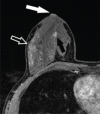
Objective: We aimed to investigate the distinction between Paget's disease of the breast (PDB) and malignant tumor invasion of nipple-areolar complex (MTION) with Magnetic resonance imaging (MRI) findings without the need for skin punch biopsy.
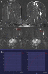
Materials and methods: MRI findings of 16 patients with pathologically proven PDB and 11 patients with pathologically proven MTION were reviewed retrospectively. MRI images were assessed for nipple morphological changes; areolar-periareolar skin changes; thickness, classification, and kinetic characteristics of the nipple-areolar complex (NAC) enhancement; morphological pattern, size, and pathological diagnosis of concomitant malignant lesions; kinetic characteristics of the concomitant malignant lesions enhancement; continuity of enhancement between the nipple and closest concomitant malignant lesion; similarity of enhancement kinetics of the NAC and concomitant malignant lesions; and nipple-to-malignant lesion distance in both patient groups.
Results: Areolar-periareolar skin thickening was statistically different between the patient groups. Enhancement kinetic pattern was classified as persistent in four patients with MTION and plateau in seven patients with PDB. Moreover, NAC enhancement kinetic characteristics were statistically different between the groups. Invasive ductal carcinoma was detected in three patients with PDB and five patients with MTION. A statistically significant difference in malignant lesion pathological types was detected between the patient groups.
Conclusion: The significant MRI findings in patients with MTION diagnosed as invasive ductal carcinoma were areolar-periareolar skin thickening and asymmetric NAC enhancement with persistent kinetics pattern. In patients diagnosed with ductal carcinoma in situ, a plateau pattern of asymmetric NAC enhancement without any areolar-periareolar skin changes on MRI may indicate PDB.
Keywords: MRI; breast cancer; breast imaging; nipple-areola complex.
©Copyright 2021 by Turkish Federation of Breast Diseases Associations.
Conflict of interest statement
Conflict of Interest: The authors declare that they have no conflict of interest.
Figures


References
-
- Da Costa D, Taddese A, Cure ML, Gerson D, Poppiti R, Esserman LE. Common and unusual diseases of the nipple-areolar complex. Radiographics. 2007;27(Suppl 1):S65–S77. - PubMed
-
- Stone K, Wheeler A. A review of anatomy, physiology, and benign pathology of the nipple. Ann Surg Oncol. 2015;22:3236–3240. - PubMed
-
- Nicholson BT, Harvey JA, Cohen MA. Nipple-areolar complex: normal anatomy and benign and malignant processes. Radiographics. 2009;29:509–523. - PubMed
-
- Friedman EP, Hall-Craggs MA, Mumtaz H, Schneidau A. Breast MR and the appearance of the normal and abnormal nipple. Clin Radiol. 1997;52:854–861. - PubMed
-
- Cho J, Chung J, Cha ES, Lee JE, Kim JH. Can preoperative 3-T MRI predict nipple-areolar complex involvement in patients with breast cancer? Clin Imaging. 2016;40:119–124. - PubMed
LinkOut - more resources
Full Text Sources
