Cyclisation strategies for stabilising peptides with irregular conformations
- PMID: 34263169
- PMCID: PMC8230030
- DOI: 10.1039/d1md00098e
Cyclisation strategies for stabilising peptides with irregular conformations
Abstract
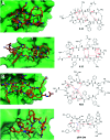
Cyclisation is a common synthetic strategy for enhancing the therapeutic potential of peptide-based molecules. While there are extensive studies on peptide cyclisation for reinforcing regular secondary structures such as α-helices and β-sheets, there are remarkably few reports of cyclising peptides which adopt irregular conformations in their bioactive target-bound state. In this review, we highlight examples where cyclisation techniques have been successful in stabilising irregular conformations, then discuss how the design of cyclic constraints for irregularly structured peptides can be informed by existing β-strand stabilisation approaches, new computational design techniques, and structural principles extracted from cyclic peptide library screening hits. Through this analysis, we demonstrate how existing peptide cyclisation techniques can be adapted to address the synthetic design challenge of stabilising irregularly structured binding motifs.
This journal is © The Royal Society of Chemistry.
Conflict of interest statement
There are no conflicts to declare.
Figures


Similar articles
-
Stabilisation of a short α-helical VIP fragment by side chain to side chain cyclisation: a comparison of common cyclisation motifs by circular dichroism.J Pept Sci. 2013 Jul;19(7):423-32. doi: 10.1002/psc.2515. Epub 2013 May 27. J Pept Sci. 2013. PMID: 23712909
-
Structurally diverse cyclisation linkers impose different backbone conformations in bicyclic peptides.Chembiochem. 2012 May 7;13(7):1032-8. doi: 10.1002/cbic.201200049. Epub 2012 Apr 11. Chembiochem. 2012. PMID: 22492661
-
Self-cyclisation as a general and efficient platform for peptide and protein macrocyclisation.Commun Chem. 2023 Mar 4;6(1):48. doi: 10.1038/s42004-023-00841-5. Commun Chem. 2023. PMID: 36871076 Free PMC article.
-
Constraining cyclic peptides to mimic protein structure motifs.Angew Chem Int Ed Engl. 2014 Nov 24;53(48):13020-41. doi: 10.1002/anie.201401058. Epub 2014 Oct 6. Angew Chem Int Ed Engl. 2014. PMID: 25287434 Review.
-
Modulating Protein-Protein Interactions by Cyclic and Macrocyclic Peptides. Prominent Strategies and Examples.Molecules. 2021 Jan 16;26(2):445. doi: 10.3390/molecules26020445. Molecules. 2021. PMID: 33467010 Free PMC article. Review.
Cited by
-
Addressing Challenges of Macrocyclic Conformational Sampling in Polar and Apolar Solvents: Lessons for Chameleonicity.J Chem Inf Model. 2023 Nov 27;63(22):7107-7123. doi: 10.1021/acs.jcim.3c01123. Epub 2023 Nov 9. J Chem Inf Model. 2023. PMID: 37943023 Free PMC article.
-
Synthesis, Pharmacological Evaluation, and Computational Studies of Cyclic Opioid Peptidomimetics Containing β3-Lysine.Molecules. 2021 Dec 28;27(1):151. doi: 10.3390/molecules27010151. Molecules. 2021. PMID: 35011383 Free PMC article.
-
Bypassing the Need for Cell Permeabilization: Nanobody CDR3 Peptide Improves Binding on Living Bacteria.Bioconjug Chem. 2023 Jul 19;34(7):1234-1243. doi: 10.1021/acs.bioconjchem.3c00116. Epub 2023 Jul 7. Bioconjug Chem. 2023. PMID: 37418494 Free PMC article.
-
Advancement from Small Peptide Pharmaceuticals to Orally Active Piperazine-2,5-dion-Based Cyclopeptides.Int J Mol Sci. 2023 Aug 31;24(17):13534. doi: 10.3390/ijms241713534. Int J Mol Sci. 2023. PMID: 37686336 Free PMC article. Review.
-
Exploration of bioactive variants of the BMP7-derived p[63-82] peptide for ameliorating the OA-associated chondrocyte phenotype.Arthritis Res Ther. 2025 Jul 30;27(1):161. doi: 10.1186/s13075-025-03599-4. Arthritis Res Ther. 2025. PMID: 40739530 Free PMC article.
References
-
- Lau J. L. Dunn M. K. Bioorg. Med. Chem. 2018;26:2700–2707. - PubMed
-
- Valeur E. Gueret S. M. Adihou H. Gopalakrishnan R. Lemurell M. Waldmann H. Grossmann T. N. Plowright A. T. Angew. Chem., Int. Ed. 2017;56:10294–10323. - PubMed
-
- Kaspar A. A. Reichert J. M. Drug Discovery Today. 2013;18:807–817. - PubMed
-
- Nevola L. Giralt E. Chem. Commun. 2015;51:3302–3315. - PubMed
Publication types
LinkOut - more resources
Full Text Sources

