. 2021 Jul;26(7):075002.
doi: 10.1117/1.JBO.26.7.075002.

In vivo two-photon-excited cellular fluorescence of melanin, NAD(P)H, and keratin enables an accurate differential diagnosis of seborrheic keratosis and pigmented cutaneous melanoma

Affiliations

In vivo two-photon-excited cellular fluorescence of melanin, NAD(P)H, and keratin enables an accurate differential diagnosis of seborrheic keratosis and pigmented cutaneous melanoma

Łukasz Szyc et al. J Biomed Opt. 2021 Jul.

Abstract

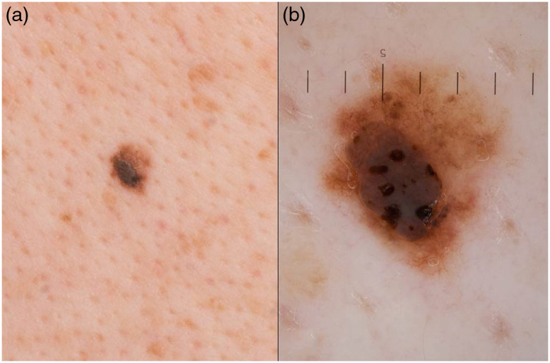
Significance: Seborrheic keratoses (SKs) are harmless pigmented skin lesions (PSLs) that may be confused clinically not only with other benign conditions but also with cutaneous melanoma (CM). As SKs are one of the most common neoplasms in adults, the importance of their correct diagnosis is high. Misclassifying SK as malignant is not rare and leads to a high number of unnecessary biopsies. On the other hand, misdiagnosing CM as SK may have a large impact on prognosis or therapy.

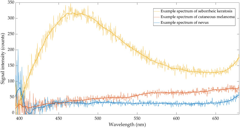
Aim: In the non-invasive technique of dermatofluoroscopy, the fluorophores in melanocytes and keratinocytes are excited in vivo with nanosecond laser pulses and the resulting spectrally resolved, melanin-dominated fluorescence signals are used to differentiate between pigmented benign lesions and CM.

Approach: In this single-center, non-interventional study, 33 PSLs of 20 patients were scanned with dermatofluoroscopy in vivo. For all included cases, dermatofluoroscopic signals were compared to pathology classification.

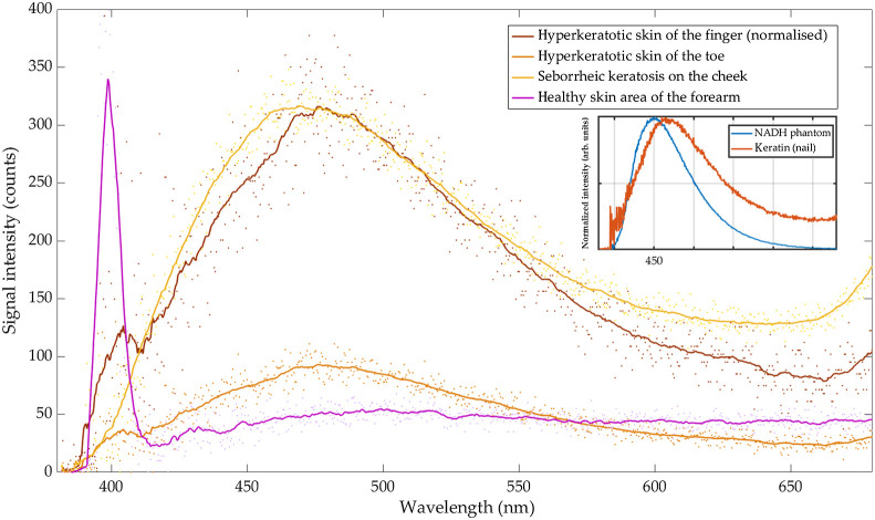
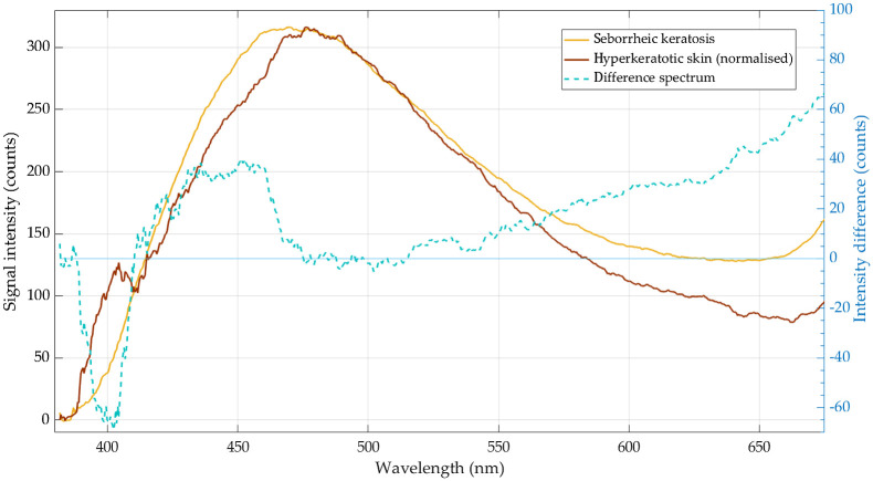
Results: The characteristic spectral features of SK were identified, where the signals are dominated by keratin, NAD(P)H, and melanin. The fluorescence spectra of SKs differed substantially from those of CM: a characteristic spectrum of SK has been identified in 27 of 28 SKs.

Conclusions: The high-accuracy differential diagnosis between CM and SK is possible with dermatofluoroscopy.

Keywords: fluorescence; malignant melanoma; melanin; seborrheic keratosis.


Figures

Fig. 1
Fig. 1
Representative pigmented lesion of this study displayed as (a) overview and (b) dermoscopic image. Histopathology confirmed the clinical diagnosis of a SK and ruled out CM.
Fig. 2
Fig. 2
Representative spectra of a SK, CM, and a nevus. Note relatively high intensity of collagen second harmonic at 400 nm in melanocytic nevus (nevus type spectrum, blue) and low-signal intensity of other skin fluorophores. In contrast, a low SHG-signal and very high intensities at 450 to 490 nm, peaking at 475 nm are characteristic for SK (SK type spectrum, yellow). In melanoma (CM type spectrum, red), the intensity of second harmonic at 400 nm is lower than in nevus, whereas the fluorescence intensity in the range between 550 and 650 nm is higher.
Fig. 3
Fig. 3
Smoothed (solid line) and raw (dots) example dermatofluoroscopic spectra of: hyperkeratotic skin of the finger (brown, normalized to SK’s maximum intensity), hyperkeratotic skin of the toe (orange), SK (yellow), and healthy skin area of the forearm (purple). Inset: the pure fluorescence spectra of: keratin of human’s nail (red line) and NADH coenzyme in a silicone phantom (blue line).
Fig. 4
Fig. 4
Smoothed spectrum of hyperkeratotic skin (brown line) normalized to the SK maximum intensity and subtracted from the smoothed spectrum of SK (yellow line). The difference spectrum is marked with dashed cyan line.

References

    1. Fink C., Haenssle H. A., “Non-invasive tools for the diagnosis of cutaneous melanoma,” Ski. Res. Technol. 23(3), 261–271 (2017).10.1111/srt.12350 - DOI - PubMed
    1. Stevenson A., et al. , “Systematic review of diagnostic accuracy of reflectance confocal microscopy for melanoma diagnosis in patients with clinically equivocal skin lesions,” Dermatol. Pract. Concept. 3, 19–27 (2013).10.5826/dpc.0304a05 - DOI - PMC - PubMed
    1. Dimitrow E., et al. , “Spectral fluorescence lifetime detection and selective melanin imaging by multiphoton laser tomography for melanoma diagnosis,” Exp. Dermatol. 18(6), 509–515 (2009).EXDEEY10.1111/j.1600-0625.2008.00815.x - DOI - PubMed
    1. Leupold D., et al. , “The stepwise two-photon excited melanin fluorescence is a unique diagnostic tool for the detection of malignant transformation in melanocytes,” Pigment Cell Melanoma Res. 24(3), 438–445 (2011).10.1111/j.1755-148X.2011.00853.x - DOI - PubMed
    1. Leupold D., Giering H. G., Bildgebende Diagnostik in der Dermatologie, Stolz W., et al., Eds., Georg Thieme Verlag, Stuttgart: (2018).