. 2021 Nov;226(8):2497-2509.
doi: 10.1007/s00429-021-02268-x. Epub 2021 Jul 15.

Diffusion MRI data, sulcal anatomy, and tractography for eight species from the Primate Brain Bank

Affiliations

Diffusion MRI data, sulcal anatomy, and tractography for eight species from the Primate Brain Bank

Katherine L Bryant et al. Brain Struct Funct. 2021 Nov.

Abstract

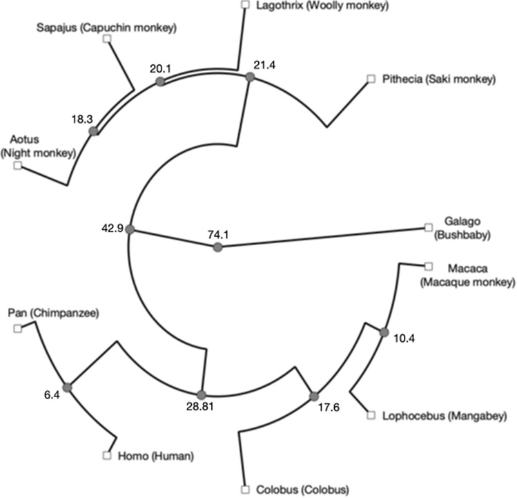
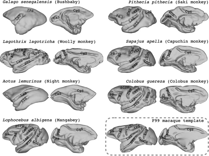
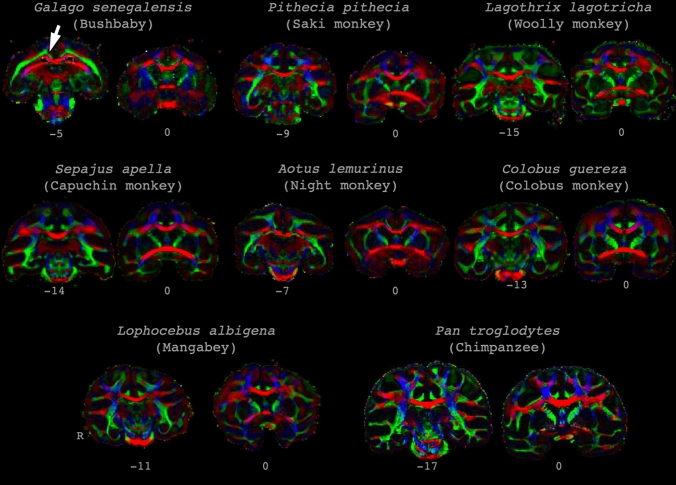
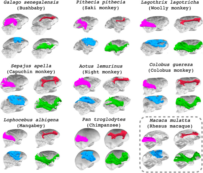
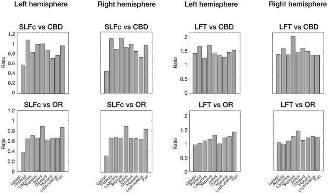
Large-scale comparative neuroscience requires data from many species and, ideally, at multiple levels of description. Here, we contribute to this endeavor by presenting diffusion and structural MRI data from eight primate species that have not or rarely been described in the literature. The selected samples from the Primate Brain Bank cover a prosimian, New and Old World monkeys, and a great ape. We present preliminary labelling of the cortical sulci and tractography of the optic radiation, dorsal part of the cingulum bundle, and dorsal parietal-frontal and ventral temporal-frontal longitudinal white matter tracts. Both dorsal and ventral association fiber systems could be observed in all samples, with the dorsal tracts occupying much less relative volume in the prosimian than in other species. We discuss the results in the context of known primate specializations and present hypotheses for further research. All data and results presented here are available online as a resource for the scientific community.

Keywords: Anthropoid elaboration; Cingulum bundle; Comparative; Connectivity; Cortex; Optic radiation.


Conflict of interest statement

None.

Figures

Fig. 1
Fig. 1
Phylogenetic relationships between the species in this study. Numbers indicate median divergence times (in millions of years) based on genomic data (Hedges et al. 2015)
Fig. 2
Fig. 2
Preliminary sulcal labelling on the lateral surface of the cortical hemisphere of the prosimian and monkey species in this study. All surfaces were reconstructed from the scanned sample except for the macaque, for which we used the F99 template brain (Van Essen et al. 2012). Left hemisphere lateral sulcus on the left, medial surface on the right. AS arcuate sulcus, CaS calcarine sulcus, CeS central sulcus, CgS cingulate sulcus, LOCS lateral occipital sulcus, IPS intra-parietal sulcus, LaS lateral sulcus, LuS lunate sulcus, POS parietal–occipital sulcus, PS principal sulcus, pSFD posterior superior frontal dimple, STS superior temporal sulcus. Note that brains are not displayed to scale
Fig. 3
Fig. 3
Principal diffusion direction maps. Images show coronal sections of the samples in this study at the level of the anterior commissure (y = 0) and the level approximately 25% from the posterior edge of the corpus callosum. As can be seen, the temporal white matter of bushbaby consists of a single sheet, rather than the more extensive pattern seen in more encephalized species, including the woolly monkey. Similarly, in the bushbaby and night monkey, the cingulum bundle consists of a white matter sheet that is difficult to distinguish from the superior longitudinal fascicle complex, as indicated for the bushbaby by the white arrow. Colors indicate principal directions, red left–right, green anterior–posterior, blue dorsal–ventral. Note that brains are not displayed to scale
Fig. 4
Fig. 4
Course of reconstructed white matter tracts. 3d representations of the course of cingulum bundle (red), optic radiation (green), longitudinal fronto-temporal tract (blue), and SLF complex (purple) through the brains of all studied species
Fig. 5
Fig. 5
Relative size of the SLF complex (SLFc) and longitudinal frontotemporal tract (LFT) in all studied species. Relative sizes of SLFc and LFT are displayed with respect to the dorsal cingulum bundle (CBD) and optic radiation (OR) for both hemispheres

References

    1. Amiez C, Kostopoulos P, Champod A, Petrides M. Local morphology predicts functional organization of the dorsal premotor region in the human brain. J Neurosci. 2006;26:2724–2731. - PMC - PubMed
    1. Amiez C, Sallet J, Hopkins WD, Meguerditchian A, Hadj-Bouziane F, Ben Hamed S, Wilson CRE, Procyk E, Petrides M. Sulcal organization in the medial frontal cortex provides insights into primate brain evolution. Nat Commun. 2019;10:1–14. - PMC - PubMed
    1. Amiez C, Sallet J, Novek J, Hadj-Bouziane F, Giacometti C, Andersson J, Hopkins WD, Petrides M. Chimpanzee histology and functional brain imaging show that the paracingulate sulcus is not human-specific. Commun Biol. 2021;4:54. - PMC - PubMed
    1. Ardesch DJ, Scholtens LH, Li L, Preuss TM, Rilling JK, van den Heuvel MP. Evolutionary expansion of connectivity between multimodal association areas in the human brain compared with chimpanzees. Proc Natl Acad Sci USA. 2019;116:7101–7106. - PMC - PubMed
    1. Assaf Y, Bouznach A, Zomet O, Marom A, Yovel Y. Conservation of brain connectivity and wiring across the mammalian class. Nat Neurosci. 2020;23:805–808. - PubMed