Review
. 2021 Jul 15;12(1):101.
doi: 10.1186/s13244-021-01042-1.

A pragmatic evidence-based approach to post-mortem perinatal imaging

Affiliations
Review

A pragmatic evidence-based approach to post-mortem perinatal imaging

Susan C Shelmerdine et al. Insights Imaging. .

Abstract

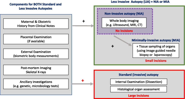
Post-mortem imaging has a high acceptance rate amongst parents and healthcare professionals as a non-invasive method for investigating perinatal deaths. Previously viewed as a 'niche' subspecialty, it is becoming increasingly requested, with general radiologists now more frequently asked to oversee and advise on appropriate imaging protocols. Much of the current literature to date has focussed on diagnostic accuracy and clinical experiences of individual centres and their imaging techniques (e.g. post-mortem CT, MRI, ultrasound and micro-CT), and pragmatic, evidence-based guidance for how to approach such referrals in real-world practice is lacking. In this review, we summarise the latest research and provide an approach and flowchart to aid decision-making for perinatal post-mortem imaging. We highlight key aspects of the maternal and antenatal history that radiologists should consider when protocolling studies (e.g. antenatal imaging findings and history), and emphasise important factors that could impact the diagnostic quality of post-mortem imaging examinations (e.g. post-mortem weight and time interval). Considerations regarding when ancillary post-mortem image-guided biopsy tests are beneficial are also addressed, and we provide key references for imaging protocols for a variety of cross-sectional imaging modalities.

Keywords: Autopsy; Diagnostic imaging; Foetus; Pregnancy loss; Radiology.


Conflict of interest statement

The authors declare that they have no competing interests.

Figures

Fig. 1
Fig. 1
The benefits for conducting a perinatal autopsy for different stakeholders
Fig. 2
Fig. 2
Components of different types of perinatal autopsy. A ‘less invasive autopsy’ (LIA) is an umbrella term for all procedures that use imaging instead of dissection for internal examination at autopsy. Where tissue sampling is also performed in a less invasive way (e.g. image-guided biopsy or laparoscopically assisted biopsy), the study is referred to as a ‘minimally invasive autopsy’ (MIA). Where no incisions are made to the body, and only imaging is acquired, this is termed a ‘non-invasive autopsy’ (NIA) [6]
Fig. 3
Fig. 3
Typical estimated gestational ages and post-mortem weights where various post-mortem imaging modalities could be used to provide diagnostic quality examinations. ** Technically, radiographs and CT can be performed at any age after 8-week gestation (when the foetal skeleton beings to ossify), but in practice they are best reserved for specific clinical situations, such as for suspected skeletal abnormalities or trauma [57]
Fig. 4
Fig. 4
Recommended post-mortem imaging flowchart for non-invasive investigation of perinatal loss. Small foetuses present a challenge for post-mortem imaging, and care should be taken when interpreting imaging results in this cohort. A foetus weighing over 500 g provides the greatest likelihood for a diagnostic quality 1.5 T MRI study [14], and those weighing < 300 g are best suited for micro-CT or high-field MRI [32]. Where neither MRI nor micro-CT is available, ultrasound may be attempted but there is a higher likelihood of a false or non-diagnostic result [26, 58]. Foetuses weighing between 300 and 500 g have been reported to take > 7 days to iodinate and therefore delay micro-CT imaging. If available, 3 T MRI could be attempted for this foetal cohort [22]
Fig. 5
Fig. 5
Perinatal post-mortem imaging for less invasive autopsy. This infographic summarises the perinatal post-mortem cross-sectional imaging options and levels of evidence for their usage

References

    1. Nese N, Bulbul Y. Diagnostic value of perinatal autopsies: analysis of 486 cases. J Perinat Med. 2018;46:175–181. doi: 10.1515/jpm-2016-0396. - DOI - PubMed
    1. Gordijn SJ, Erwich JJ, Khong TY. Value of the perinatal autopsy: critique. Pediatr Dev Pathol. 2002;5:480–488. doi: 10.1007/s10024-002-0008-y. - DOI - PubMed
    1. Widmann R, Caduff R, Giudici L, et al. Value of postmortem studies in deceased neonatal and pediatric intensive care unit patients. Virchows Arch. 2017;470:217–223. doi: 10.1007/s00428-016-2056-0. - DOI - PubMed
    1. Nayak SS, Shukla A, Lewis L, et al. Clinical utility of fetal autopsy and its impact on genetic counseling. Prenat Diagn. 2015;35:685–691. doi: 10.1002/pd.4592. - DOI - PubMed
    1. Rodriguez MA, Prats P, Rodriguez I, Cusi V, Comas C. Concordance between prenatal ultrasound and autopsy findings in a tertiary center. Prenat Diagn. 2014;34:784–789. doi: 10.1002/pd.4368. - DOI - PubMed

LinkOut - more resources