. 2021 Jun 21;29(13):20105-20120.
doi: 10.1364/OE.425632.

Multispectral fluorescence lifetime imaging device with a silicon avalanche photodetector

Multispectral fluorescence lifetime imaging device with a silicon avalanche photodetector

Xiangnan Zhou et al. Opt Express. .

Abstract

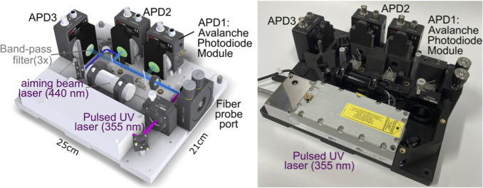
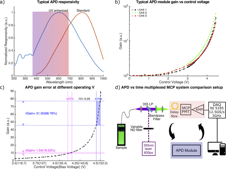
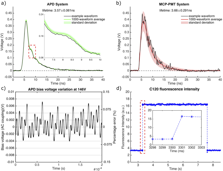
We report the design, development, and characterization of a novel multi-spectral fluorescence lifetime measurement device incorporating solid-state detectors and automated gain control. For every excitation pulse (∼1 µJ, 600 ps), this device records complete fluorescence decay from multiple spectral channels simultaneously within microseconds, using a dedicated UV enhanced avalanche photodetector and analog to digital convert (2.5 GS/s) in each channel. Fast (<2 ms) channel-wise dynamic range adjustment maximizes the signal-to-noise ratio. Fluorophores with known lifetime ranging from 0.5-6.0 ns were used to demonstrate the device accuracy. Current results show the clear benefits of this device compared to existing devices employing microchannel-plate photomultiplier tubes. This is demonstrated by 5-fold reduction of lifetime measurement variability in identical conditions, independent gain adjustment in each spectral band, and 4-times faster imaging speed. The use of solid-state detectors will also facilitate future improved performance and miniaturization of the instrument.


Conflict of interest statement

The authors declare no conflicts of interest.

Figures

Fig. 1.
Fig. 1.
Schematics and picture of the 3 channel APD-based fluorescence lifetime device.
Fig. 2.
Fig. 2.
a) Spectral response of UV enhanced APD and non-UV enhanced APD module. Shaded rainbow area represents the typical emission spectrum range of biological tissue excited by 355 nm laser. Adapted with permission from Thorlabs. b) Typical gain (multiplication factor) curve of the 3 APD modules. Unit-to-unit variation of the gain is observed. c) Due to APD modules’ nonlinear behavior, same error in bias voltage leads to drastic different error of gain at different operating voltage. d) Schematics of the multi-APDs device vs time multiplexing MCP device used in the SNR comparison experiment.
Fig. 3.
Fig. 3.
Fluorescence background removal process. The background waveform is normalized (scaled) based on the fluorescence signal generated from the optical fiber probe, which should remain constant during image acquisition, and subtracted from the acquired raw waveform. The normalization (scaling) factor is computed as the ratio of area under the curve (AUC) between the red dotted lines of the raw waveform and background waveform. As the method is based on the constant signal of the fiber probe fluorescence, no knowledge of the detector gain is required making it a fast and simple but robust method.
Fig. 4.
Fig. 4.
FLIm signal processing pipeline. The raw signal acquired at 2.5 GS/s is first re-sampled to equivalent of 5 GS/s by interpolation to match the sampling rate of system IRF. After re-sampling the fluorescence waveforms from all measurements were temporally aligned using a constant fraction discriminator to compensate for laser jitter. The signal is then deconvolved using Laguerre expansion-based method to extract fluorescence lifetime and intensity. A fluorescence lifetime map can be generated for scanning application.
Fig. 5.
Fig. 5.
Investigation of measured fluorescence waveforms as a function of signal amplitude and APD gain, characterized using 1000 measurements of Coumarin 120 solution. a) Mean and standard deviation of fluorescence waveforms at different APD gain. b) Display of normalized averaged waveforms highlights some difference of temporal response that may affect estimated lifetime. c) Mean and standard deviation of fluorescence waveforms at different optical signal intensity (obtained by varying the laser pulse energy). d) Display of normalized waveforms highlights some difference in temporal response.
Fig. 6.
Fig. 6.
Distribution of Coumarin 120 fluorescence lifetime under (a) increasing optical signal intensity. (b) increasing APD gain. (c) constant signal peak voltage (1 V) where each increase of APD gain is compensated by a decrease of optical signal intensity to maintain a constant 1 V peak voltage.
Fig. 7.
Fig. 7.
Back-to-back SNR comparison of muti-APDs FLIm device (a) and time multiplexing single PMT FLIm device (b). Mean fluorescence waveform, one raw waveform and standard deviation (shaded area) were shown. c) Variation of APD bias voltage @146 V. Adapted with permission from Thorlabs. d) Response of the APD-based detection to a step change of bias voltage. The response time is less than 2 ms. No overshoot was detectable at either rising or falling edge. The time resolution of the measurement was 1 ms.
Fig. 8.
Fig. 8.
Representative FLIm images obtained via the freehand scanning of ex vivo fresh lamb tissue. a) white light image of lamb tissue sample. b) and c) FLIm points measurement locations for different 355 nm laser configuration. d) and e) white light images of the lamb tissue augmented with lifetime maps from 470/28 nm spectral band under different 355 nm laser repetition rate.

References

    1. Berezin M. Y., Achilefu S., “Fluorescence lifetime measurements and biological imaging,” Chem. Rev. 110(5), 2641–2684 (2010).10.1021/cr900343z - DOI - PMC - PubMed
    1. Monici M., “Cell and tissue autofluorescence research and diagnostic applications,” Biotechnol. Annu. Rev. 11, 227–256 (2005).10.1016/S1387-2656(05)11007-2 - DOI - PubMed
    1. Croce A. C., Bottiroli G., “Autofluorescence spectroscopy and imaging: a tool for biomedical research and diagnosis,” Eur. J. Histochem. 58, 2461 (2014).10.4081/ejh.2014.2461 - DOI - PMC - PubMed
    1. Coda S., Thompson A. J., Kennedy G. T., Roche K. L., Ayaru L., Bansi D. S., Stamp G. W., Thillainayagam A. V., French P. M., Dunsby C., “Fluorescence lifetime spectroscopy of tissue autofluorescence in normal and diseased colon measured ex vivo using a fiber-optic probe,” Biomed. Opt. Express 5(2), 515–538 (2014).10.1364/BOE.5.000515 - DOI - PMC - PubMed
    1. Malik B. H., Lee J., Cheng S., Cuenca R., Jabbour J. M., Cheng Y. S., Wright J. M., Ahmed B., Maitland K. C., Jo J. A., “Objective Detection of Oral Carcinoma with Multispectral Fluorescence Lifetime Imaging In Vivo,” Photochem. Photobiol. 92(5), 694–701 (2016).10.1111/php.12627 - DOI - PMC - PubMed