. 2021 Nov 23;60(46):3497-3506.
doi: 10.1021/acs.biochem.1c00370. Epub 2021 Jul 15.

Mechanisms of O2 Activation by Mononuclear Non-Heme Iron Enzymes

Affiliations

Mechanisms of O2 Activation by Mononuclear Non-Heme Iron Enzymes

Edward I Solomon et al. Biochemistry. .

Abstract

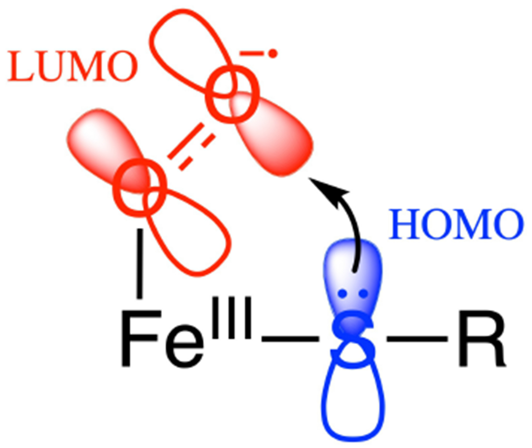
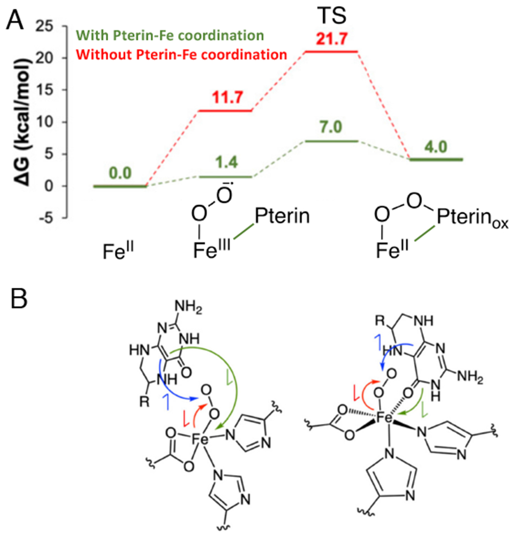
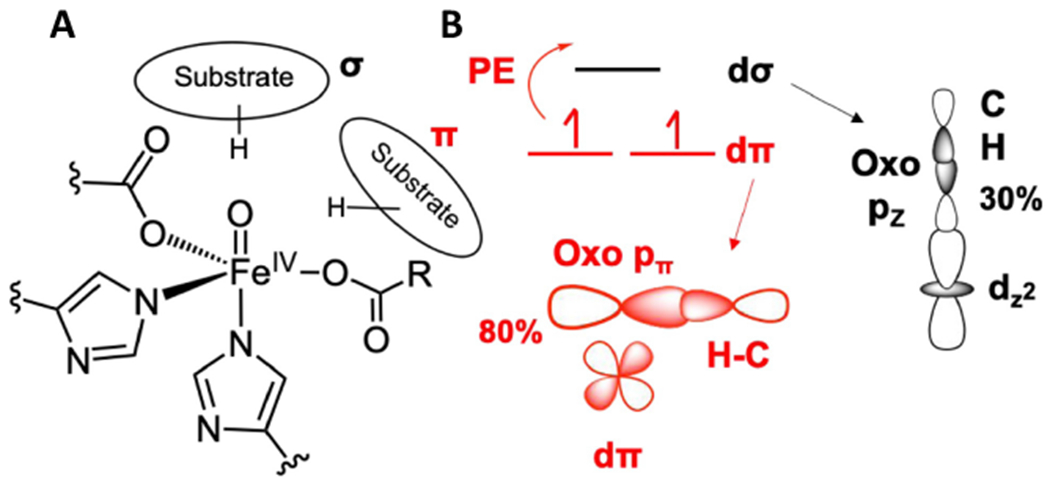
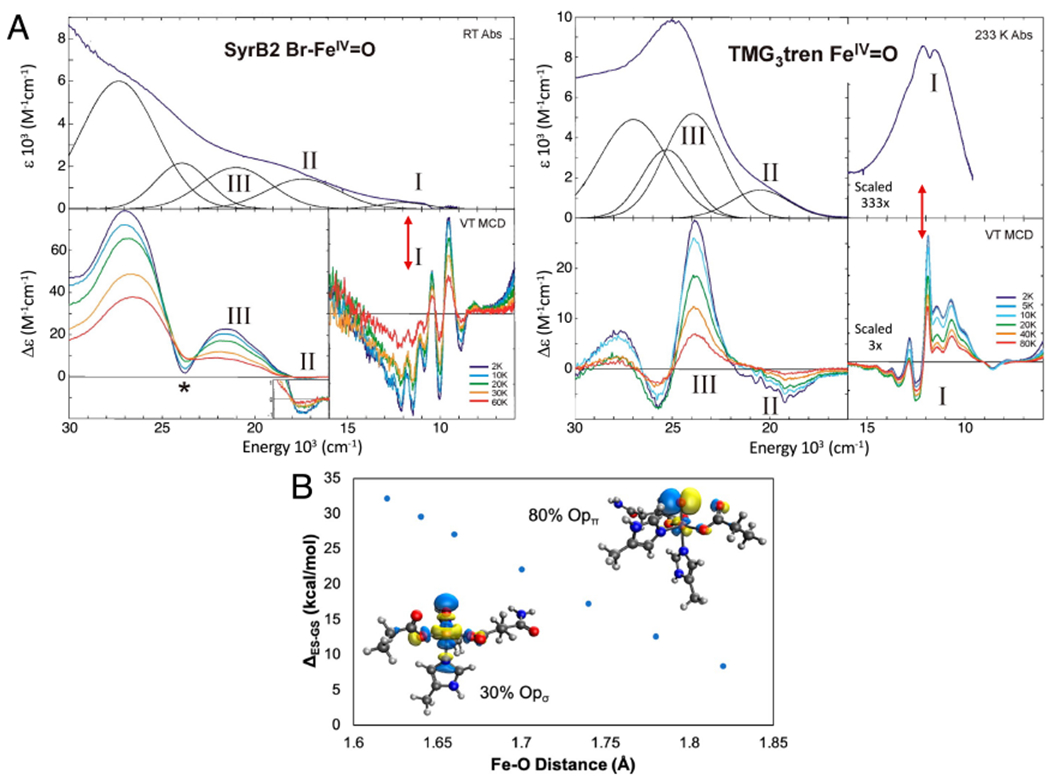
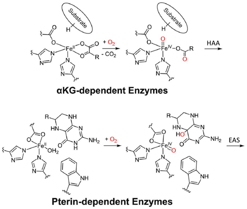
Two major subclasses of mononuclear non-heme ferrous enzymes use two electron-donating organic cofactors (α-ketoglutarate or pterin) to activate O2 to form FeIV═O intermediates that further react with their substrates through hydrogen atom abstraction or electrophilic aromatic substitution. New spectroscopic methodologies have been developed, enabling the study of the active sites in these enzymes and their oxygen intermediates. Coupled to electronic structure calculations, the results of these spectroscopies provide fundamental insight into mechanism. This Perspective summarizes the results of these studies in elucidating the mechanism of dioxygen activation to form the FeIV═O intermediate and the geometric and electronic structure of this intermediate that enables its high reactivity and selectivity in product formation.


Figures

Figure 1:
Figure 1:
FeIII-superoxo π* FMO (i.e. the LUMO) for electrophilic attack on substrate.
Figure 2:
Figure 2:
Near IR MCD spectra of FeII sites in FIH (A) and DAOCS (B) for resting enzyme (top), substrate bound (right second down), akg bound (left middle and right third down) and both akg and substrate bound (bottom). Ferrous ligand field (LF) transitions and charge transfer (CT) transitions. Figure adapted from [Iyer, S. R. et al. (2018) O2 activation by non-heme FeII α-ketoglutarate-dependent enzyme variants: Elucidating the role of the facial triad carboxylate in FIH. Journal of the American Chemical Society 140, 11777–11783.] and [Goudarzi, S. et al. (2020) Evaluation of a concerted vs. sequential oxygen activation mechanism in α-ketoglutarate—dependent nonheme ferrous enzymes. Proceedings of the National Academy of Sciences 117, 5152–5159]. Copyright [2018] and [2020] respectively.
Figure 3:
Figure 3:
Substrate binding (red) to pterin-bound (blue) FeII tryptophan hydroxylase (TPH). A. Abs (top) and MCD (bottom, near-IR MCD inset). B. Resonance Raman spectra (excitation at 334.5 nm). Adapted from [Iyer, S. R. et al. (2021) Direct coordination of pterin to FeII enables neurotransmitter biosynthesis in the pterin-dependent hydroxylases. Proceedings of the National Academy of Sciences 118.] Copyright [2021].
Figure 4:
Figure 4:
Reaction of substrate and pterin bound FeII-TPH with O2. A. Kinetics of first 175 ms of the reaction. B. Formation and decay kinetics at 442 nm in H2O and D2O. C. Mössbauer spectrum of 442 nm intermediate. D. Resonance Raman spectrum of 442 nm intermediate (excitation at 457.9 nm). Adapted from [Iyer, S. R. et al. (2021) Direct coordination of pterin to FeII enables neurotransmitter biosynthesis in the pterin-dependent hydroxylases. Proceedings of the National Academy of Sciences 118.] Copyright [2021].
Figure 5:
Figure 5:
Reaction Coordinate for FeII-pterin TPH. A. Effect of pterin binding to FeII on reaction energetics (red to green). B. Schematics of electronic structure contributions of pterin carbonyl coordination on the energetics in A. Red arrow indicates e-donation from FeII to O2, blue arrow indicates e- donation from pterin to O2; the green arrow indicates donation of a second e- from pterin to Fe (to compensate for the Fe donation to the O2). The green electron donation is facilitated by pterin coordination to the Fe (depicted on the right). Adapted from [Iyer, S. R. et al. (2021) Direct coordination of pterin to FeII enables neurotransmitter biosynthesis in the pterin-dependent hydroxylases. Proceedings of the National Academy of Sciences 118.] Copyright [2021].
Figure 6:
Figure 6:
NRVS spectrum of the FeIV=O intermediate of TauD. Adapted from [Srnec, M. et al. (2020) Nuclear Resonance Vibrational Spectroscopic Definition of the Facial Triad FeIV=O Intermediate in Taurine Dioxygenase: Evaluation of Structural Contributions to Hydrogen Atom Abstraction. Journal of the American Chemical Society 142, 18886–18896.] Copyright [2020].
Figure 7:
Figure 7:
FeIV=O structure and FMOs. A. Schematic of FeIV=O intermediate with possible substrate orientations along (σ) and perpendicular (π) to the Fe-oxo bond. B. σ (black) and π (red) FMOs enabling HAA along (σ) and perpendicular to (π) the Fe-oxo bond. PE indicates promotion energy required to reach the dπ FMO. Adapted from [Srnec, M. et al. (2020) Nuclear Resonance Vibrational Spectroscopic Definition of the Facial Triad FeIV=O Intermediate in Taurine Dioxygenase: Evaluation of Structural Contributions to Hydrogen Atom Abstraction. Journal of the American Chemical Society 142, 18886–18896.] Copyright [2020].
Figure 8:
Figure 8:
Experimental insight into dπ* FMO. A. Abs (top) and MCD (bottom) definition of FeIV=O FMOs in SyrB2 (left) and its TMG3tren model complex (right). Red double arrows indicate dπ* to dσ* LF excitation at an Fe=O bond length of 1.62 Å. B. Dependence of the dπ* to dσ* excitation energy (the promotion energy (PE) in Fig. 7B) on Fe=O bond length (1.82 Å at the TS). Insets show the σ and π FMOS at an Fe=O distance of 1.62 Å. Adapted from [Srnec, M. et al. (2020) Nuclear Resonance Vibrational Spectroscopic Definition of the Facial Triad FeIV=O Intermediate in Taurine Dioxygenase: Evaluation of Structural Contributions to Hydrogen Atom Abstraction. Journal of the American Chemical Society 142, 18886–18896.] and [Srnec, M. et al. (2016) Electronic Structure of the Ferryl Intermediate in the alpha-Ketoglutarate Dependent Non-Heme Iron Halogenase SyrB2: Contributions to H Atom Abstraction Reactivity. Journal of the American Chemical Society 138, 5110–5122.] Copyright [2020] and [2016] respectively.
Figure 9:
Figure 9:
Rebound halogenation vs hydroxylation. A. After HAA by SyrB2 the resultant FeIII-OH has the substrate radical oriented above the HO-FeIII-Cl plane. Energetics of rebound hydroxylation in red and rebound halogenation in green. B. FMOs for rebound, dπ* of HO-FeIII is 4.5 kcal/mol higher than dπ* of Cl-FeIII in energy. Adapted from [Srnec, M. et al. (2017) Frontier Molecular Orbital Contributions to Chlorination versus Hydroxylation Selectivity in the Non-Heme Iron Halogenase SyrB2. Journal of the American Chemical Society 139, 2396–2407.] Copyright [2017].
Scheme 1:
Scheme 1:
Reaction mechanisms for cofactor (αkg and pterin) dependent non-heme iron enzymes. Adapted from [Iyer, S. R. et al. (2021) Direct coordination of pterin to FeII enables neurotransmitter biosynthesis in the pterin-dependent hydroxylases. Proceedings of the National Academy of Sciences 118.] Copyright [2021].
Scheme 2:
Scheme 2:
General mechanistic strategy for cofactor dependent NHFe enzymes.
Scheme 3:
Scheme 3:
FeII-DAOCS/αkg reaction with O2 in the absence (A) and presence (B) of bound substrate (penicillin G). Adapted from [Goudarzi, S. et al. (2020) Evaluation of a concerted vs. sequential oxygen activation mechanism in α-ketoglutarate—dependent nonheme ferrous enzymes. Proceedings of the National Academy of Sciences 117, 5152–5159.] Copyright [2020].

References

    1. Solomon EI, Light KM, Liu LV, Srnec M, and Wong SD (2013) Geometric and Electronic Structure Contributions to Function in Non-heme Iron Enzymes. Accounts of Chemical Research 46, 2725–2739. - PMC - PubMed
    1. Jasniewski AJ, and Que L Jr. (2018) Dioxygen Activation by Nonheme Diiron Enzymes: Diverse Dioxygen Adducts, High-Valent Intermediates, and Related Model Complexes. Chemical Reviews 118, 2554–2592. - PMC - PubMed
    1. Poulos TL (2014) Heme Enzyme Structure and Function. Chemical Reviews 114, 3919–3962. - PMC - PubMed
    1. Baldwin JE, and Abraham E (1988) The Biosynthesis of Penicillins and Cephalosporins. Natural Product Reports 5, 129–145. - PubMed
    1. Busby RW, and Townsend CA (1996) A single monomeric iron center in clavaminate synthase catalyzes three nonsuccessive oxidative transformations. Bioorganic Medicinal Chemistry 4, 1059–1064. - PubMed

Publication types

MeSH terms