

Altered hippocampal functional connectivity patterns in patients with cognitive impairments following ischaemic stroke: A resting-state fMRI study
- PMID: 34266772
- PMCID: PMC8527045
- DOI: 10.1016/j.nicl.2021.102742
Altered hippocampal functional connectivity patterns in patients with cognitive impairments following ischaemic stroke: A resting-state fMRI study
Abstract
Background: Ischemic stroke with cognitive impairment is a considerable risk factor for developing dementia. Identifying imaging markers of cognitive impairment following ischemic stroke will help to develop prevention strategies against post-stroke dementia.
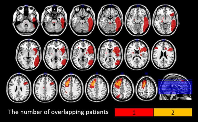
Methods: We investigated the hippocampal functional connectivity (FC) pattern following ischemic stroke, using resting-state fMRI (rs-fMRI). Thirty-three cognitively impaired patients after ischemic stroke and sixteen age-matched controls with no known history of neurological disorder were recruited for the study. No patient had a direct ischaemic insult to hippocampus on the examination of brain imaging. Seven subfields of hippocampus were used as seeds region for FC analyses.
Results: Across all hippocampal subfields, FC with the inferior parietal lobule was reduced in stroke patients as compared with healthy controls. This decreased FC included both supramarginal gyrus and angular gyrus. The FC of hippocampal subfields with cerebellum was increased. Importantly, the degree of the altered FC between hippocampal subfields and inferior parietal lobule was associated with their impaired memory function.
Conclusion: Our results demonstrated that decreased hippocampal-inferior parietal lobule connectivity was associated with cognitive impairment in patients with ischemic stroke. These findings provide novel insights into the role of hippocampus in cognitive impairment following ischemic stroke.
Keywords: Cognitive impairment; Functional connectivity; Hippocampus; Ischemic stroke; Resting-state functional magnetic resonance imaging.
Copyright © 2021. Published by Elsevier Inc.
Conflict of interest statement
The authors declare that they have no known competing financial interests or personal relationships that could have appeared to influence the work reported in this paper.
Figures


References
-
- Amunts K., Kedo O., Kindler M., Pieperhoff P., Mohlberg H., Shah N.J., Habel U., Schneider F., Zilles K. Cytoarchitectonic mapping of the human amygdala, hippocampal region and entorhinal cortex: intersubject variability and probability maps. Anat Embryol (Berl) 2005;210:343–352. - PubMed
-
- Ashburner J. A fast diffeomorphic image registration algorithm. Neuroimage. 2007;38:95–113. - PubMed
-
- Bai F., Zhang Z., Watson D.R., Yu H., Shi Y., Yuan Y., Zang Y., Zhu C., Qian Y. Abnormal functional connectivity of hippocampus during episodic memory retrieval processing network in amnestic mild cognitive impairment. Biol Psychiatry. 2009;65:951–958. - PubMed
Publication types
MeSH terms
Grants and funding
LinkOut - more resources
Full Text Sources
Medical
