Clinical Trial
. 2021 Nov;11(11):2748-2763.
doi: 10.1158/2159-8290.CD-21-0407. Epub 2021 Jul 15.

A Phase I Trial of Regional Mesothelin-Targeted CAR T-cell Therapy in Patients with Malignant Pleural Disease, in Combination with the Anti-PD-1 Agent Pembrolizumab

Affiliations
Clinical Trial

A Phase I Trial of Regional Mesothelin-Targeted CAR T-cell Therapy in Patients with Malignant Pleural Disease, in Combination with the Anti-PD-1 Agent Pembrolizumab

Prasad S Adusumilli et al. Cancer Discov. 2021 Nov.

Abstract

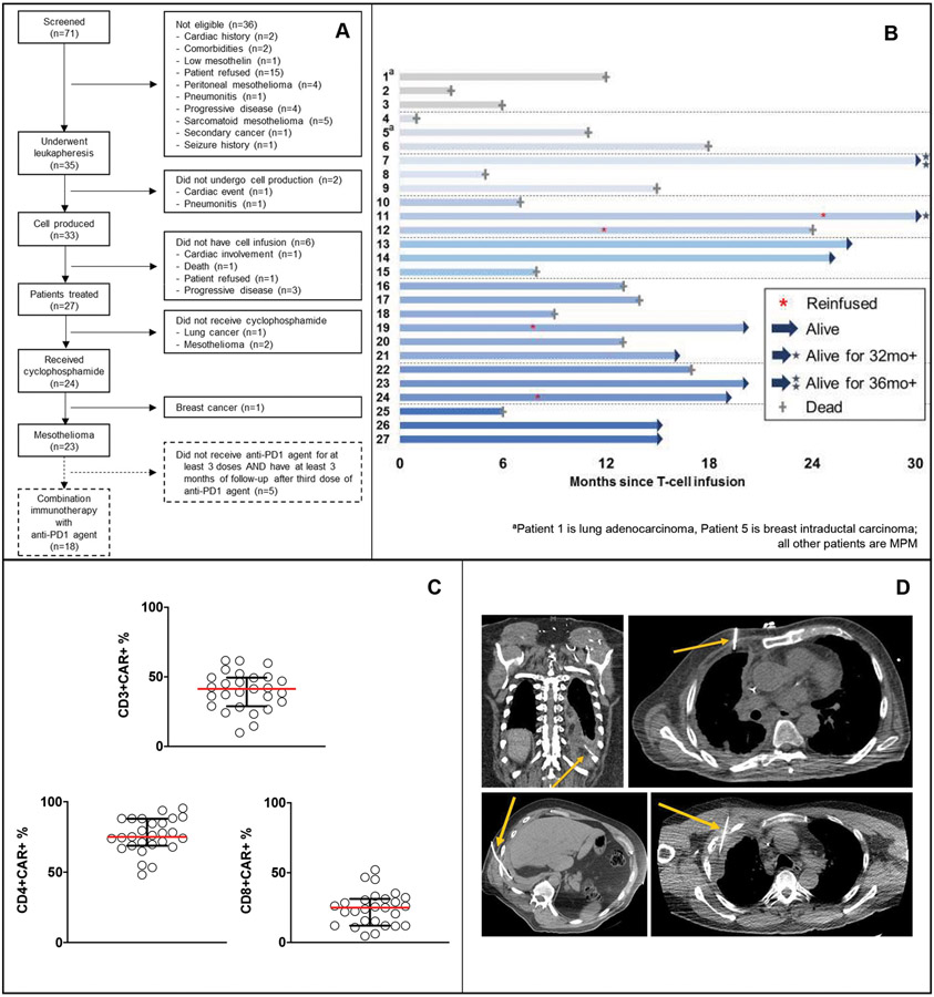
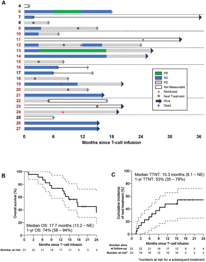
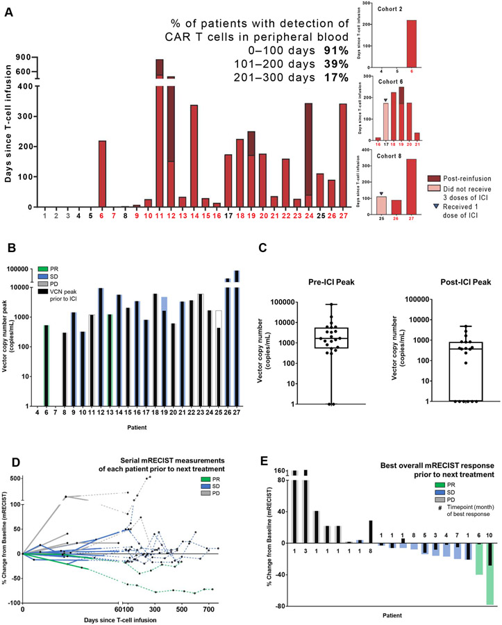
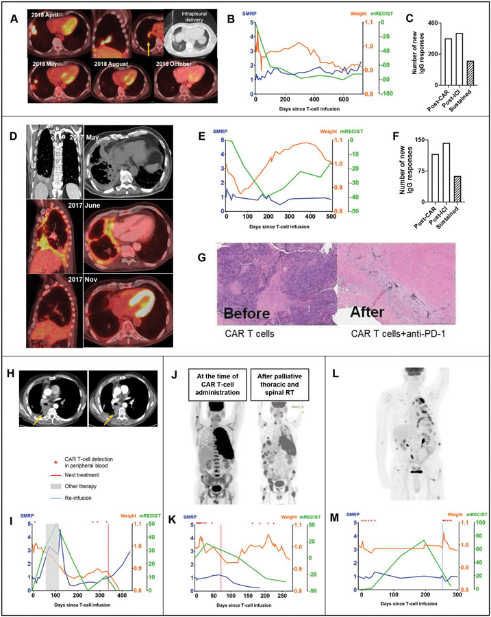
Malignant pleural diseases, comprising metastatic lung and breast cancers and malignant pleural mesothelioma (MPM), are aggressive solid tumors with poor therapeutic response. We developed and conducted a first-in-human, phase I study of regionally delivered, autologous, mesothelin-targeted chimeric antigen receptor (CAR) T-cell therapy. Intrapleural administration of 0.3M to 60M CAR T cells/kg in 27 patients (25 with MPM) was safe and well tolerated. CAR T cells were detected in peripheral blood for >100 days in 39% of patients. Following our demonstration that PD-1 blockade enhances CAR T-cell function in mice, 18 patients with MPM also received pembrolizumab safely. Among those patients, median overall survival from CAR T-cell infusion was 23.9 months (1-year overall survival, 83%). Stable disease was sustained for ≥6 months in 8 patients; 2 exhibited complete metabolic response on PET scan. Combination immunotherapy with CAR T cells and PD-1 blockade agents should be further evaluated in patients with solid tumors. SIGNIFICANCE: Regional delivery of mesothelin-targeted CAR T-cell therapy followed by pembrolizumab administration is feasible, safe, and demonstrates evidence of antitumor efficacy in patients with malignant pleural diseases. Our data support the investigation of combination immunotherapy with CAR T cells and PD-1 blockade agents in solid tumors.See related commentary by Aldea et al., p. 2674.This article is highlighted in the In This Issue feature, p. 2659.


Conflict of interest statement

Conflicts of Interest: P.S.A. has received research funding from ATARA Biotherapeutics, has served on the Scientific Advisory Board or as consultant to ATARA Biotherapeutics, Bayer, Carisma Therapeutics, Imugene, ImmPACT Bio, and Takeda Therapeutics, and has patents, royalties, and intellectual property on mesothelin-targeted CARs and T-cell therapies, method for detection of cancer cells using virus, and pending patent applications on T-cell therapies. D.R.J. serves as a consultant for AstraZeneca and Merck.

Figures

Figure 1.
Figure 1.. Clinical details of patients with primary or metastatic pleural malignancy treated in the phase I clinical trial (N=27).
A, A single dose of mesothelin-targeted CAR T cells was administered by intrapleural administration without (cohort 1, patients 1-3) or with (cohorts 2-8, patients 4-27) cyclophosphamide preconditioning. All patients had histologically proven pleural malignancy (patient 1, non-small cell lung cancer; patient 5, human epidermal growth factor receptor 2–negative breast cancer; all others, MPM). B, The swimmer plot demonstrates each patient as a bar, with alive or dead status indicated as of June 2020. Four patients received reinfusion of CAR T cells (*). C, CAR T-cell transduction (median, 41% [range, 10%-62%]) was successful in both CD4 and CD8 T cells – percentages of CD4+ CAR+ (median, 75%; range, 48%-95%) and CD8+ CAR+ (median, 25%; range, 5%-52%). Circles represent individual data points corresponding to a single patient across all dose cohorts. Horizontal lines indicate median values, with interquartile ranges shown for the group. D, CAR T cells were administered intrapleurally either through a pleural catheter (n=11) or via intervention radiology–guided imaging (n=16).
Figure 2.
Figure 2.. Outcomes of patients with MPM (n=23).
A, Therapy responses in patients with MPM (3 did not have measurable disease, 1 had no subsequent scan available) or disease stabilization or progression until next treatment during a period (0 to 36 months), as monitored by mRECIST on CT scan, are shown—PR (green), SD (blue), and PD (gray) are represented in relation to time in months. Solid line indicates survival post-next treatment. Patients who received combination immunotherapy are represented in red type. B, The Kaplan-Meier curve reports overall survival (OS) of patients with malignant pleural mesothelioma after CAR T-cell infusion (median survival, 17.7 months [95% confidence interval, 13.2 months to not estimable {NE}]). C, The time-to-next-treatment (TTNT) curve shows the proportion of patients receiving next treatment over time (median time to next treatment, 15.3 months [95% confidence interval, 8.1 months to NE]).
Figure 3.
Figure 3.. Outcomes analyses of patients with MPM (n=23).
A, Presence of CAR T cells in serially collected peripheral blood samples from each patient was detected and quantified using a multiplex real-time PCR assay. CAR T cells (as quantified by vector copy number per milliliter) can be detected in peripheral blood as early as 1 to 5 days after intrapleural administration and as late as 100 to 300 days in a majority of patients. Duration of detection of CAR T cells by PCR in the peripheral blood is shown. No CAR T cells were detected in the peripheral blood of patients treated in cohort 1. Patients who received combination therapy are represented in red color on the X-axis; patients who received reinfusion are represented by stacked bar graphs. B, Following intrapleural administration, CAR T cells can be detected in peripheral blood as early as 1 to 5 days in a majority of patients and as late as 100 to 300 days in some patients. Peak vector copy number is shown; colored bars indicate best overall response via mRECIST, and black bars indicate best response prior to ICI start. C, Box-plot shows peak vector copy number pre- and post-ICI. D, Therapy response of patients with MPM (3 patients did not have measurable disease, 1 had no subsequent scan available) before starting next treatment, defined by mRECIST measurement on CT scan as partial response (PR), stable disease (SD), or progressive disease (PD) (within 0-770 days of CAR T-cell infusion). Solid line indicates measurements pre-ICI; dotted line indicates measurements post-ICI. E, Best overall response as measured by mRECIST, along with the time point of best response in months, is shown for 23 patients with MPM (3 patients did not have measurable disease, 1 had no subsequent scan available). The median time point of initiation of the best response among patients without progression (PR or SD) was 4 months, compared with 1 month for patients with progression (PD). Colored bars indicate best overall response via mRECIST; black bars indicate best response prior to ICI start.
Figure 4.
Figure 4.. Clinical responses and correlative analyses of representative patients.
A, A 76-year-old patient diagnosed with stage 3A epithelioid MPM (TMB, 2.6 mt/Mb; mesothelin expression, 100%; PD-L1 expression, 0%) following platinum-based chemotherapy with disease burden in the right pleura, fissure, diaphragm, and hilar lymph nodes received a single intrapleural dose of 6M mesothelin-targeted CAR T cells/kg following cyclophosphamide preconditioning. A CT scan performed 4 weeks later showed a 28% reduction in the fissural tumor nodules. The patient received pembrolizumab starting at week 6 after CAR T-cell administration. A CT performed at 3 months showed a 62% reduction in the target disease by mRECIST and complete metabolic response on PET scan. B, At 26 months, the patient remained functionally well, with stable mRECIST, weight, and serum SMRP values (SMRP and weight fold change from baseline, mRECIST percent change from baseline; represented on the Y-axis), and did not require any additional therapies. C, Proteomic analyses showed new IgG responses, compared with baseline, following CAR T cells alone and after initiation of pembrolizumab—total responses sustained at both time points are indicated by the shaded bar. D, A 72-year-old patient with biphasic MPM (stage 3A; TMB, 4.9 mt/Mb; mesothelin expression, 25%; PD-L1 expression, 1%) underwent platinum-based chemotherapy with resulting SD that was unresectable. The patient was administered a single dose of 0.3M mesothelin-targeted CAR T cells/kg intrapleurally after cyclophosphamide (cohort 2). A PET scan showed metabolically active right MPM encircling the pleural cavity. Six weeks after CAR T-cell administration, the patient received pembrolizumab. Three months after pembrolizumab, a PET scan showed complete metabolic response (D), and a CT scan showed 40% PR by mRECIST (E). For 16 months after CAR T-cell administration, the patient remained functionally normal, gained and maintained weight, maintained baseline serum SMRP level (E), and had evidence on peripheral blood evaluation of new IgG responses following CAR T cells and pembrolizumab (some responses sustained) (F)—the patient did not require any additional treatments during this time. Peripheral blood and tumor biopsy evaluation documented presence of CAR T cells at 32 weeks. A repeat biopsy of the suspected tumor relapse showed cancer cells embedded in a bed of stroma (G), compared with abundant tumor noticed before administration of CAR T cells. H, A 66-year-old patient with epithelioid MPM with tumor relapse (TMB, 2 mt/Mb; 3 prior lines of therapy) received a single intrapleural dose of 1M CAR T cells/kg following cyclophosphamide. Reduction in the target lesion can be seen on CT scans after pembrolizumab (right panel), compared with after CAR T-cell therapy (left panel). I, Serial measurements of response by mRECIST (green), serum SMRP level (blue), and patient weight reflecting the general condition (orange) are shown during a period of 500 days. Following CAR T-cell administration, a minimal reduction in serum SMRP level was noted. The patient went on to receive another treatment, with PD indicated by an increase in serum SMRP level and mRECIST measurement associated with weight loss. No CAR T cells were detected in peripheral blood at the time of the patient’s return to the institution. On day 121, the patient started to receive pembrolizumab, with a subsequent decrease in serum SMRP level and mRECIST measurement and with stabilization of weight. More importantly, CAR T cells were redetected in the peripheral blood, as indicated by the red asterisk. J, A patient with epithelioid MPM with advanced, stage 4 disease received hemithoracic and spine palliative radiation therapy for progressive disease 71 days after intrapleural CAR T cells and pembrolizumab. The patient’s further clinical course showed a reduction in the thoracic tumor associated with redetection of CAR T cells in peripheral blood 100 days later. K, Serial tumor measurements (percent change from baseline) by mRECIST, fold change in serum SMRP level, and fold change in weight are shown during a period of 250 days after CAR T-cell infusion. L, A 70-year-old patient with epithelioid MPM with disease relapse with extensive metastases (4 previous lines of therapy) received CAR T cells and subsequent pembrolizumab. M, A second dose of CAR T cells was administered intrapleurally in August 2019. CAR T cells were detectable in peripheral blood for 4 weeks after the first infusion and for 13 weeks after reinfusion, at which time the patient stopped follow-up at our institution. Reduction in the target lesion was seen by mRECIST following reinfusion, although the patient developed new metastatic lesions.
Figure 5.
Figure 5.. Peripheral blood and pleural fluid clonality and protein analyses.
A-C, Peripheral blood mononuclear cells collected from each patient at different time points were subjected to gDNA isolation to study T-cell receptor beta clonality using ImmunoSEQ analysis. A, Shown are the numbers of expanded clones at 1, 2, and 3 to 5 weeks after CAR T-cell infusion followed by (B) maximum fold change in clone number delineated by the response relative to the value at baseline; horizontal lines indicate medians, with interquartile ranges. C, Simpson clonality index (lower value indicates diverse clonal population, an increase indicates clonal expansion) before CAR T-cell infusion and at 3 to 5 weeks after CAR T-cell infusion in the peripheral blood of patients with PR, SD, or PD, as measured by mRECIST, is shown; horizontal lines indicate medians, with interquartile ranges. D-F, Proteomic analyses showed evidence of new IgG responses in the peripheral blood (epitope spreading) after administration of both CAR T cells and pembrolizumab. Serum samples collected from each patient at baseline, after CAR T-cell infusion, and after administration of pembrolizumab were subjected to a ProtoArray assay to identify anti-IgG responses against 24,000 proteins at each time point. Shown are (D) medians (interquartile ranges) of new IgG responses, (E) the percentage of new IgG responses among total IgG responses in each patient, (F) prevalent (>3.5-fold expansion, compared with baseline) IgG responses after administration of CAR T cells and pembrolizumab and sustained IgG responses at both time points, and (G) the percentage of unique IgG responses among total prevalent IgG responses in each patient; horizontal lines indicate medians, with interquartile ranges. H, Presence and expansion of CAR T cells was noted in the pleural effusions collected at early time points (week 0-4), even when the peripheral blood analyses either showed low levels or did not show CAR T cells (n=4 patients with both pleural effusion and peripheral blood samples available). I, Pleural fluid and serum profiles of 68 proteins assessed using Luminex assays calculated as an average at 0, 1, 2, and 3 weeks after CAR T-cell infusion time points are shown as normalized values across time points. The majority of proteins measured were expressed at a maximal level at 2 weeks in the pleural fluid. J, Bar chart of proteins with >2-fold increase at week 2, compared with their baseline values, in pleural fluid (green bars) and their corresponding values in serum (gray bars). K, Clonal expansion was evident in the pleural fluid by week 2. In pleural fluid, there was an 84% to 100% overlap of expanded clones and a 66% to 82% overlap of all clones found in the peripheral blood. L, The peripheral clonality of each patient was determined at several time points and was compared with the infused product clonality to dynamically track changes in the identified clones. White squares show the fraction of clones found in the total repertoire also seen in the infusion product; black squares show the fraction of expanded clones that overlapped with the infusion product at several time points (up to 13 months) after CAR T-cell infusion, including the baseline sample collected before CAR T-cell infusion, is shown. Horizontal lines represent medians and interquartile ranges.

Comment in

References

    1. Vogelzang NJ, Rusthoven JJ, Symanowski J, Denham C, Kaukel E, Ruffie P, et al. Phase III study of pemetrexed in combination with cisplatin versus cisplatin alone in patients with malignant pleural mesothelioma. J Clin Oncol 2003;21(14):2636–44. - PubMed
    1. Zalcman G, Mazieres J, Margery J, Greillier L, Audigier-Valette C, Moro-Sibilot D, et al. Bevacizumab for newly diagnosed pleural mesothelioma in the Mesothelioma Avastin Cisplatin Pemetrexed Study (MAPS): a randomised, controlled, open-label, phase 3 trial. Lancet 2016;387(10026):1405–14. - PubMed
    1. Ujiie H, Kadota K, Nitadori JI, Aerts JG, Woo KM, Sima CS, et al. The tumoral and stromal immune microenvironment in malignant pleural mesothelioma: A comprehensive analysis reveals prognostic immune markers. Oncoimmunology 2015;4(6):e1009285. - PMC - PubMed
    1. Bograd AJ, Suzuki K, Vertes E, Colovos C, Morales EA, Sadelain M, et al. Immune responses and immunotherapeutic interventions in malignant pleural mesothelioma. Cancer Immunol Immunother 2011;60(11):1509–27. - PMC - PubMed
    1. Klampatsa A, O'Brien SM, Thompson JC, Rao AS, Stadanlick JE, Martinez MC, et al. Phenotypic and functional analysis of malignant mesothelioma tumor-infiltrating lymphocytes. Oncoimmunology 2019;8(9):e1638211. - PMC - PubMed

Publication types