

Dynamic mechanical interaction between injection liquid and human tissue simulant induced by needle-free injection of a highly focused microjet
- PMID: 34267280
- PMCID: PMC8282861
- DOI: 10.1038/s41598-021-94018-6
Dynamic mechanical interaction between injection liquid and human tissue simulant induced by needle-free injection of a highly focused microjet
Abstract
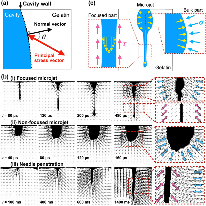
This study investigated the fluid-tissue interaction of needle-free injection by evaluating the dynamics of the cavity induced in body-tissue simulant and the resulting unsteady mechanical stress field. Temporal evolution of cavity shape, stress intensity field, and stress vector field during the injection of a conventional injection needle, a proposed highly focused microjet (tip diameter much smaller than capillary nozzle), and a typical non-focused microjet in gelatin were measured using a state-of-the-art high-speed polarization camera, at a frame rate up to 25,000 f.p.s. During the needle injection performed by an experienced nurse, high stress intensity lasted for an order of seconds (from beginning of needle penetration until end of withdrawal), which is much longer than the order of milliseconds during needle-free injections, causing more damage to the body tissue. The cavity induced by focused microjet resembled a funnel which had a narrow tip that penetrated deep into tissue simulant, exerting shear stress in low intensity which diffused through shear stress wave. Whereas the cavity induced by non-focused microjet rebounded elastically (quickly expanded into a sphere and shrank into a small cavity which remained), exerting compressive stress on tissue simulant in high stress intensity. By comparing the distribution of stress intensity, tip shape of the focused microjet contributed to a better performance than non-focused microjet with its ability to penetrate deep while only inducing stress at lower intensity. Dynamic mechanical interaction revealed in this research uncovered the importance of the jet shape for the development of minimally invasive medical devices.
© 2021. The Author(s).
Conflict of interest statement
The authors declare no competing interests.
Figures


References
-
- Rodríguez-Palomares JF, Dux-Santoy L, Guala A, Kale R, Maldonado G, Teixido-Tura G, Galian L, Huguet M, Valente F, Gutierrez L, et al. Aortic flow patterns and wall shear stress maps by 4dflow cardiovascular magnetic resonance in the assessment of aortic dilatation in bicuspid aortic valve disease. J. Cardiovasc. Magn. Reson. 2018;20(1):1–15. doi: 10.1186/s12968-018-0451-1. - DOI - PMC - PubMed
-
- Sass LR, Khani M, Romm J, Daners MS, McCain K, Freeman T, Carter GT, Weeks DL, Petersen B, Aldred J, et al. Non-invasive MRI quantification of cerebrospinal fluid dynamics in amyotrophic lateral sclerosis patients. Fluids Barriers CNS. 2020;17(1):1–14. doi: 10.1186/s12987-019-0164-3. - DOI - PMC - PubMed
Publication types
LinkOut - more resources
Full Text Sources
