. 2021 Jul 15;4(1):874.
doi: 10.1038/s42003-021-02399-1.

DeepEMhancer: a deep learning solution for cryo-EM volume post-processing

Affiliations

DeepEMhancer: a deep learning solution for cryo-EM volume post-processing

Ruben Sanchez-Garcia et al. Commun Biol. .

Abstract

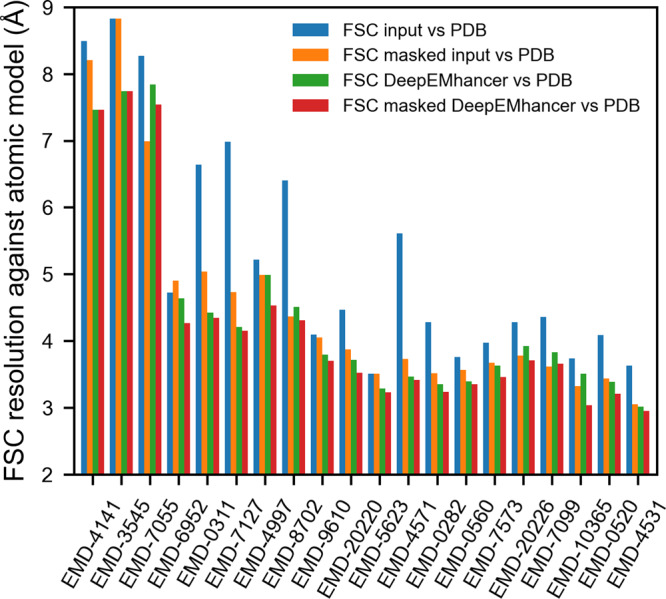
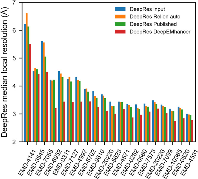
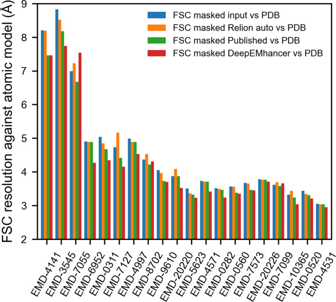
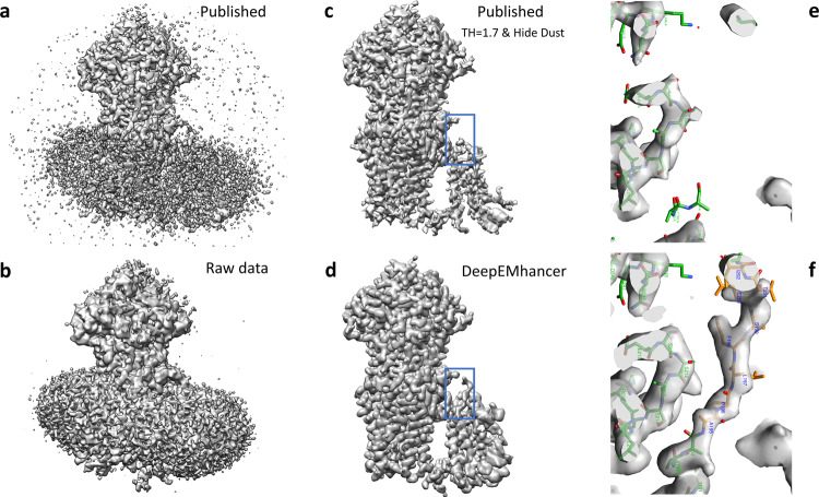
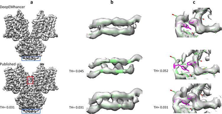
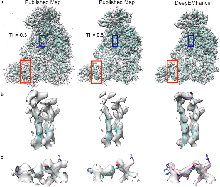
Cryo-EM maps are valuable sources of information for protein structure modeling. However, due to the loss of contrast at high frequencies, they generally need to be post-processed to improve their interpretability. Most popular approaches, based on global B-factor correction, suffer from limitations. For instance, they ignore the heterogeneity in the map local quality that reconstructions tend to exhibit. Aiming to overcome these problems, we present DeepEMhancer, a deep learning approach designed to perform automatic post-processing of cryo-EM maps. Trained on a dataset of pairs of experimental maps and maps sharpened using their respective atomic models, DeepEMhancer has learned how to post-process experimental maps performing masking-like and sharpening-like operations in a single step. DeepEMhancer was evaluated on a testing set of 20 different experimental maps, showing its ability to reduce noise levels and obtain more detailed versions of the experimental maps. Additionally, we illustrated the benefits of DeepEMhancer on the structure of the SARS-CoV-2 RNA polymerase.


Conflict of interest statement

The authors declare no competing interests.

Figures

Fig. 1
Fig. 1. DeepEMhancer produces maps that are more similar to the atomic models.
Resolution (determined by Fourier shell correlation coefficient, FSC) between the reference maps obtained from the atomic model and (1) the input maps (blue), (2) the input maps tightly masked (orange), (3) the post-processed maps by DeepEMhancer (green) and (4) the post-processed maps by DeepEMhancer tightly masked (red). EMDB entries are sorted by published global resolution.
Fig. 2
Fig. 2. DeepEMhancer produces better quality maps.
DeepRes median local resolution estimation for (1) the input maps (blue), (2) the post-processed maps obtained with Relion postprocessing automatic B-factor (orange), (3) the post-processed maps deposited in EMDB (green) and (4) the post-processed maps obtained with DeepEMhancer (Red). EMDB entries are sorted by published global resolution.
Fig. 3
Fig. 3. DeepEMhancer produces better results than global B-factor-based methods.
Resolution (determined by Fourier shell correlation coefficient, FSC) between the reference maps obtained from the atomic model and (1) the input maps (blue), (2) the post-processed maps obtained with Relion postprocessing automatic B-factor (orange), (3) the post-processed maps deposited in EMDB (green), and (4) the post-processed maps obtained with DeepEMhancer (red). EMDB entries are sorted by published global resolution.
Fig. 4
Fig. 4. DeepEMhancer results on testing map EMD-7099.
a Lateral view of the published map (B-factor sharpened, shown at the threshold recommended by the authors). b Lateral view of the raw data map obtained from the half maps that was used as input for DeepEMhancer. c Lateral view of the published map after rising the threshold and removing the small connected components so that the signal coming from the lipids was suppressed. As a collateral consequence, some densities corresponding to the protein were also lost. d Lateral view of the map obtained with DeepEMhancer. e Zoom-in of the region marked with a blue box in c. f Zoom-in of the region marked with a blue box in d, in which DeepEMhancer post-processed map, contrary to the published map, shows the densities corresponding to a missing loop in PDB 6bhu chain A. As a result, the residues A195 to I203 have been de novo modeled (new residues depicted in yellow, published in green).
Fig. 5
Fig. 5. DeepEMhancer results on testing map EMD-4997.
a Overview of the published map (B-factor sharpened, shown at the threshold recommended by the authors), bottom, and the map obtained with DeepEMhancer, top. Red box highlights an artifact that has been automatically removed by DeepEMhancer. Blue box delimits the region showed in b. b Zoom-in of the region marked with a blue box that contains the β-sheet R7-A10, chains A and B. The published volume is shown at the recommended threshold and at the threshold at which the backbone begins to look discontinuous. As it can be appreciated, the DeepEMhancer solution resolves better than the published map of the two strands of the sheet. c Zoom-in of the region centered at chain B residues H121 and Y361 (colored in magenta). The published volume is shown at the recommended threshold and at the smaller threshold at which the density that connects the two residues disappears. As it can be appreciated, DeepEMhancer post-processed map resolves better than the published map of the two residues.
Fig. 6
Fig. 6. Use case EMD-30178 from SARS-CoV-2 RNA-dependent RNA polymerase.
a Overview of the published map displayed with two different thresholds 0.3 (recommended by the authors, left) and 0.5 (middle panel) and processed with DeepEMhacer (right). PDB 7btf is shown in ribbon, red squares designated the zoomed areas in b panel and blue squares the zoomed areas in c. b Zoom-in and extraction of the density from the 3D reconstruction of the published map at different thresholds and DeepEMhacer map corresponding to the red squares in a, chain D from residues V115–I132. Newly traced residues in the DeepEMhancer map are shown in pink. c Zoom-in and extraction of the density from the 3D reconstruction of the published map at different thresholds and DeepEMhacer map corresponding to the blue boxes.

References

    1. Rosenthal PB, Henderson R. Optimal determination of particle orientation, absolute hand, and contrast loss in single-particle electron cryomicroscopy. J. Mol. Biol. 2003;333:721–745. doi: 10.1016/j.jmb.2003.07.013. - DOI - PubMed
    1. Kimanius D, Forsberg BO, Scheres SH, Lindahl E. Accelerated cryo-EM structure determination with parallelisation using GPUs in RELION-2. Elife. 2016;5:e18722. doi: 10.7554/eLife.18722. - DOI - PMC - PubMed
    1. Zivanov J, et al. New tools for automated high-resolution cryo-EM structure determination in RELION-3. Elife. 2018;7:e42166. doi: 10.7554/eLife.42166. - DOI - PMC - PubMed
    1. Terwilliger TC, Sobolev OV, Afonine PV, Adams PD. Automated map sharpening by maximization of detail and connectivity. Acta Crystallogr. Sect. D. 2018;74:545–559. doi: 10.1107/S2059798318004655. - DOI - PMC - PubMed
    1. Vilas JL, et al. Re-examining the spectra of macromolecules. Current practice of spectral quasi B-factor flattening. J. Struct. Biol. 2020;209:107447. doi: 10.1016/j.jsb.2020.107447. - DOI - PubMed

Publication types

MeSH terms

Substances