

Three-Dimensional Surface Imaging and Printing in Anatomic Pathology
- PMID: 34267987
- PMCID: PMC8274305
- DOI: 10.4103/jpi.jpi_8_21
Three-Dimensional Surface Imaging and Printing in Anatomic Pathology
Abstract
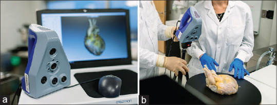
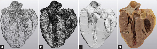
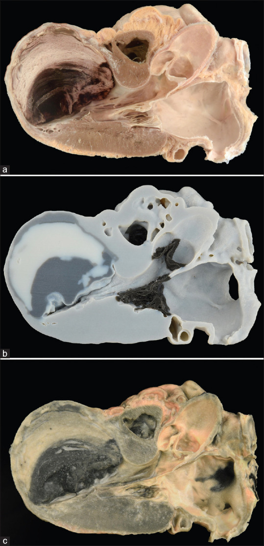
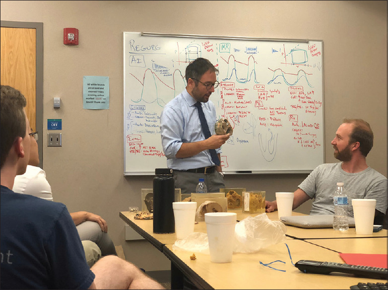
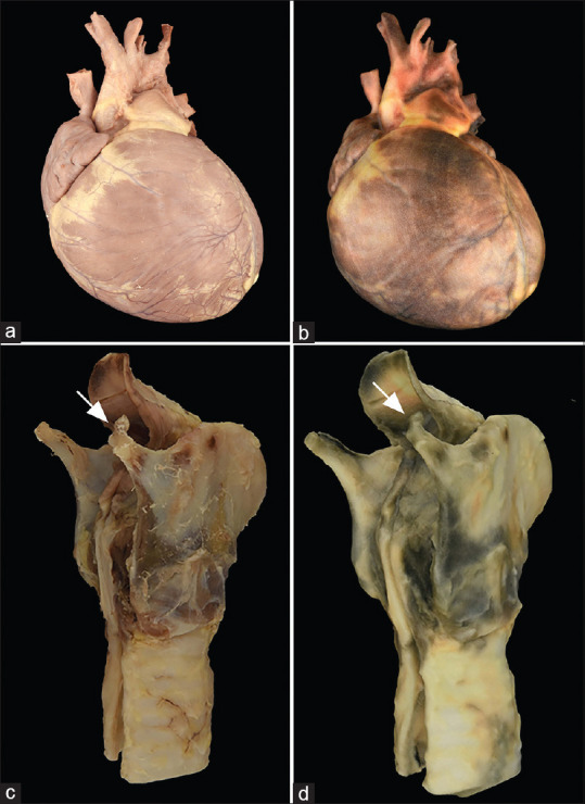
Three-dimensional (3D) imaging is increasingly being incorporated into a variety of medical specialties: surgery and radiology being but two prominent examples. Image-intensive disciplines, such as anatomic pathology (AP), represent excellent potential candidates for further exploration of this innovative technology. Multiple potential use cases exist within AP, involving patient care, education, and research. These use cases broadly include direct utilization of the 3D digital assets for viewing on a 2D screen, populating 3D extended reality platforms (virtual reality, augmented reality, and mixed reality) as well as generation of 3D printed photorealistic specimen models. Herein, these use cases are explored with specific regard to our experiences and yet unrealized potential. Future directions and considerations are also discussed.
Keywords: Augmented reality; photogrammetry; printing; scanning; virtual reality.
Copyright: © 2021 Journal of Pathology Informatics.
Conflict of interest statement
There are no conflicts of interest.
Figures

References
-
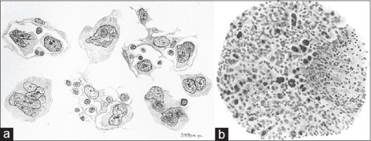
- Dawson PJ. The original illustrations of Hodgkin's disease. Ann Diagn Pathol. 1999;3:386–93. - PubMed
-
- Reed DM. On the pathological changes in Hodgkin's disease, with especial reference to its relation to tuberculosis. Johns Hopkins Hosp Rep. 1902;10:113–96.
-
- Sternberg C. There is also a peculiar tuberculosis of the lymphatic apparatus which appears as a pseudoleukimie. Z Heilk. 1898;19:21–91.
-
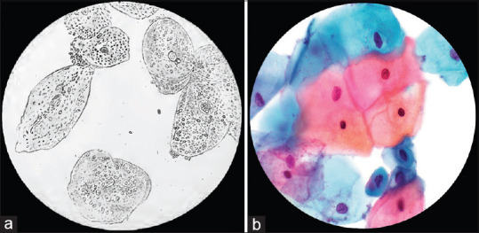
- Diamantis A, Magiorkinis E, Androutsos G. Alfred francois donné (1801-78): A pioneer of microscopy, microbiology and haematology. J Med Biogr. 2009;17:81–7. - PubMed
LinkOut - more resources
Full Text Sources
