. 2021 Jun 29:11:668247.
doi: 10.3389/fonc.2021.668247. eCollection 2021.

PI3K Promotes Basal Cell Carcinoma Growth Through Kinase-Induced p21 Degradation

Affiliations

PI3K Promotes Basal Cell Carcinoma Growth Through Kinase-Induced p21 Degradation

Rachel Y Chow et al. Front Oncol. .

Abstract

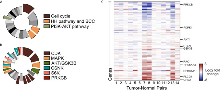
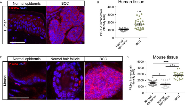
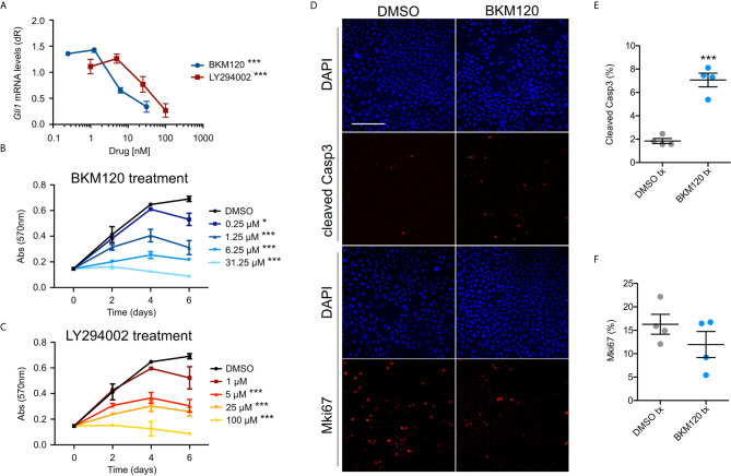
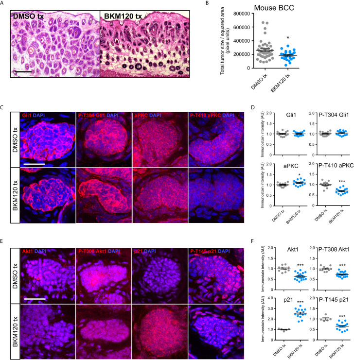
Basal cell carcinoma (BCC) is a locally invasive epithelial cancer that is primarily driven by the Hedgehog (HH) pathway. Advanced BCCs are a critical subset of BCCs that frequently acquire resistance to Smoothened (SMO) inhibitors and identifying pathways that bypass SMO could provide alternative treatments for patients with advanced or metastatic BCC. Here, we use a combination of RNA-sequencing analysis of advanced human BCC tumor-normal pairs and immunostaining of human and mouse BCC samples to identify a PI3K pathway expression signature in BCC. Pharmacological inhibition of PI3K activity in BCC cells significantly reduces cell proliferation and HH signaling. However, treatment of Ptch1fl/fl ; Gli1-CreERT2 mouse BCCs with the PI3K inhibitor BKM120 results in a reduction of tumor cell growth with no significant effect on HH signaling. Downstream PI3K components aPKC and Akt1 showed a reduction in active protein, whereas their substrate, cyclin-dependent kinase inhibitor p21, showed a concomitant increase in protein stability. Our results suggest that PI3K promotes BCC tumor growth by kinase-induced p21 degradation without altering HH signaling.

Keywords: PI3K - AKT pathway; atypical PKCι; basal cell carcinoma; hedgehog; p21.


Conflict of interest statement

The authors declare that the research was conducted in the absence of any commercial or financial relationships that could be construed as a potential conflict of interest.

Figures

Figure 1
Figure 1
PI3K/AKT pathway is upregulated in advanced BCC tumors. (A) KEGG analysis of the upregulated genes in advance BCC tumors highlighting the significant indicated terms. Cell cycle, p = 3.10 x 10⁻⁸; BCC, p = 1.03 x 10⁻⁴; HH signaling pathway, p = 2.49 x 10⁻⁴; PI3K-AKT signaling pathway, p = 0.00675. (B) Kinase Enrichment Analysis of differentially expressed genes showing significant kinases as indicated. In descending significance to color codes: CDK2, p = 4.80 x 10⁻¹²; CDK1, p = 1.13 x 10⁻⁸; MAPK14, p = 2.59 x 10⁻⁶; GSK3B, p = 5.42 x 10⁻⁶; CDK15, p = 3.88 x 10⁻⁴; CDK14, p = 4.39 x 10⁻⁴; CDK18, p = 4.94 x 10⁻⁴; CDK11A, p = 6.23 x 10⁻⁴; MAPK1, p = 0.00460; AKT1, p = 0.00534; MAP3K10, p = 0.00641; CSNK2A1, p = 0.00796; MAPK9, p = 0.00828; RPS6KA5, p = 0.0123; CSNK2A2, p = 0.0165; CSNK1E, p = 0.0268; CSNK1D, p = 0.0275; PRKCB, p = 0.0281; RPS6KA1, p = 0.0332. (C) Heat map of the differentially expressed PI3K pathway genes in advanced human BCCs compared to patient-matched normal skin. X mark, absence of data.
Figure 2
Figure 2
PIK3CA is upregulated in human and mouse BCC tumors. (A) Immunofluorescence staining of PI3KCA (red) and DAPI counterstain (blue) in human normal epidermis and nodular BCC tumors. Scale bar, 50 μm. (B) Quantification of PI3KCA immunofluorescence intensity (five points of measurement per sample, n=4 samples). AU, arbitrary unit. Error bar, SEM. (C) Immunofluorescence staining of Pi3kca (red) and DAPI counterstain (blue) in mouse normal epithelium, normal hair follicle, and BCC tumors. Scale bar, 25 μm. (D) Quantification of Pi3kca immunofluorescence intensity (five points of measurement per animal, n = 5 mice). AU, arbitrary unit. Error bar, SEM. Significance was determined by unpaired two-tailed t test. *p < 0.05; ***p < 0.001.
Figure 3
Figure 3
Inhibition of PI3K suppresses BCC cell growth and HH signaling. (A) Gli1 mRNA expression in ASZ001 cells treated with DMSO or varying concentrations of BKM120 or LY294002 (n = 3 experiments). dR, delta reporter gene normalized to passive reference dye. Error bar, SEM. Significance was determined by one-way ANOVA test. ***P < 0.001. (B, C) MTT assay of ASZ001 cells treated with DMSO or varying concentrations of (B) BKM120 or (C) LY294002 (n = 3 experiments). Abs, absorbance. Error bar, SEM. Significance was determined by two-way ANOVA test. *p < 0.05; ***p < 0.001. (D) Immunofluorescence staining of the indicated markers in ASZ001 cells treated with DMSO or BKM120. Scale bar, 200 µm. (E) Quantification of cleaved Casp3 signal (n = 4 experiments). (F) Quantification of Mki67 signal (n = 4 experiments). Error bars, SEM. Significance was determined by unpaired two-tailed t test. ***p < 0.001.
Figure 4
Figure 4
PI3K inhibition suppresses murine BCC growth and stabilizes p21. (A) Hematoxylin and eosin staining of dorsal back skin collected from Ptchfl/fl; Gli1-CreERT2 mice treated with DMSO or BKM120. Tx, treatment. Scale bar, 50 μm. (B) Quantification of total tumor size per square area (n>250 tumors from 5 mice). Tx, treatment. (C) Immunofluorescence staining of indicated markers (red) and DAPI counterstain (blue) in Ptch1fl/fl; Gli1-CreERT2 tumors treated with DMSO or BKM120. Scale bar, 25 μm. (D) Quantification of immunofluorescence intensity of indicated markers (five points of measurement per animal, n=3 mice). AU, arbitrary unit. (E) Immunofluorescence staining of indicated markers (red) and DAPI counterstain (blue) in Ptch1fl/fl; Gli1-CreERT2 tumors treated with DMSO or BKM120. Scale bar, 25 μm. (F) Quantification of immunofluorescence intensity of indicated markers (five points of measurement per animal, n=3 mice). Error bars, SEM. Significance was determined by unpaired two-tailed t test. *p < 0.05; ***p < 0.001.

References

    1. Armas-López L, Zúñiga J, Arrieta O, Ávila-Moreno F. The Hedgehog-GLI Pathway in Embryonic Development and Cancer: Implications for Pulmonary Oncology Therapy. Oncotarget (2017) 8(36):60684–703. 10.18632/oncotarget.19527 - DOI - PMC - PubMed
    1. Taylor MD, Liu L, Raffel C, Hui CC, Mainprize TG, Zhang X, et al. . Mutations in SUFU Predispose to Medulloblastoma. Nat Genet (2002) 31(3):306–10. 10.1038/ng916 - DOI - PubMed
    1. Raffel C, Jenkins RB, Frederick L, Hebrink D, Alderete B, Fults DW, et al. . Sporadic Medulloblastomas Contain PTCH Mutations. Cancer Res (1997) 47(5):842–5. - PubMed
    1. Tostar U, Malm CJ, Meis-Kindblom JM, Kindblom LG, Toftgård R, Undén AB. Deregulation of the Hedgehog Pathway: A Possible Role for the PTCH and SUFU Genes in Human Rhabdomyoma and Rhabdomyosarcoma Development. J Pathol (2006) 208(1):17–25. 10.1002/path.1882 - DOI - PubMed
    1. Reifenberger J, Wolter M, Knobbe CB, Köhler B, Schönicke A, Scharwächter C, et al. . Somatic Mutations in PTCH, Smoh, SUFUH, and TP53 Genes in Sporadic Basal Cell Carcinomas. Br J Dermatol (2005) 152(1):43–51. 10.1111/j.1365-2133.2005.06353.x - DOI - PubMed