Multicenter Study
. 2021 Sep;56(9):814-828.
doi: 10.1007/s00535-021-01813-z. Epub 2021 Jul 15.

Gastric epithelial neoplasm of fundic-gland mucosa lineage: proposal for a new classification in association with gastric adenocarcinoma of fundic-gland type

Affiliations
Multicenter Study

Gastric epithelial neoplasm of fundic-gland mucosa lineage: proposal for a new classification in association with gastric adenocarcinoma of fundic-gland type

Hiroya Ueyama et al. J Gastroenterol. 2021 Sep.

Abstract

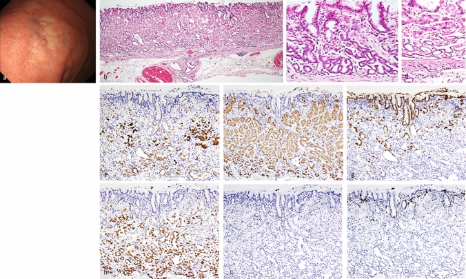
Background: Gastric adenocarcinoma of fundic-gland type (GA-FG) is a rare variant of gastric neoplasia. However, the etiology, classification, and clinicopathological features of gastric epithelial neoplasm of fundic-gland mucosa lineage (GEN-FGML; generic term of GA-FG related neoplasm) are not fully elucidated. We performed a large, multicenter, retrospective study to establish a new classification and clarify the clinicopathological features of GEN-FGML.

Methods: One hundred GEN-FGML lesions in 94 patients were collected from 35 institutions between 2008 and 2019. We designed a new histopathological classification of GEN-FGML using immunohistochemical analysis and analyzed via clinicopathological, immunohistochemical, and genetic evaluation.

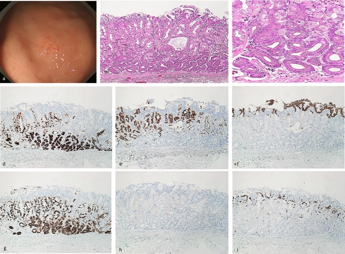
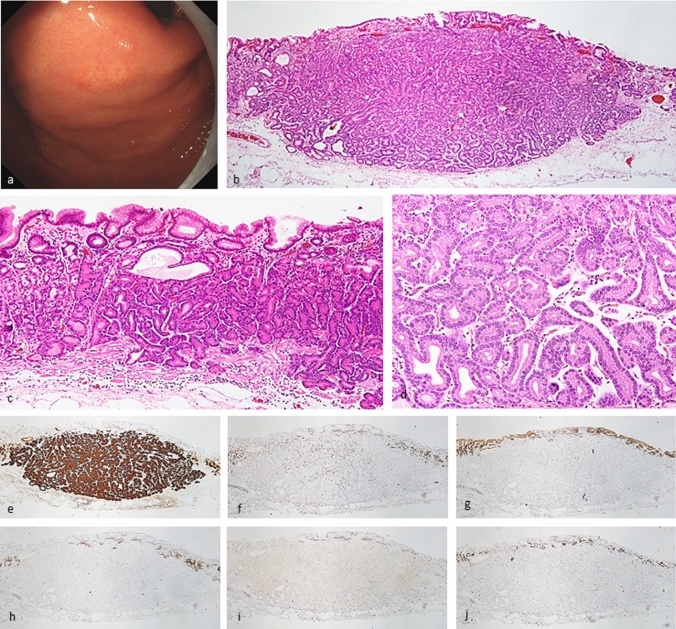
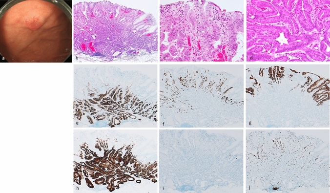
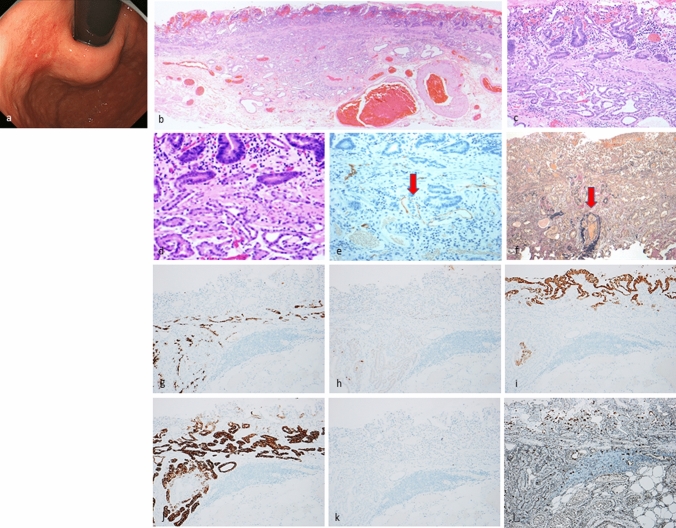
Results: GEN-FGML was classified into 3 major types; oxyntic gland adenoma (OGA), GA-FG, and gastric adenocarcinoma of fundic-gland mucosa type (GA-FGM). In addition, GA-FGM was classified into 3 subtypes; Type 1 (organized with exposure type), Type 2 (disorganized with exposure type), and Type 3 (disorganized with non-exposure type). OGA and GA-FG demonstrated low-grade epithelial neoplasm, and GA-FGM should be categorized as an aggressive variant of GEN-FGML that demonstrated high-grade epithelial neoplasm (Type 2 > 1, 3). The frequent presence of GNAS mutation was a characteristic genetic feature of GEN-FGML (7/34, 20.6%; OGA 1/3, 33.3%; GA-FG 3/24, 12.5%; GA-FGM 3/7, 42.9%) in mutation analysis using next-generation sequencing.

Conclusions: We have established a new histopathological classification of GEN-FGML and propose a new lineage of gastric epithelial neoplasm that harbors recurrent GNAS mutation. This classification will be useful to estimate the malignant potential of GEN-FGML and establish an appropriate standard therapeutic approach.

Keywords: Gastric adenocarcinoma of fundic-gland mucosa type; Gastric adenocarcinoma of fundic-gland type; Gastric epithelial neoplasm of fundic-gland mucosa lineage; Oxyntic gland adenoma.


Conflict of interest statement

All authors have no financial relationships relevant to this publication.

Figures

Fig. 1
Fig. 1
Oxyntic gland adenoma. Endoscopic image by white light endoscopy; a The flatly elevated lesion with submucosal tumor shape was located at the greater curvature of the fornix and had a whitish color and dilated vessels with branch architecture. The background mucosa had no atrophic change. Histological features (b, c); b Oxyntic gland adenoma was located at only deep area. The surface of the lesion was covered with normal foveolar epithelium. c The tumor cells were mainly composed of highly differentiated columnar cells mimicking fundic gland cells, predominantly chief cells, with pale gray-blue, basophilic cytoplasms, and mildly enlarged nuclei. Immunohistochemical results (di); d pepsinogen-I ( +), e H + /K + -ATPase (focally +), f MUC5AC (−, MUC5AC is only positive for superficial non-neoplastic foveolar cells), g MUC6 ( +), h p53 (−), i Ki-67 Labeling Index = 1%. Pathological diagnosis; U, 0-IIb, 5 × 4 mm, Oxyntic gland adenoma, UL0, Ly0, V0, HM0, VM0
Fig. 2
Fig. 2
Gastric adenocarcinoma of fundic-gland type. Endoscopic image by white light endoscopy; a The flatly elevated lesion with submucosal tumor shape was located at the greater curvature of the cardia and had a whitish color and dilated vessels with branch architecture. The background mucosa had no atrophic change. Histological features (bd); b, c Gastric adenocarcinoma resembling fundic-gland cells was located from beneath surface to deep area (SM600μm). The surface of the lesion was covered with normal foveolar epithelium, whereas the deep area of the tumor showed irregular branching and dilatation. d The tumor cells were mainly composed of highly differentiated columnar cells mimicking fundic-gland cells, predominantly chief cells, with pale gray-blue, basophilic cytoplasms, and mildly enlarged nuclei. Immunohistochemical results (ej); e pepsinogen-I ( +), f H + /K + -ATPase (focally +), g MUC5AC (−), h MUC6 (−), i p53 (−), j Ki-67 Labeling Index = 1%. Pathological diagnosis; U, 0-IIa, 9 × 6 mm, gastric adenocarcinoma of fundic-gland type, pT1b/SM2(600 µm), UL0, Ly0, V0, HM0, VM0
Fig. 3
Fig. 3
Gastric adenocarcinoma of fundic-gland mucosa type: type 1 (organized with exposure type). Endoscopic image by white light endoscopy; a The flatly elevated lesion with submucosal tumor shape was located at the posterior wall side of the fornix and had a reddish color. The background mucosa had no atrophic change. Histological features (bd); b Gastric adenocarcinoma resembling fundic-gland cells was located at deep area (SM700μm) and showed irregular branching and dilatation. The surface area of the lesion was composed of foveolar type well-differentiated adenocarcinoma followed by the component of GA-FG. Tissue construct of foveolar epithelium and fundic gland is maintained, and tumor is exposed on the surface. c (surface area) The tumor cells were composed of foveolar type well-differentiated adenocarcinoma with low-grade atypia. d (deep area) The tumor cells were mainly composed of highly differentiated columnar cells mimicking fundic-gland cells, predominantly chief cells, with pale gray-blue, basophilic cytoplasms, and mildly enlarged nuclei. Immunohistochemical results (ej); e pepsinogen-I ( +), f H + /K + -ATPase (focally +), g MUC5AC (surface, +), h MUC6 ( +), i p53 (−), j Ki-67 Labeling Index = 10%. Pathological diagnosis; U, 0-IIa, 15 × 13 mm, gastric adenocarcinoma of fundic-gland mucosa type, pT1b/SM2(700 µm), UL0, Ly0, V0, HM0, VM0
Fig. 4
Fig. 4
Gastric adenocarcinoma of fundic-gland mucosa type: type 2 (disorganized with exposure type). Endoscopic image by white light endoscopy; a The reddish depressed lesion was located at the posterior wall side of the cardia. The background mucosa had no atrophic change. Histological features (bd); b, c, d Gastric adenocarcinoma resembling fundic-gland cells was located at deep area (SM1000μm) and showed irregular branching and dilatation. The surface area of the lesion was composed of foveolar type well-differentiated adenocarcinoma with low-grade atypia. However, the layered architecture was destroyed, and tissue construct of foveolar epithelium and fundic gland is collapsed, and tumor is exposed on the surface. Immunohistochemical/histochemical results (el); e Lymphatic invasion (red arrow) demonstrated by D2-40, f Venous invasion (red arrow) demonstrated by EVG, g pepsinogen-I ( +), h H + /K + -ATPase (focally +), i MUC5AC (surface and deep area +), j MUC6 ( +), k p53 (−), l Ki-67 Labeling Index = 10%). Pathological diagnosis; U, 0-IIc, 25 × 20 mm, Adenocarcinoma of fundic-gland mucosa type, T1b/SM2(1000 µm), UL1, Ly1, V1, pHM0, pVM0
Fig. 5
Fig. 5
Gastric adenocarcinoma of fundic-gland mucosa type: Type 3 (disorganized with non-exposure type). Endoscopic image by white light endoscopy; a The flatly elevated lesion with submucosal tumor shape was located at the greater curvature of the cardia and had a whitish color and dilated vessels with branch architecture. The background mucosa had no atrophic change. Histological features (bd); b, c Gastric adenocarcinoma resembling fundic-gland cells was located at deep area and showed irregular branching and dilatation. The surface of the lesion was covered with normal foveolar epithelium. d In the middle of the mucosal layer, the tumor cells were composed of highly differentiated columnar cells mimicking fundic-gland cells, predominantly parietal cells. Immunohistochemical results (ej); e pepsinogen-I ( +), f H + /K + -ATPase (diffuse +), g MUC5AC ( +), h MUC6 ( +), i p53 (−), j Ki-67 Labeling Index = 3%). The tumor cells exhibited differentiation both gastric foveolar epithelium and fundic-gland differentiation in the deep area. Tissue construct of foveolar epithelium and fundic gland is collapsed, and tumor is not exposed on the surface. Pathological diagnosis; U, 0-IIc, 27 × 23 mm, Adenocarcinoma of fundic-gland mucosa type, T1b/SM1(100 µm), UL0, Ly0, V0, pHM0, pVM0

References

    1. Ueyama H, Yao T, Nakashima Y, et al. Gastric adenocarcinoma of fundic gland type (chief cell predominant type): proposal for a new entity of gastric adenocarcinoma. Am J Surg Pathol. 2010;34:609–619. doi: 10.1097/PAS.0b013e3181d94d53. - DOI - PubMed
    1. Ueyama H, Matsumoto K, Nagahara A, et al. Gastric adenocarcinoma of the fundic gland type (chief cell predominant type) Endoscopy. 2014;46:153–157. doi: 10.1055/s-0034-1364955. - DOI - PubMed
    1. Ueyama H, Yao T, Matsumoto K, et al. Establishment of endoscopic diagnosis for gastric adenocarcinoma of fundic gland type (chief cell predominant type) using magnifying endoscopy with narrow-band imaging. Stomach Intest. 2015;50:1533–1547.
    1. Ueyama H, Matsumoto K, Nagahara A, et al. Core tips for the endoscopic diagnosis of gastric adenocarcinoma of fundic gland type (chief cell predominant type) Gastrointest Endosc. 2016;58:1169–1177.
    1. Ueyama H, Yao T, Matsumoto K, et al. Gastric adenocarcinoma of fundic gland type. Stomach Intest. 2018;53:753–767.

Publication types

Supplementary concepts