

Early Postnatal Oxygen Exposure Predicts Choroidal Thinning in Neonates
- PMID: 34269816
- PMCID: PMC8297422
- DOI: 10.1167/iovs.62.9.23
Early Postnatal Oxygen Exposure Predicts Choroidal Thinning in Neonates
Abstract
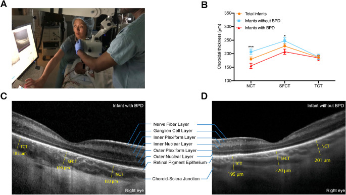
Purpose: To evaluate whether choroidal thickness (CT) using arm-mounted optical coherence tomography (OCT) in infants screened for retinopathy of prematurity (ROP) correlates with oxygen exposure in neonates.
Methods: OCT images were obtained in infants screened for ROP in a single level IV neonatal intensive care unit. CT was measured at three different locations: the subfoveal center and 1.5 mm from the fovea center in each direction. Correlation and regression analyses were performed to determine the relationship between clinical factors and CT. Clinical factors included gestational age, birth weight, presence of bronchopulmonary dysplasia (BPD), and fraction of inspired oxygen (FiO2) at defined time points: 30 weeks postmenstrual age (PMA), 36 weeks PMA, and on day of imaging.
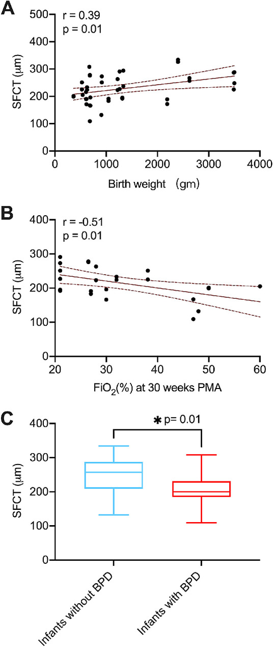
Results: Mean subfoveal, nasal, and temporal choroidal thicknesses CT (SFCT, NCT, and TCT, respectively) were 228.0 ± 51.4 µm, 179.7 ± 50.3 µm, and 186.4 ± 43.8 µm, respectively. SFCT was found to be significantly thicker than NCT and TCT (P < 0.0001 and P = 0.0002, respectively), but no significant difference was found between NCT and TCT (P = 0.547). Compared with infants without BPD, infants with BPD had thinner SFCT and NCT (P = 0.01 and P = 0.0008, respectively). Birth weight was positively correlated with SFCT (r = 0.39, P = 0.01) and NCT (r = 0.33, P = 0.045) but not TCT. Gestational age and ROP stage were not significantly associated with CT. SFCT was found to be significantly thinner with higher average FiO2 supplementation levels at 30 weeks PMA (r = -0.51, P = 0.01) but not at 36 weeks PMA. Regression analysis revealed that FiO2 at 30 weeks PMA was an independent predictor of SFCT in infants screened for ROP (P = 0.01).
Conclusions: Early postnatal exposure (<32 weeks PMA) to higher oxygen supplementation in premature neonates statistically predicts choroidal thinning.
Conflict of interest statement
Disclosure:
Figures
References
-
- Ying GS, Bell EF, Donohue P, Tomlinson LA, Binenbaum G.. Perinatal risk factors for the retinopathy of prematurity in postnatal growth and ROP study. Ophthalmic Epidemiol. 2019; 26(4): 270–278. - PubMed
Publication types
MeSH terms
Substances
LinkOut - more resources
Full Text Sources
