

Long splenic flexure carcinoma requiring laparoscopic extended left hemicolectomy with CME and transverse-rectal anastomosis: technique for a modified partial Deloyers in 5 steps to achieve enough reach and preserving middle colic vessels
- PMID: 34269879
- PMCID: PMC8847254
- DOI: 10.1007/s00423-021-02240-7
Long splenic flexure carcinoma requiring laparoscopic extended left hemicolectomy with CME and transverse-rectal anastomosis: technique for a modified partial Deloyers in 5 steps to achieve enough reach and preserving middle colic vessels
Abstract
Introduction: This How-I-Do-It article presents a modified Deloyers procedure by mean of the case of a 67-year-old female with adenocarcinoma extending for a long segment and involving the splenic flexure and proximal descending colon who underwent a laparoscopic left extended hemicolectomy (LELC) with derotation of the right colon and primary colorectal anastomosis.
Background: While laparoscopic extended right colectomy is a well-established procedure, LELC is rarely used (mainly for distal transverse or proximal descending colon carcinomas extending to the area of the splenic flexure). LELC presents several technical challenges which are demonstrated in this How-I-Do-It article.
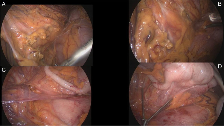
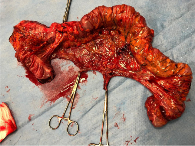
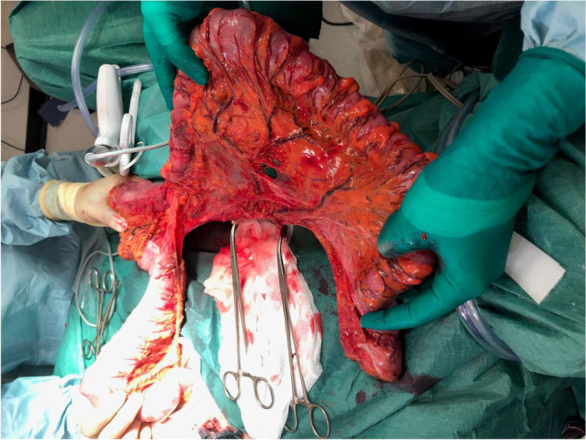
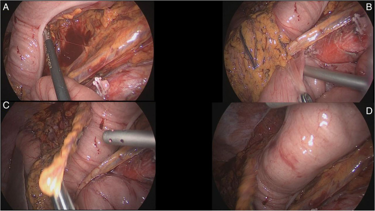
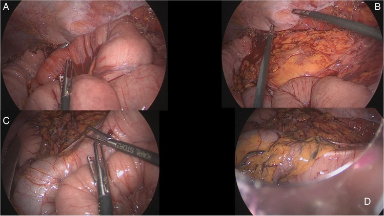
Technique and methods: Firstly, the steps needed to mobilize the left colon and procure a safe approach to the splenic flexure are described, especially when a tumor is closely related to it. This is achieved by mobilization and resection of the descending colon, while maintaining a complete mesocolic excision to the level of the duodenojejunal ligament for the inferior mesenteric vein and flush to the aorta for the inferior mesenteric artery. Subsequently, we depict the adjuvant steps required to enable a primary anastomosis by trying to mobilize the transverse colon and release as much of the mesocolic attachments at the splenic flexure area. Finally, we present the rare instance when a laparoscopic derotation of the ascending colon is required to provide a tension-free anastomosis. The resection is completed by delivery of the fully derotated ascending colon and hepatic flexure through a suprapubic mini-Pfannenstiel incision. The primary colorectal anastomosis is subsequently fashioned in a tension-free way and provides for a quick postoperative recovery of the patient.
Results: This modified Deloyers procedure preserves the middle colic since the fully mobilized mesocolon allows for a tension-free anastomosis while maintaining better blood supply to the mobilized stump. Also, by eliminating the need for a mesenteric window and the transposition of the caecum, we allow the small bowel to rest over the anastomosis and the mobilized transverse colon and reduce the possibility of an internal herniation of the small bowel into the mesentery.
Conclusions: Laparoscopic derotation of the right colon and a partial, modified Deloyers procedure preserving the middle colic vessels are feasible techniques in experienced hands to provide primary anastomosis after LELC with improved functional outcome. Nevertheless, it is important to consider anatomical aspects of the left hemicolectomy along with oncological considerations, to provide both a safe oncological resection along with good postoperative bowel function.
Keywords: Colonic derotation; Complete mesocolic excision; Deloyers procedure; Embryology; Left extended colectomy; Splenic flexure carcinoma.
© 2021. The Author(s).
Conflict of interest statement
The authors declare no competing interests.
Figures


References
-
- Mesenteric and peritoneal anatomy: basic and applied science–chapter 12, Taylor & Fancis, March 2017, 10.1201/9781315381565-2, In book: Mesenteric principles of gastrointestinal surgery
-
- Degiuli M, Reddavid R, Ricceri F, Di Candido F, Ortenzi M, Elmore U, Belluco C, Rosati R, Guerrieri M, Spinelli A, Members of the Italian Society of Surgical Oncology Colorectal Cancer Network (SICO-CCN) Collaborative Group Segmental colonic resection is a safe and effective treatment option for colon cancer of the splenic flexure: a nationwide retrospective study of the Italian Society of Surgical Oncology-Colorectal Cancer Network Collaborative Group. Dis Colon Rectum. 2020;63(10):1372–1382. doi: 10.1097/DCR.0000000000001743. - DOI - PubMed
-
- Rega D, Pace U, Scala D, Chiodini P, Granata V, Fares Bucci A, Pecori B, Delrio P. Treatment of splenic flexure colon cancer: a comparison of three different surgical procedures: Experience of a high volume cancer center. Sci Rep. 2019;9(1):10953. doi: 10.1038/s41598-019-47548-z. - DOI - PMC - PubMed
Publication types
MeSH terms
LinkOut - more resources
Full Text Sources
