. 2022 Feb;15(1):15-26.
doi: 10.1007/s12265-021-10155-3. Epub 2021 Jul 16.

Renal Revascularization Attenuates Myocardial Mitochondrial Damage and Improves Diastolic Function in Pigs with Metabolic Syndrome and Renovascular Hypertension

Affiliations

Renal Revascularization Attenuates Myocardial Mitochondrial Damage and Improves Diastolic Function in Pigs with Metabolic Syndrome and Renovascular Hypertension

Rahele A Farahani et al. J Cardiovasc Transl Res. 2022 Feb.

Abstract

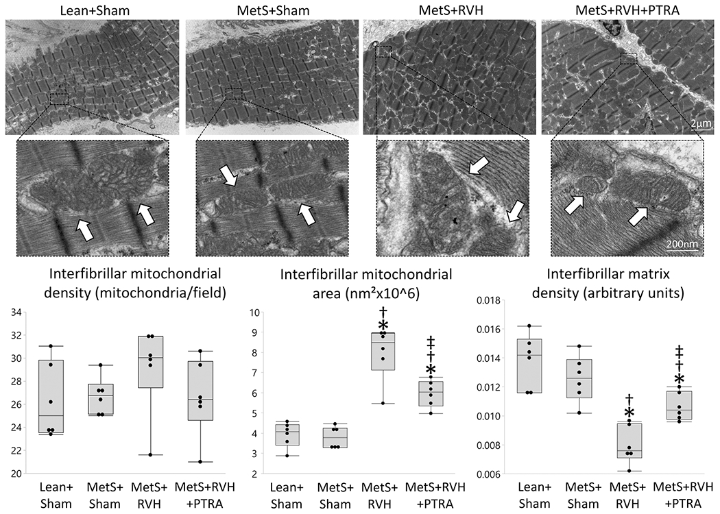
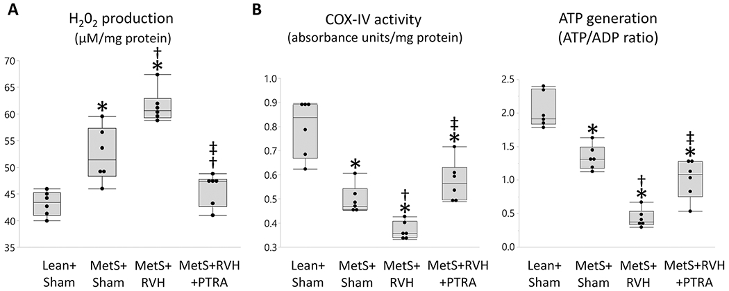
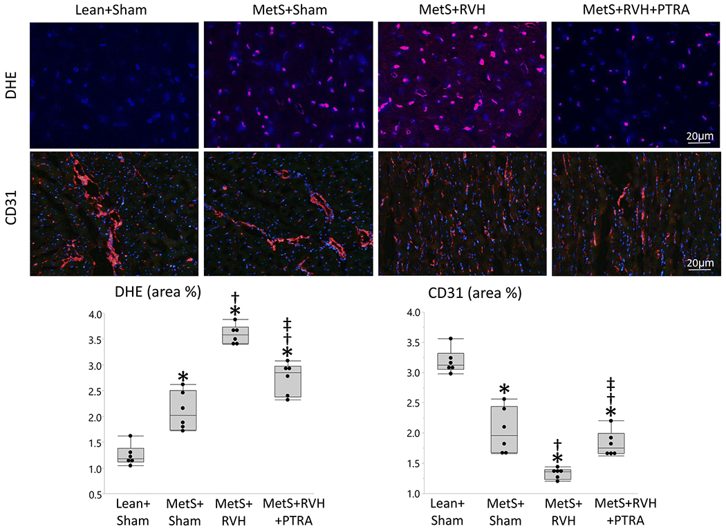
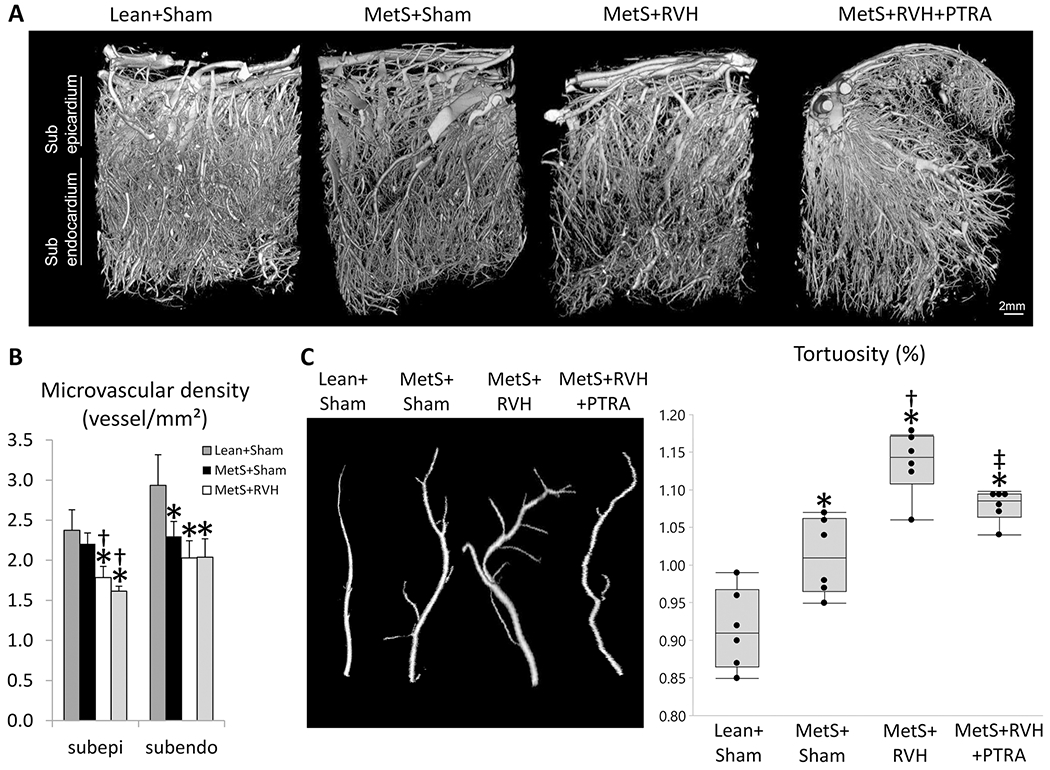
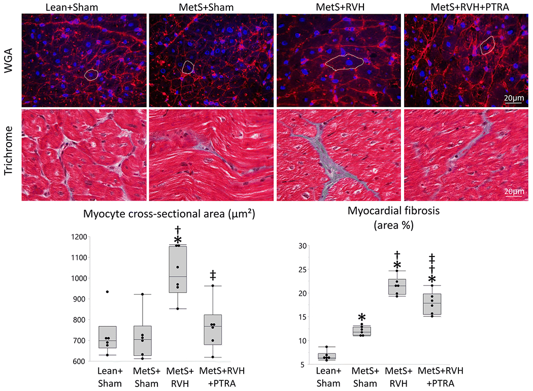
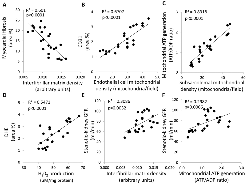
Percutaneous transluminal renal angioplasty (PTRA) may improve cardiac function in renovascular hypertension (RVH), but its effect on the biological mechanisms implicated in cardiac damage remains unknown. We hypothesized that restoration of kidney function by PTRA ameliorates myocardial mitochondrial damage and preserves cardiac function in pigs with metabolic syndrome (MetS) and RVH. Pigs were studied after 16 weeks of MetS+RVH, MetS+RVH treated 4 weeks earlier with PTRA, and Lean and MetS Sham controls (n=6 each). Cardiac function was assessed by multi-detector CT, whereas cardiac mitochondrial morphology and function, microvascular remodeling, and injury pathways were assessed ex vivo. PTRA attenuated myocardial mitochondrial damage, improved capillary and microvascular maturity, and ameliorated oxidative stress and fibrosis, in association with attenuation of left ventricular remodeling and diastolic dysfunction. Myocardial mitochondrial damage correlated with myocardial injury and renal dysfunction. Preservation of myocardial mitochondria with PTRA can enhance cardiac recovery, underscoring its therapeutic potential in experimental MetS+RVH.

Keywords: Cardiac dysfunction; Metabolic syndrome; Mitochondria; Renovascular hypertension; Revascularization.


Conflict of interest statement

Conflict of Interest

The authors declare no competing interests.

Figures

Figure 1.
Figure 1.
PTRA attenuated myocardial mitochondrial structural damage. Representative transmission electron microscopy images of interfibrillar mitochondria (arrows) and quantification of mitochondrial density, area, and matrix density in study groups (n=6/group each). *p<0.05 vs. Lean+Sham; †p<0.05 vs. MetS+Sham; ‡p<0.05 vs. MetS+RVH.
Figure 2.
Figure 2.
Renal revascularization improved cardiac mitochondrial function. Quantification of myocardial mitochondrial hydrogen peroxide (H202) production (A), cytochrome-c oxidase (COX)-IV activity (B), and ATP/ADP ratio (C) in study groups (n=6/group each). *p<0.05 vs. Lean+Sham; †p<0.05 vs. MetS+Sham; ‡p<0.05 vs. MetS+RVH.
Figure 3.
Figure 3.
PTRA attenuated cardiac oxidative stress and improved capillary density. Representative myocardial dihydroethidium (DHE, red) and immunofluorescence staining of CD31, and quantification of myocardial production of superoxide anion (DHE area %) and myocardial capillary density (CD31 area %) (n=6/group each). *p<0.05 vs. Lean+Sham; †p<0.05 vs. MetS+Sham; ‡p<0.05 vs. MetS+RVH.
Figure 4.
Figure 4.
Renal revascularization improved myocardial microvascular maturity. A Representative 3D micro-computed tomography images of the left ventricle in study groups. B Quantification of spatial density of microvessels in the subepicardium and subendocardium. C Quantification of microvascular tortuosity in study groups. *p<0.05 vs. Lean+Sham; †p<0.05 vs. MetS+Sham; ‡p<0.05 vs. MetS+RVH.
Figure 5.
Figure 5.
Renal revascularization ameliorated LV remodeling and fibrosis. Representative LV sections stained with wheat germ agglutinin (WGA) and trichrome, and quantification of myocyte cross-sectional area and myocardial fibrosis in Lean+Sham, MetS+Sham, MetS+RVH, and MetS+RVH+PTRA. *p<0.05 v Lean+Sham; †p<0.05 vs. MetS+Sham; ‡p<0.05 vs. MetS+RVH.
Figure 6.
Figure 6.
Cardiac mitochondrial damage correlated with myocardial injury. A Myocardial mitochondrial matrix density correlated inversely with myocardial fibrosis. B Endothelial cell mitochondrial density correlated directly with myocardial capillary density (CD31 area %). C Subsarcolemmal mitochondrial density correlated directly with cardiac mitochondrial ATP generation. D Myocardial mitochondrial H202 production ATP generation correlated directly with myocardial production of superoxide anion (DHE area %). E Myocardial mitochondrial matrix density correlated directly with stenotic kidney GFR. F Myocardial mitochondrial ATP generation correlated directly with stenotic kidney GFR.

References

    1. Herrmann SM and Textor SC, Renovascular hypertension. Endocrinol Metab Clin North Am, 2019. 48(4): p. 765–778. - PMC - PubMed
    1. Green D and Kalra PA, The heart in atherosclerotic renovascular disease. Front Biosci (Elite Ed), 2012. 4: p. 856–64. - PubMed
    1. Textor SC and Lerman LO, Paradigm shifts in atherosclerotic renovascular disease: Where are we now? J Am Soc Nephrol, 2015. 26(9): p. 2074–80. - PMC - PubMed
    1. Kane GC, et al., Renal artery revascularization improves heart failure control in patients with atherosclerotic renal artery stenosis. Nephrol Dial Transplant, 2010. 25(3): p. 813–20. - PubMed
    1. Holmuhamedov EL, et al., Cardiac subsarcolemmal and interfibrillar mitochondria display distinct responsiveness to protection by diazoxide. PLoS One, 2012. 7(9): p. e44667. - PMC - PubMed

Publication types