. 2021 Aug 9;39(8):1115-1134.e12.
doi: 10.1016/j.ccell.2021.06.016. Epub 2021 Jul 21.

Binary pan-cancer classes with distinct vulnerabilities defined by pro- or anti-cancer YAP/TEAD activity

Affiliations

Binary pan-cancer classes with distinct vulnerabilities defined by pro- or anti-cancer YAP/TEAD activity

Joel D Pearson et al. Cancer Cell. .

Abstract

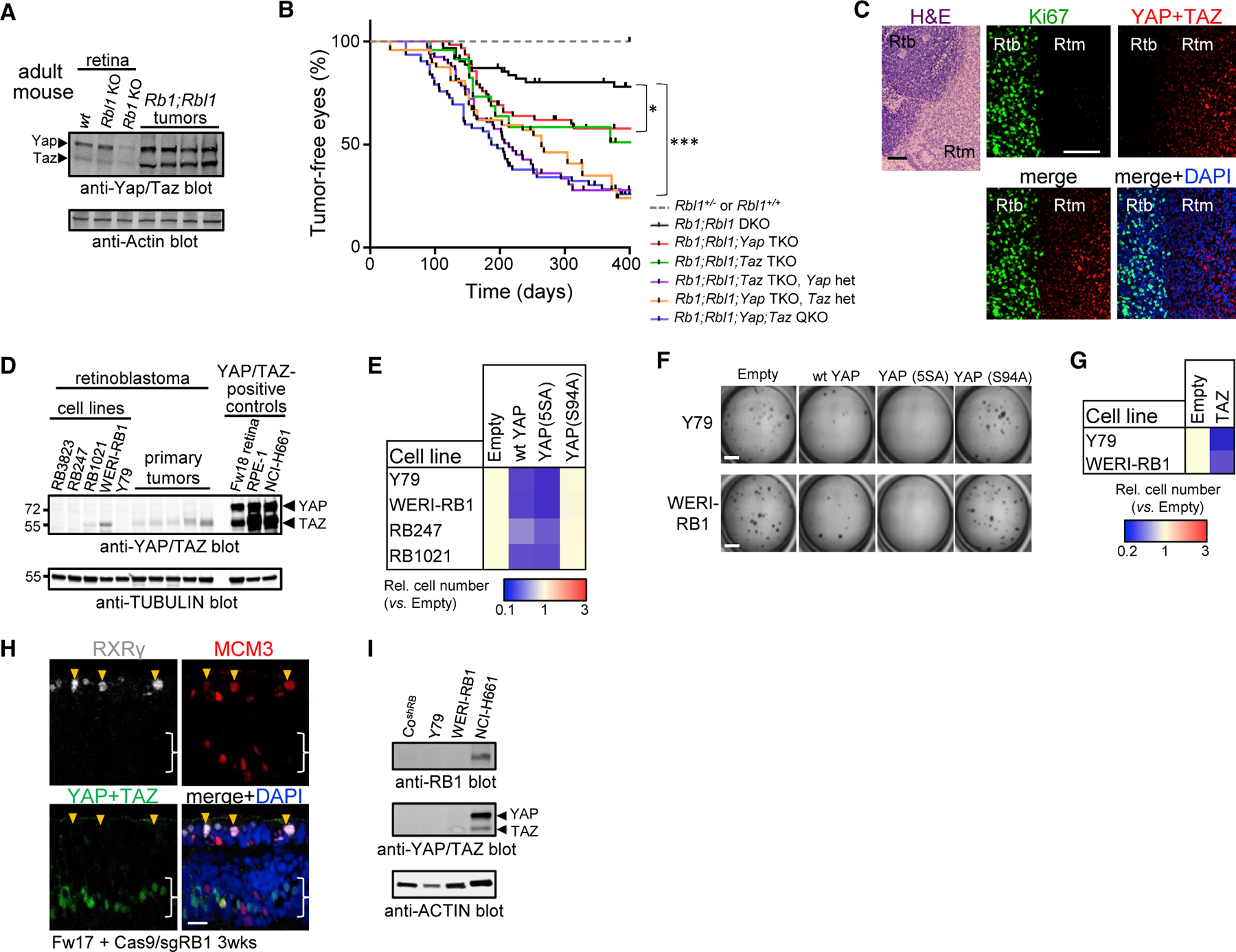
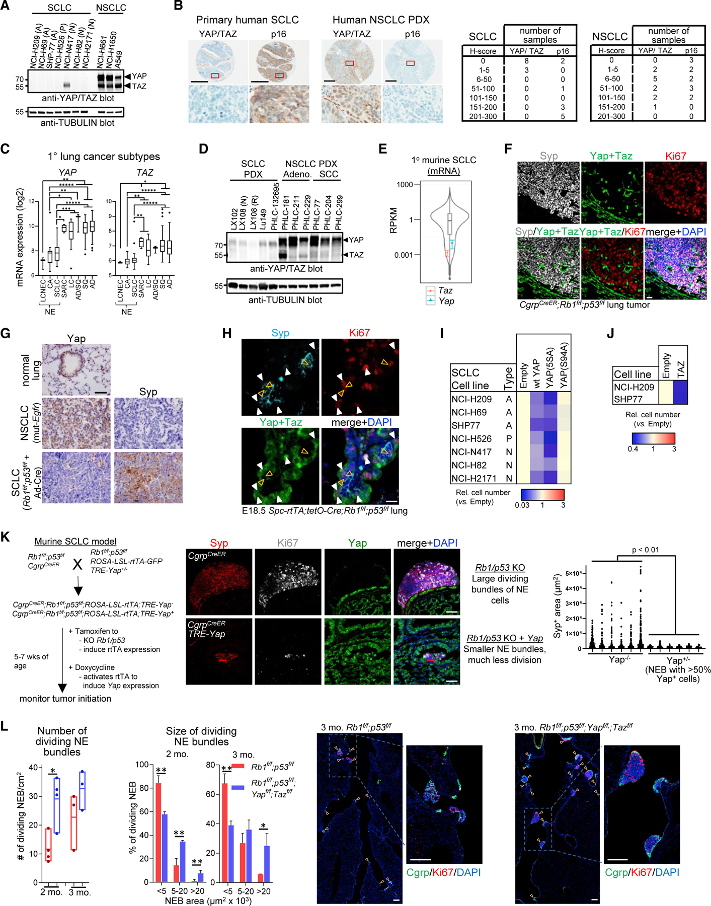
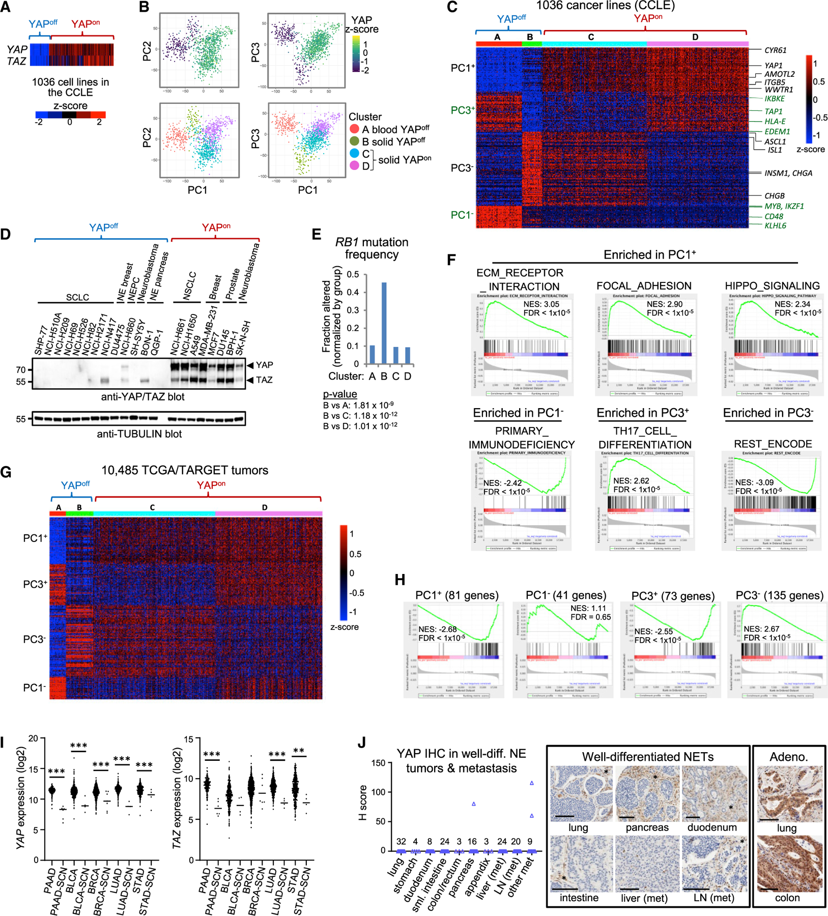
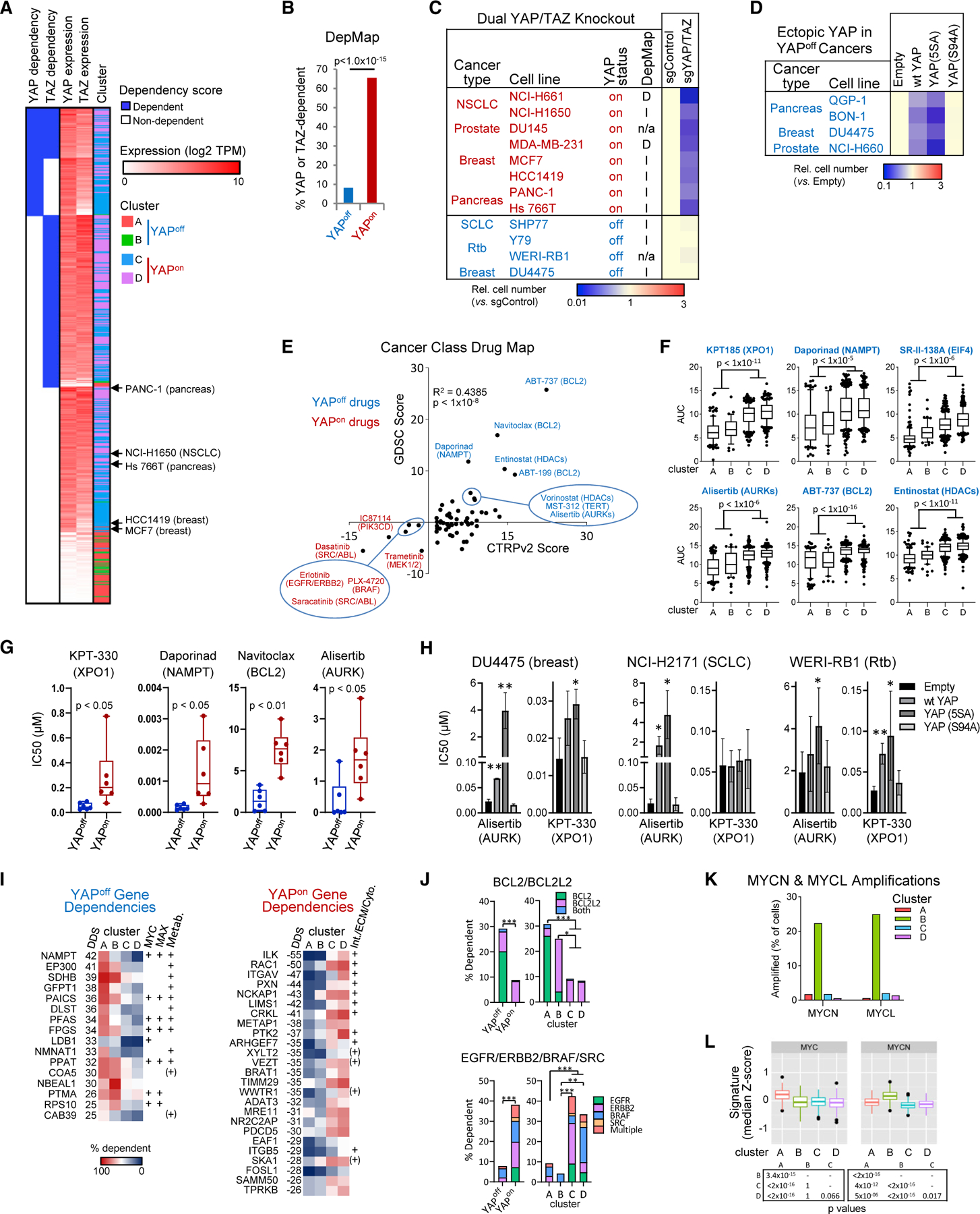
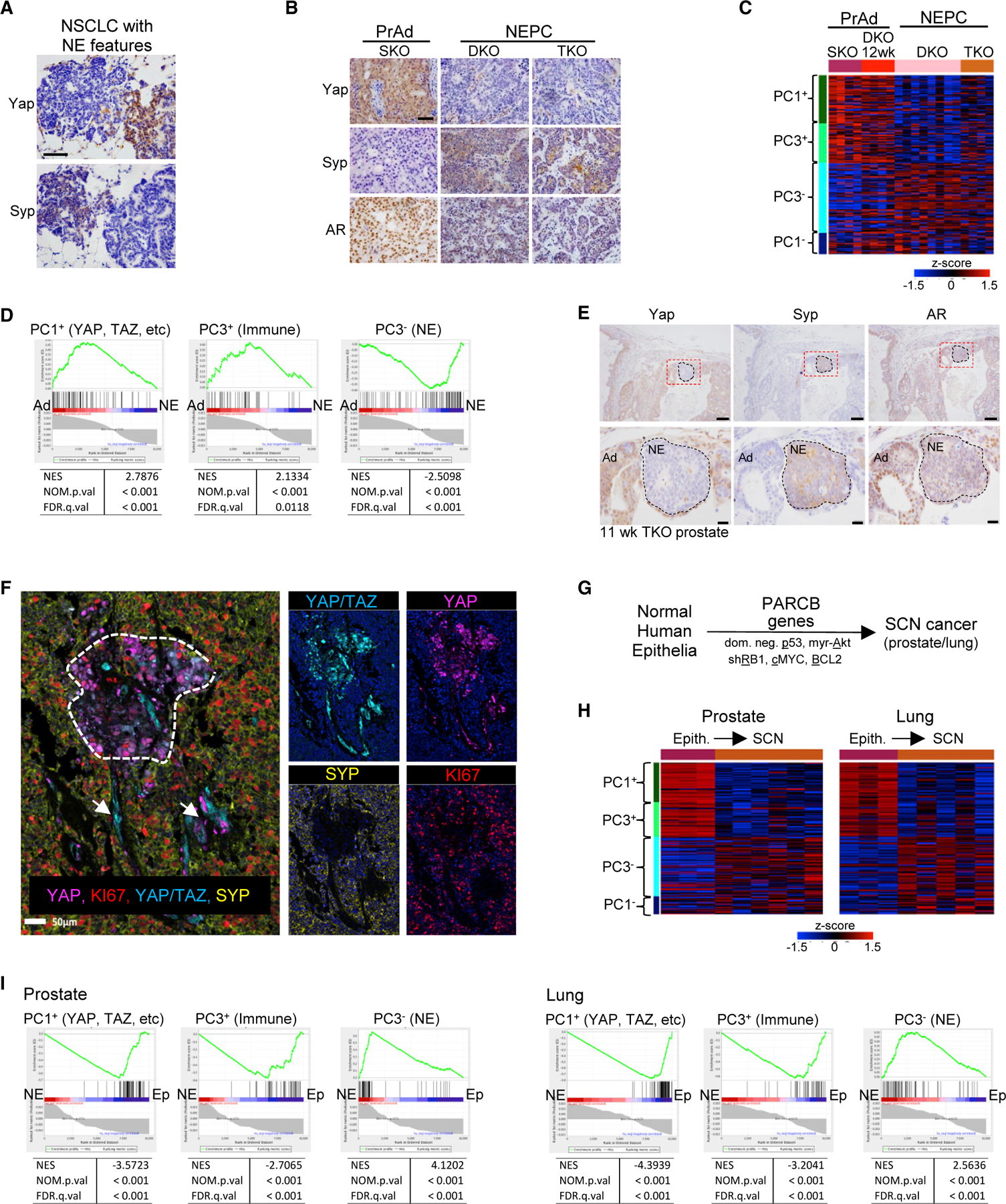
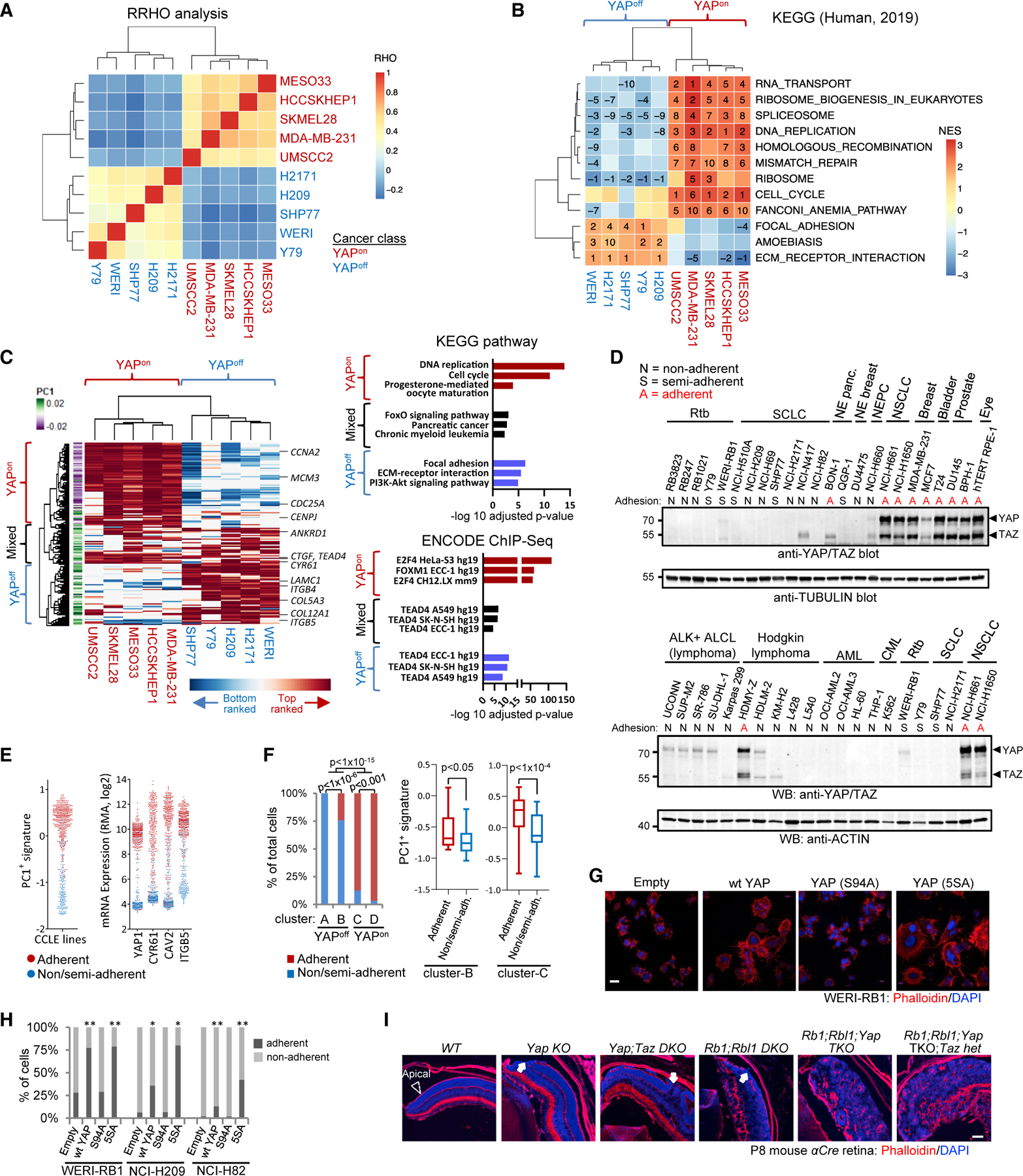
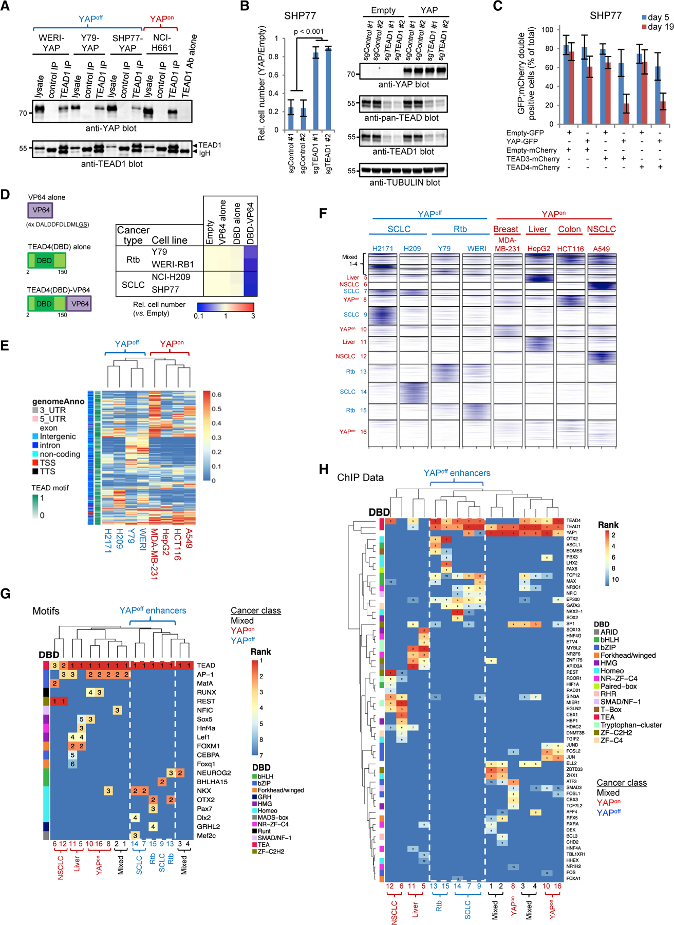
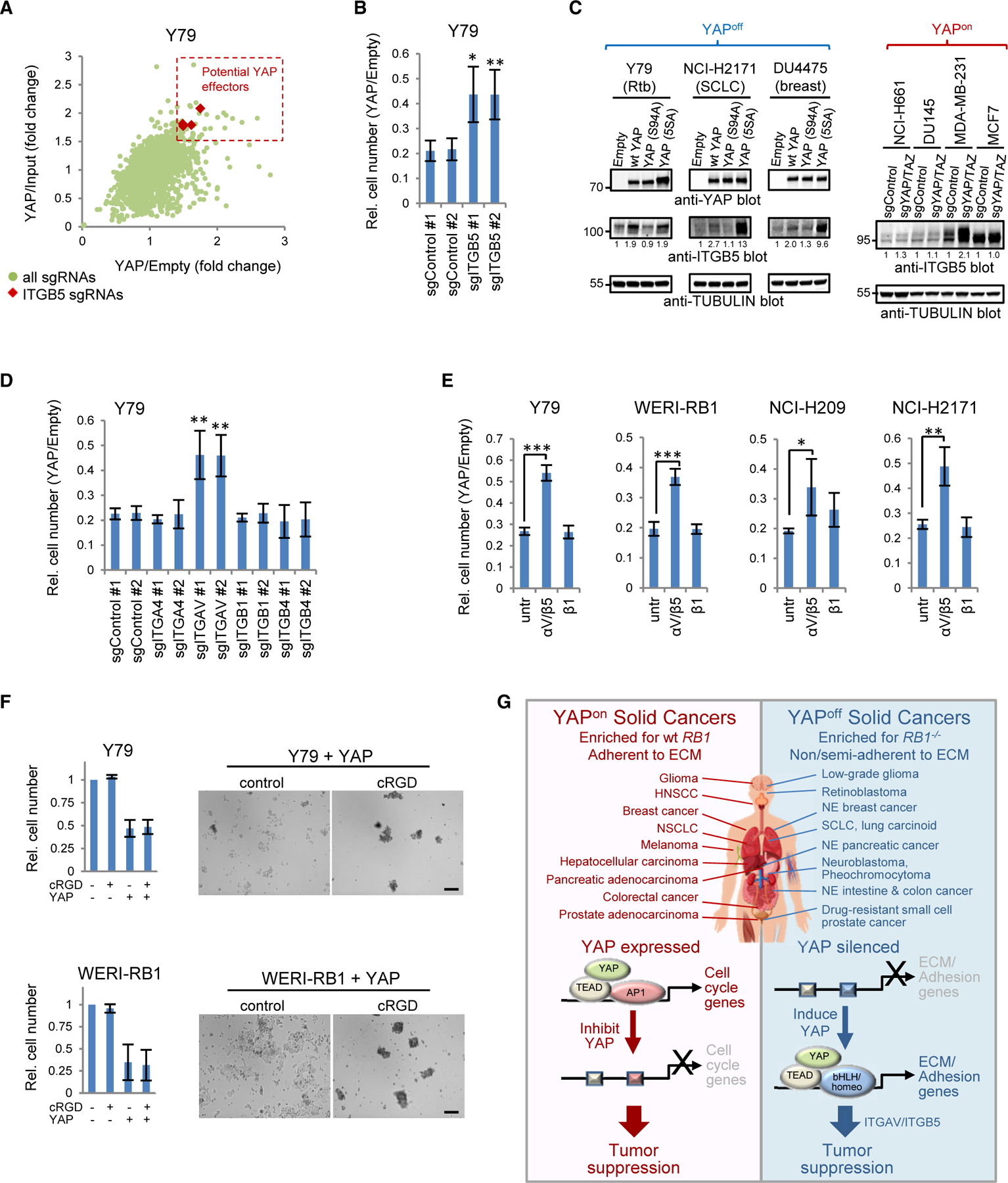
Cancer heterogeneity impacts therapeutic response, driving efforts to discover over-arching rules that supersede variability. Here, we define pan-cancer binary classes based on distinct expression of YAP and YAP-responsive adhesion regulators. Combining informatics with in vivo and in vitro gain- and loss-of-function studies across multiple murine and human tumor types, we show that opposite pro- or anti-cancer YAP activity functionally defines binary YAPon or YAPoff cancer classes that express or silence YAP, respectively. YAPoff solid cancers are neural/neuroendocrine and frequently RB1-/-, such as retinoblastoma, small cell lung cancer, and neuroendocrine prostate cancer. YAP silencing is intrinsic to the cell of origin, or acquired with lineage switching and drug resistance. The binary cancer groups exhibit distinct YAP-dependent adhesive behavior and pharmaceutical vulnerabilities, underscoring clinical relevance. Mechanistically, distinct YAP/TEAD enhancers in YAPoff or YAPon cancers deploy anti-cancer integrin or pro-cancer proliferative programs, respectively. YAP is thus pivotal across cancer, but in opposite ways, with therapeutic implications.

Keywords: RB1; TAZ/WWTR1; TEAD; YAP; cancer plasticity; cancer stratification; neuroendocrine cancer; retinoblastoma; retinoma; small cell cancers.


Conflict of interest statement

Declaration of interests B.H.L. reports honoraria and non-financial support from AstraZeneca, and has received research funding from Pfizer and AstraZeneca not related to this work.

Figures

Figure 1.
Figure 1.. Obligate YAP/TAZ silencing in retinoblastoma
(A) Western blots of murine retinal or tumor lysates. (B) Kaplan-Meyer from αCre mice of the indicated genotypes. *p < 0.01, ***p < 0.0001, log rank (Mantel-Cox) test. DKO/TKO/QKO, double/triple/quadruple knockout; het, heterozygous. (C) H&E and immunofluorescence (IF) in human retinoblastoma (Rtb) and retinoma (Rtm). Scale bar, 100 mm. (D) Western blots of Rtb cell lines and tumors. (E) Ectopic YAP or hyperactive YAP5SA, but not YAPS94A, impedes growth of Rtb lines (n = 3, p < 0.01). (F) Soft agar assays of Rtb lines expressing the indicated YAP constructs. Scale bars, 3 mm. (G) Ectopic TAZ impedes growth of Rtb lines (n = 3, p < 0.001). (H) IF in human Fw17 RB1/ retinal explants. Orange arrowheads, YAP dividing (MCM3+) cones (RXRγ+); white brackets, YAP+ progenitors/Müller glia. Scale bar, 20 mm. (I) Western blots of human RB1-depleted cone line (CoshRB). NCI-H661, control for RB1, YAP/TAZ. See also Figure S1.
Figure 2.
Figure 2.. Obligate YAP/TAZ silencing in SCLC
(A) Western blots of SCLC, NSCLC cell lines. (B) YAP/TAZ and p16 (marker for RB1 loss) IHC in primary human SCLC and NSCLC PDXs (left) and scoring (right). Scale bars, 300 µm. (C) YAP and TAZ mRNA in primary human lung tumors. *p < 0.05, **p < 0.01, ***p < 0.001, *****p < 1 × 10⁻⁵. (D) Western blots of YAP/TAZ in SCLC and NSCLC PDXs. PDXs are from different donors, except the paired treatment naive (N) and cisplatin/etoposide-resistant (R) LX108 samples. (E) Violin plot of low Yap and Taz mRNA in murine SCLC. (F) Yap/Taz IF in CgrpCreER;Rb1f/f;p53f/f SCLC. Neighboring airway/alveolar cells express Yap/Taz but not dividing Syp+ NE cells. Note rare Syp;Ki67;Yap/Taz+ cells within tumor. Scale bar, 20 µm. (G) IHC for Ccsp-rtTA;tetO-EGFRL858R NSCLC and Ad-CMV-Cre-Rb1f/f;p53f/f;Ptenf/+ SCLC. Scale bar, 100 µm. (H) IF on embryonic day 18.5 (E18.5) Spc-rtTA;tetO-Cre;Rb1f/f;p53f/f lungs. Orange arrows, Yap dividing (Ki67+) NE cells (Syp+); white arrows, Yap+ dividing non-NE cells (Syp). Scale bar, 20 µm. (I) Ectopic YAP or hyperactive YAP5SA (n = 3, p < 0.05) but not YAPS94A impede growth of SCLC lines. (J) Ectopic TAZ suppresses growth of SCLC lines (n = 3, p < 0.0001). (K) Outline for murine SCLC +/– Yap (left), images (middle), and area (right) of control and Yap+ SCLC 3 months after tam/dox. NEB, NE bundle. t test with Welch’s correction. Scale bars, 50 µm. (L) Graphs show number (left) and size (middle) of dividing NEB in Rb1f/f;p53f/f or Rb1f/f;p53f/f;Yapf/f;Tazf/f mice 2 or 3 months after Ad-CMV-Cre administration. Mean ± SD, n = 3 (3 months) or 4 (2 months) mice/genotype; *p < 0.05, **p < 0.01. Representative images (right) are from 3 month mice. Scale bars, 200 µm. See also Figure S2.
Figure 3.
Figure 3.. YAP, TAZ, and their integrin/ECM/adhesion targets stratify binary cancer classes
(A) YAP/TAZ mRNA across the CCLE. (B) PCA on CCLE transcriptome data. (C) k-Means clustering of the top (>1.8%) PC1/PC3 genes. (D) Western blots of YAPoff and YAPon lines. (E) RB1 mutation frequency (clusters from C); Fisher’s exact test. (F) GSEA of KEGG pathways or ENCODE/ChEA TF binding to ranked PC1/PC3 gene lists. (G) Expression of the top 330 PC1/3 genes in TCGA/TARGET cancers. (H) GSEA of the top PC1/3 gene sets from (C) versus an SCN cancer signature (Balanis et al., 2019). (I) YAP/TAZ mRNA expression in the indicated TCGA cancers. (J) YAP IHC in lung and gastrointestinal well-differentiated NETs and metastases (met). Similar results with a YAP/TAZ antibody. LN, lymph node. *YAP+ stroma/ endothelia. Scale bars, 100 µm. See also Figure S3 and Tables S1 and S2.
Figure 4.
Figure 4.. Binary cancer classes exhibit contrasting YAP/TAZ function and vulnerabilities
(A) YAP/TAZ dependency and mRNA levels in DepMap cell lines. The indicated YAPon lines were tested in dual KO assays (see panel C). (B) YAP or TAZ dependency in YAPoff or YAPon lines; Fisher’s test. (C) YAP/TAZ KO suppresses growth of YAPon (n = 3, p < 0.05) but not YAPoff cells. D, I, n/a, YAP/TAZ-dependent, -independent, or not available in DepMap. (D) Ectopic YAP or YAP5SA, but not YAPS94A, impede growth of indicated YAPoff lines (n = 3, p < 0.05). (E) Drug map of YAPoff solid cancer-selective (blue) or YAPon cancer-selective (red) drugs in CTRPv2 and GDSC databases. (F) Area under the curve (AUC) of select top scoring drugs in CTRPv2 database. (G) Validation of indicated YAPoff-selective drugs. (H) Effect of the indicated expression vectors on the sensitivity of YAPoff lines to YAPoff-selective drugs. Mean ± SD, n ≥ 3; *p < 0.05, **p < 0.01. (I) Top dependency genes selective for cluster A/B (YAPoff) or cluster C/D (YAPon) cell lines. MYC/MAX-bound targets and metabolic (Metab.) genes (YAPoff), or genes related to integrin signaling, ECM, or cytoskeletal regulation (Int./ECM/Cyto., YAPon) are indicated; +, from Enrichr; (+), from the literature. (J) DepMap-defined dependency of different cancer classes on select gene combinations. Fisher’s test: *p < 0.05, **p < 0.01, ***p < 0.0001. (K) MYCN or MYCL amplification in cancer classes. (L) MYC and MYCN signature scores in cancer classes. Pairwise Wilcoxon test, Bonferroni correction. See also Figure S4 and Tables S3 and S4.
Figure 5.
Figure 5.. Epithelial-to-SCN cancer is a YAPon to YAPoff switch
(A) IHC on a murine lung model with mixed NSCLC (Syp/Yap+) and NE (Syp+/Yap) histology. Scale bar, 100 µm. (B) IHC in murine PrAd (SKO: Pten/) or NEPC (DKO: Pten/;Rb1/ or TKO:Pten/;Rb1–/–;p53–/–). Scale bar, 100 µm. (C) PC1/3 gene levels in murine prostate cancer (cf. Figure 3C). (D) PC1/3 gene enrichment in a ranked list (Table S5) distinguishing murine PrAd and NEPC confirms these prostate tumors are YAPon and YAPoff cancers, respectively. (E) IHC on emerging NE tumors in 11-week murine TKO prostate. Scale bars, 100 µm (top), 20 µm (bottom). (F) Multispectral IHC of mixed human NEPC (SYP+) and PrAd (SYP). Only PrAd (dotted area) and blood vessels (arrows) stain for YAP or YAP/TAZ. (G) PARCB gene code to reprogram human primary epithelia to SCN cancer (Park et al., 2018). (H) PC1/3 gene levels in primary human epithelia or SCN cancer derived as in (G) (cf. Figure 3C). (I) PC1/3 gene enrichment in a ranked list (Table S5) distinguishing the two states in (G) confirms the PARCB genes induce YAPon to YAPoff reprogramming. See also Table S5.
Figure 6.
Figure 6.. Distinct YAP targets drive opposite adhesive behavior of binary cancer classes
(A) Hierarchical clustering of RHO coefficients from rank-rank hypergeometric overlap (RRHO) to compare YAP targets in YAPon and YAPoff lines. (B) Hierarchical clustering of KEGG pathways enriched in YAP targets. Numbers: rank (stats in Table S6). (C) Unsupervised clustering of the top YAP-induced genes (left). Graphs of the top enriched KEGG pathways and ENCODE TFs (right). (D) YAP/TAZ western blots and adhesion type for indicated cell lines. (E) PC1+ signature scores (left) and levels of select genes (right) in CCLE lines with indicated adhesive behavior. (F) Proportion of adherent and non/semi-adherent cell lines in cancer classes (left; Fisher’s test); PC1+ signature score in the adherent and non/semi-adherent cell lines from clusters B and C (right). (G) Phalloidin (F-actin) staining of WERI-RB1 Rtb cells transduced with the indicated vectors. (H) YAP increases adhesion in YAPoff cell lines. n R 3; Fisher’s test: *p < 0.05, **p < 0.01. (I) Phalloidin staining of retina from aCre mice of the indicated genotypes (abbreviations, Figure 1B). Arrowhead, apical surface; arrows, apical breaks. See also Figure S5 and Table S6.
Figure 7.
Figure 7.. Enhancer usage dictates opposite YAP/TEAD effects
(A) TEAD1/YAP coimmunoprecipitation in YAPon and YAP-transduced YAPoff cells. (B) TEAD1 deletion prevents YAP-induced cytostasis. Mean ± SD, n = 4. (C) Ectopic TEAD3 or TEAD4 enhances YAP-meditated cytostasis. Mean ± SD, n = 3. (D) TEAD4-VP64 impedes YAPoff cell line growth (n = 3, p < 0.001). (E) Hierarchical clustering of the top 1,000 TEAD4 ChIP-seq peaks from each YAPon or YAP-transduced YAPoff line. (F) k-Means clustering of TEAD4 peaks defines 16 enhancer groups. (G) Unsupervised hierarchical clustering of enriched TF motifs in the 16 TEAD4-enhancer groups. Numbers: motif rank. (H) Hierarchical clustering of co-bound TFs in the 16 TEAD enhancer groups. See also Figures S6 and S7 and Table S7.
Figure 8.
Figure 8.. ITGAV/ITGB5 are effectors of YAP-induced cytostasis
(A) Scatterplot of fold difference between YAP- and empty vector-transduced or input cells for each single-guide RNA in the CRISPR screen. (B) ITGB5 knockout rescues YAP-induced cytostasis. (C) Western blots in YAP-transduced YAPoff or YAP/TAZ-knockout YAPon lines. (D) Knockout of ITGAV, but not other integrins, alleviates YAP-induced cytostasis. (E) ITGAV/B5, but not ITGB1, blocking antibody alleviates YAP-induced cytostasis. All panels: mean ± SD, n ≥ 3. *p < 0.05, **p < 0.01, ***p < 0.001 versus singleguide control or untreated (untr) cells. (F) Cyclic RGD peptide (cRGD) blocks YAP-induced adhesion (images), but not cytostasis (graphs). Scale bars, 200 µm. (G) Summary of contrasting YAP/TEAD mechanisms of action in solid YAPon and YAPoff cancers. See also Figure S8.

References

    1. Aggarwal R, Huang J, Alumkal JJ, Zhang L, Feng FY, Thomas GV, Weinstein AS, Friedl V, Zhang C, Witte ON, et al. (2018). Clinical and genomic characterization of treatment-emergent small-cell neuroendocrine prostate cancer: a multi-institutional prospective study. J. Clin. Oncol 36, 2492–2503. - PMC - PubMed
    1. Akeno N, Miller AL, Ma X, and Wikenheiser-Brokamp KA (2015). p53 suppresses carcinoma progression by inhibiting mTOR pathway activation. Oncogene 34, 589–599. - PMC - PubMed
    1. Akeno N, Reece AL, Callahan M, Miller AL, Kim RG, He D, Lane A, Moulton JS, and Wikenheiser-Brokamp KA (2017). TRP53 mutants drive neuroendocrine lung cancer through loss-of-function mechanisms with gain-of-function effects on chemotherapy response. Mol. Cancer Ther 16, 2913–2926. - PMC - PubMed
    1. Amemiya HM, Kundaje A, and Boyle AP (2019). The ENCODE blacklist: identification of problematic regions of the genome. Sci. Rep 9, 9354. - PMC - PubMed
    1. Balanis NG, Sheu KM, Esedebe FN, Patel SJ, Smith BA, Park JW, Alhani S, Gomperts BN, Huang J, Witte ON, et al. (2019). Pan-cancer convergence to a small-cell neuroendocrine phenotype that shares susceptibilities with hematological malignancies. Cancer Cell 36, 17–34.e7. - PMC - PubMed

Publication types

MeSH terms

Substances