. 2021 Jul 16;136(1):88-96.
doi: 10.3171/2021.1.JNS203536. Print 2022 Jan 1.

Use of predictive spatial modeling to reveal that primary cancers have distinct central nervous system topography patterns of brain metastasis

Affiliations

Use of predictive spatial modeling to reveal that primary cancers have distinct central nervous system topography patterns of brain metastasis

Josh Neman et al. J Neurosurg. .

Abstract

Objective: Brain metastasis is the most common intracranial neoplasm. Although anatomical spatial distributions of brain metastasis may vary according to primary cancer subtype, these patterns are not understood and may have major implications for treatment.

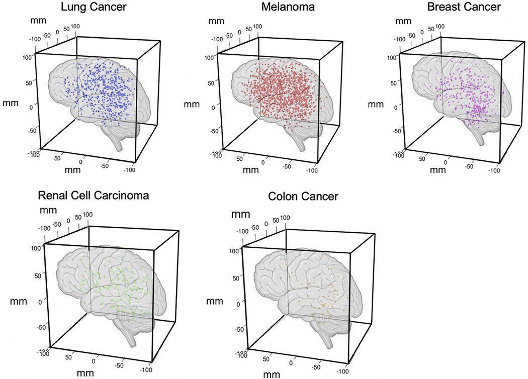
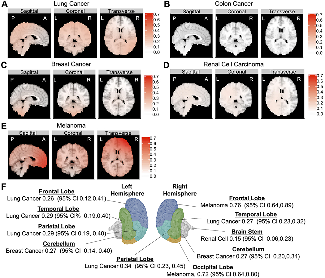
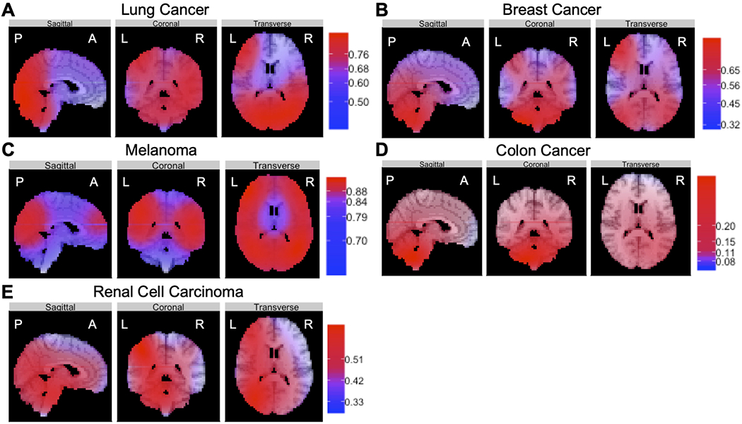
Methods: To test the hypothesis that the spatial distribution of brain metastasis varies according to cancer origin in nonrandom patterns, the authors leveraged spatial 3D coordinate data derived from stereotactic Gamma Knife radiosurgery procedures performed to treat 2106 brain metastases arising from 5 common cancer types (melanoma, lung, breast, renal, and colorectal). Two predictive topographic models (regional brain metastasis echelon model [RBMEM] and brain region susceptibility model [BRSM]) were developed and independently validated.

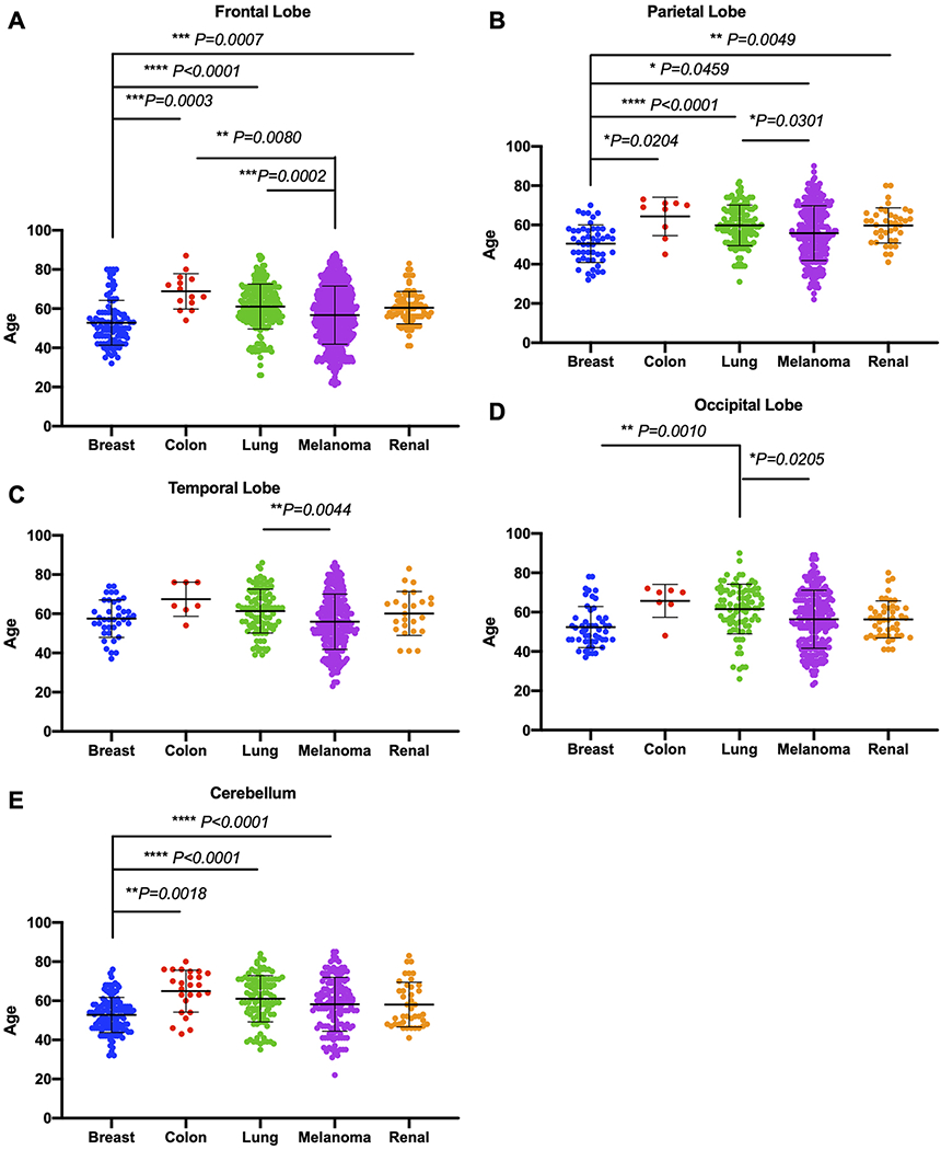
Results: RBMEM assessed the hierarchical distribution of brain metastasis to specific brain regions relative to other primary cancers and showed that distinct regions were relatively susceptible to metastasis, as follows: bilateral temporal/parietal and left frontal lobes were susceptible to lung cancer; right frontal and occipital lobes to melanoma; cerebellum to breast cancer; and brainstem to renal cell carcinoma. BRSM provided probability estimates for each cancer subtype, independent of other subtypes, to metastasize to brain regions, as follows: lung cancer had a propensity to metastasize to bilateral temporal lobes; breast cancer to right cerebellar hemisphere; melanoma to left temporal lobe; renal cell carcinoma to brainstem; and colon cancer to right cerebellar hemisphere. Patient topographic data further revealed that brain metastasis demonstrated distinct spatial patterns when stratified by patient age and tumor volume.

Conclusions: These data support the hypothesis that there is a nonuniform spatial distribution of brain metastasis to preferential brain regions that varies according to cancer subtype in patients treated with Gamma Knife radiosurgery. These topographic patterns may be indicative of the abilities of various cancers to adapt to regional neural microenvironments, facilitate colonization, and establish metastasis. Although the brain microenvironment likely modulates selective seeding of metastasis, it remains unknown how the anatomical spatial distribution of brain metastasis varies according to primary cancer subtype and contributes to diagnosis. For the first time, the authors have presented two predictive models to show that brain metastasis, depending on its origin, in fact demonstrates distinct geographic spread within the central nervous system. These findings could be used as a predictive diagnostic tool and could also potentially result in future translational and therapeutic work to disrupt growth of brain metastasis on the basis of anatomical region.

Keywords: Gamma Knife; brain metastasis; oncology; spatial distribution; stereotactic radiosurgery; topography.


Conflict of interest statement

Disclosures

The authors report no conflict of interest concerning the materials or methods used in this study or the findings specified in this paper.

Figures

FIG. 1.
FIG. 1.
3D representations of the observed distributions of brain metastases according to primary origin. Stereotactic volumetric Cartesian coordinates obtained from the Leksell surgical coordinate frame in the x, y, and z planes at the time of GKRS treatment were recorded for each individual metastatic lesion (breast [285 lesions], colon [52], lung [502], melanoma [1099], and renal cell carcinoma [168]). Figure is available in color online only.
FIG. 2.
FIG. 2.
Correlations between age and brain metastasis subtype for individual neuroanatomical lobes. Given the predicted regionalization of metastasis by cancer subtype, we further examined whether age differentiation was observed. A: Breast cancer patients with frontal lobe metastasis were significantly younger than patients with colon (p = 0.0003), lung (p < 0.0001), and renal (p = 0.0007) metastasis in the frontal lobe. Furthermore, melanoma patients with frontal lobe metastasis were significantly younger than patients with lung (p = 0.0002) and colon (p = 0.0080) metastasis in the frontal lobe. B: Among those with parietal lobe metastasis, breast cancer patients were significantly younger than patients with colon (p = 0.0204), lung (p < 0.0001), melanoma (p = 0.0459), and renal cell (p = 0.0049) tumors, and lung cancer patients were significantly older than patients with melanoma (p = 0.0301). C: Similarly, among those with temporal lobe metastasis, lung cancer patients were significantly older than melanoma patients (p = 0.0044). D: Among those with occipital lobe metastasis, lung cancer patients were significantly older than patients with breast cancer (p = 0.0010) and melanoma (p = 0.0205). E: Among those with cerebellar metastasis, breast cancer patients were significantly younger than patients with colon (p = 0.0018), lung (p < 0.0001), and melanoma (p < 0.0001) tumors. Figure is available in color online only.
FIG. 3.
FIG. 3.
The results of RBMEM indicated the hierarchical distribution of metastasis to preselected brain regions relative to other primary cancers. Multinomial analysis was used to determine which of the preselected brain regions (frontal, parietal, temporal, and occipital lobes; cerebellum; and brainstem) were most likely to be metastasized from 5 primary cancers. A: Topographic results show that lung-to-brain metastasis was most likely to occur in the left frontal cortex, right parietal lobe, left parietal lobe, and left temporal lobe. B: Colon cancer demonstrated a distribution pattern similar to random and did not have dominant representation in any CNS location. C: Breast cancer had the highest probability of metastasis to the right and left cerebellar hemispheres. D: Renal cell carcinoma had the highest probability of metastasis to the brainstem. E: Melanoma had the highest probability of metastasis to the right frontal lobe and right occipital lobe relative to other tumor subtypes. F: Topographic illustration of the CNS showing the primary cancers with the highest probabilities of metastasis to stereotactic coordinates corresponding to the center of each neuroanatomical lobe. Figure is available in color online only.
FIG. 4.
FIG. 4.
The results of BRSM revealed that primary cancers, independent of each other, had defined topographic distributions of metastasis to specific brain regions. Using fitted models to generate prediction probabilities for these brain regions, we found that lung cancer (A) had the highest propensity to metastasize to the left and right temporal lobes, breast cancer (B) had the highest proclivity for metastasis to the right cerebellar hemisphere, melanoma (C) had the highest probability of metastasis to the left temporal lobe, colon cancer (D) had the highest probability of metastasis to the right cerebellar hemisphere, and renal cell carcinoma (E) had the highest predilection for spread to the brainstem. Figure is available in color online only.

References

    1. Deshpande K, Buchanan I, Martirosian V, Neman J. Clinical perspectives in brain metastasis. Cold Spring Harb Perspect Med. 2020;10(6):a037051. - PMC - PubMed
    1. Peinado H, Zhang H, Matei IR, et al. Pre-metastatic niches: organ-specific homes for metastases. Nat Rev Cancer. 2017;17(5):302–317. - PubMed
    1. Termini J, Neman J, Jandial R. Role of the neural niche in brain metastatic cancer. Cancer Res. 2014;74(15):4011–4015. - PMC - PubMed
    1. Chang EL, Wefel JS, Hess KR, et al. Neurocognition in patients with brain metastases treated with radiosurgery or radiosurgery plus whole-brain irradiation: a randomised controlled trial. Lancet Oncol. 2009;10(11):1037–1044. - PubMed
    1. Wood SN. On confidence intervals for generalized additive models based on penalized regression splines. Aust N Z J Stat. 2006;48(4):445–464.

Publication types

MeSH terms