Review
. 2022 Oct;52(11):2149-2158.
doi: 10.1007/s00247-021-05130-8. Epub 2021 Jul 16.

Current and emerging artificial intelligence applications for pediatric musculoskeletal radiology

Affiliations
Review

Current and emerging artificial intelligence applications for pediatric musculoskeletal radiology

Amaka C Offiah. Pediatr Radiol. 2022 Oct.

Abstract

Artificial intelligence (AI) is playing an ever-increasing role in radiology (more so in the adult world than in pediatrics), to the extent that there are unfounded fears it will completely take over the role of the radiologist. In relation to musculoskeletal applications of AI in pediatric radiology, we are far from the time when AI will replace radiologists; even for the commonest application (bone age assessment), AI is more often employed in an AI-assist mode rather than an AI-replace or AI-extend mode. AI for bone age assessment has been in clinical use for more than a decade and is the area in which most research has been conducted. Most other potential indications in children (such as appendicular and vertebral fracture detection) remain largely in the research domain. This article reviews the areas in which AI is most prominent in relation to the pediatric musculoskeletal system, briefly summarizing the current literature and highlighting areas for future research. Pediatric radiologists are encouraged to participate as members of the research teams conducting pediatric radiology artificial intelligence research.

Keywords: Artificial intelligence; Bone; Children; Musculoskeletal; Pediatric radiology.


Conflict of interest statement

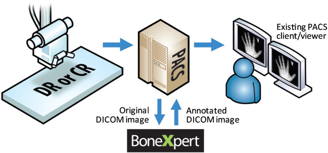
Professor Offiah has conducted research with Visiana in relation to BoneXpert software and has received funding for the development of dREAMS.

Figures

Fig. 1
Fig. 1
BoneXpert workflow. The radiographer who has performed the left hand and wrist radiograph sends it to the picture archiving and communication system (PACS) in the usual way. The reporting radiologist (or radiographer) transfers the image to BoneXpert, where the automated bone age estimation is performed. BoneXpert then returns the annotated image to the PACS. The reporting radiologist now not only has access to the BoneXpert-derived Greulich and Pyle and the Tanner and Whitehouse 3 bone age assessments, but he or she can also review the original radiograph for disease-related abnormality (e.g., evidence of a skeletal dysplasia). Image courtesy of Peter Bak and Hans Henrik Thodberg (Visiana). CR computed radiography, DICOM digital imaging and communications in medicine, DR digital radiography
Fig. 2
Fig. 2
Posteroanterior left hand and wrist radiograph in a 3-week-old boy, following interpretation of bone age by BoneXpert. The figures in the small white boxes represent the Greulich and Pyle bone ages of the individual bones. BA (GP) Greulich and Pyle bone age (gender), BA SDS Bone age standard deviation score (ethnicity), BA (TW3) Tanner and Whitehouse three-bone age, BHI bone health index (digital X-ray radiogram), BHI SDS bone health index standard deviation score (ethnicity), CauEu Caucasian European, M male, N/A not available (no ossified carpal bones in this child), y years. See also Table 2
Fig. 3
Fig. 3
Lateral spine dual-energy X-ray absorptiometry scan in a 12-year-old boy, left, with deformity results right. Morphometric vertebral fracture assessment using SpineAnalyzer identifies four mild (T4, T10, T11, L1) and three moderate (T9, T12, L3) fractures. Bicon. biconcave, SQ semi-quantitative score (of Genant et al. [56])

References

    1. van Ginneken B (2018) AI and radiologists — a painful divorce? ECR 2018 presentation. https://vimeo.com/258232453. Accessed 12 Mar 2021
    1. No authors (n.d.) AI for radiology: an implementation guide. Products. Website. https://grand-challenge.org/aiforradiology/. Accessed 10 Mar 2021
    1. Koska IO. Applications of deep learning in radiology and pediatric radiology. Pediatr Radiol. 2019;49:247–317.
    1. Van Rijn RR, Lequin MH, Thodberg HH. Automatic determination of Greulich and Pyle bone age in healthy Dutch children. Pediatr Radiol. 2009;39:591–597. doi: 10.1007/s00247-008-1090-8. - DOI - PubMed
    1. Thodberg HH, Sävendahl L. Validation and reference values of automated bone age determination for four ethnicities. Acad Radiol. 2010;17:1425–1432. doi: 10.1016/j.acra.2010.06.007. - DOI - PubMed