Review
. 2021 Sep;158(6):1394-1411.
doi: 10.1111/jnc.15471. Epub 2021 Aug 6.

Basal forebrain cholinergic system in the dementias: Vulnerability, resilience, and resistance

Affiliations
Review

Basal forebrain cholinergic system in the dementias: Vulnerability, resilience, and resistance

Changiz Geula et al. J Neurochem. 2021 Sep.

Abstract

The basal forebrain cholinergic neurons (BFCN) provide the primary source of cholinergic innervation of the human cerebral cortex. They are involved in the cognitive processes of learning, memory, and attention. These neurons are differentially vulnerable in various neuropathologic entities that cause dementia. This review summarizes the relevance to BFCN of neuropathologic markers associated with dementias, including the plaques and tangles of Alzheimer's disease (AD), the Lewy bodies of diffuse Lewy body disease, the tauopathy of frontotemporal lobar degeneration (FTLD-TAU) and the TDP-43 proteinopathy of FTLD-TDP. Each of these proteinopathies has a different relationship to BFCN and their corticofugal axons. Available evidence points to early and substantial degeneration of the BFCN in AD and diffuse Lewy body disease. In AD, the major neurodegenerative correlate is accumulation of phosphotau in neurofibrillary tangles. However, these neurons are less vulnerable to the tauopathy of FTLD. An intriguing finding is that the intracellular tau of AD causes destruction of the BFCN, whereas that of FTLD does not. This observation has profound implications for exploring the impact of different species of tauopathy on neuronal survival. The proteinopathy of FTLD-TDP shows virtually no abnormal inclusions within the BFCN. Thus, the BFCN are highly vulnerable to the neurodegenerative effects of tauopathy in AD, resilient to the neurodegenerative effect of tauopathy in FTLD and apparently resistant to the emergence of proteinopathy in FTLD-TDP and perhaps also in Pick's disease. Investigations are beginning to shed light on the potential mechanisms of this differential vulnerability and their implications for therapeutic intervention.

Keywords: Alzheimer's disease; TDP-43 proteinopathy; basal forebrain cholinergic neurons; diffuse lewy body disease; frontotemporal lobar degeneration; tauopathy.


Conflict of interest statement

Disclosure Statement

The authors have no conflicts of interest to declare.

Figures
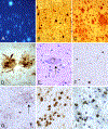
Figure 1.
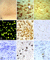
Figure 1.. Pathologic entities associated with dementias.
(A) Thioflavin-S stain visualizes mature extracellular senile plaques and intracellular neurofibrillary tangles in Alzheimer’s disease (AD) cortex. (B) Phosphotau immunoreactivity is present in neurofibrillary tangles and neuropil threads in AD. (C) Plaques in AD cortex display immunoreactivity for the amyloid-β peptide. (D) Dystrophic neurites within mature plaques in AD brains display immunoreactivity for phosphotau. (E) A substantia nigra neuron in a case of diffuse Lewy body disease (DLBD) contains a LB in a hemotoxylin & eosin stained section. (F) α-Synuclein immunoreactivity is present in neurons and neurites in DLBD cortex. (G) Phosphotau immunoreactivity in corticobasal degeneration visualizes inclusions in neurons, neurites and astrocytic plaques (arrow). (H) Cortical Pick bodies containing phosphotau immunoreactivity in Pick’s disease. (I) Phosphorylated TDP-43 immunoreactive inclusions in cortical neurons in TDP-43 proteinopathy.
Figure 2.
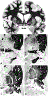
Figure 2.. Anatomical delineation of the nucleus basalis of Meynert (Ch4-nbM) in the human brain.
(Top) Myelin stained whole-brain section, showing the substantia innominata below the crossing anterior commissure within which the anterior aspects of Ch4-nbM neurons are located. (A-D) Acetylcholinesterase stained sections delineating the anterior (A), anterointermediate (B), intermediate (C) and posterior (D) sectors of Ch4-nbM. Ac – anterior commissure; ai – anterointermediate sector of Ch4-nbM; al – anterolateral subsector of Ch4-nbM; am – anteromedial subsector of Ch4-nbM; Am – amygdala; an – ansa lenticularis; ap – ansa peduncularis; bl – basolateral nucleus of amygdala; GP – globus pallidus; GPe – globus pallidus externa; GPi – globus pallidus interna; GPv – ventral globus pallidus; hc – head of caudate; Hp – hippocampus; ic – internal capsule; id – intermediodorsal subsector of Ch4-nbM; iml – internal medullary lamina or globus pallidus; iv – intermedioventral subsector of Ch4-nbM; Nhl – nucleus of the horizontal limb of the diagonal band of Broca; nst – nucleus of stria terminalis; oc – optic chiasm; ot – optic tract; p – posterior sector of Ch4-nbM; Pt – putamen; si – substantia innominata; son – supraoptic nucleus of hypothalamus; v – blood vessel; vt – temporal horn of lateral ventricle; tc – tail of caudate. A-D are reproduced with permission from Wiley and Sons: Mesulam, M. M. and Geula, C. (1988) Nucleus basalis (Ch4) and cortical cholinergic innervation in the human brain: observations based on the distribution of acetylcholinesterase and choline acetyltransferase. J.Comp.Neurol. 275, 216–240.
Figure 3.
Figure 3.. Cholinergic neurons of the nucleus basalis of Meynert (Ch4-nbM) in dementias.
Low (A) and high (B) power images of choline-acetyltransferase immunoreactive (ChAT) Ch4-nbM neurons in the normal human brain. (C) Ch4-nbM of an AD brain, demonstrating significant loss of ChAT immunoreactive Ch4-nbM neurons. (D) Thioflavin-S staining, demonstrating presence of mature globose neurofibrillary tangles in the remaining Ch4-nbM neurons in AD. (E) Phosphotau immunoreactivity in the remaining Ch4-nbM neurons in AD. (F) α-Synuclein immunoreactivity in Ch4-nbM neurons in diffuse Lewy body dementia. (G) Robust preservation of magnocellular Nissl stained Ch4-nbM neurons in Pick’s disease; Pick bodies are not observed in Ch4-nbM neurons in contrast to abundant Pick bodies in cortex (see Figure 1H). (H) Abundant accumulation of phosphotau accumulation in Ch4-nbM neurons in corticobasal degeneration, despite preservation of Ch4-nbM neurons. (I) Absence of TDP-43 mature inclusions in Ch4-nbM neurons in TDP-43 proteinopathy; only occasional pre-inclusions (arrow) are seen in Ch4-nbM neurons.

References

    1. Ahmadian SS, Rezvanian A, Peterson M, Weintraub S, Bigio EH, Mesulam MM and Geula C (2015) Loss of calbindin-D28K is associated with the full range of tangle pathology within basal forebrain cholinergic neurons in Alzheimer’s disease. Neurobiology of aging 36, 3163–3170. - PMC - PubMed
    1. Aigner TG, Mitchell SJ, Aggleton JP, Delong MR, Struble RG, Price DL, Wenk GL and Mishkin M (1987) Effects of scopolamine and physostigmine on recognition memory in monkeys with ibotenic-acid lesions of the nucleus basalis of Meynert. Psychopharmacol. 92, 292–300. - PubMed
    1. Allen SJ, MacGowan SH, Treanor JJ, Feeney R, Wilcock GK and Dawbarn D (1991) Normal beta-NGF content in Alzheimer’s disease cerebral cortex and hippocampus. Neurosci.Lett 131, 135–139. - PubMed
    1. Alzheimer A (1977) A unique illness involving the cerebral cortex. In: Neurological Classics in Modern Translation, (Rothenberg DA and Hochberg FH eds.), pp. 41–43. Hafner Press, London.
    1. Araujo DM, Lapchak PA, Robitaille Y, Gauthier S and Quirion R (1988) Differential alteration of various cholinergic markers in cortical and subcortical regions of human brain in Alzheimer’s disease. J.Neurochem 50, 1914–1923. - PubMed

Publication types

MeSH terms

Substances