Review
. 2021 Aug;54(4):5462-5506.
doi: 10.1111/ejn.15393. Epub 2021 Aug 4.

Structure, function and connectivity fingerprints of the frontal eye field versus the inferior frontal junction: A comprehensive comparison

Affiliations
Review

Structure, function and connectivity fingerprints of the frontal eye field versus the inferior frontal junction: A comprehensive comparison

Marco Bedini et al. Eur J Neurosci. 2021 Aug.

Abstract

The human prefrontal cortex contains two prominent areas, the frontal eye field and the inferior frontal junction, that are crucially involved in the orchestrating functions of attention, working memory and cognitive control. Motivated by comparative evidence in non-human primates, we review the human neuroimaging literature, suggesting that the functions of these regions can be clearly dissociated. We found remarkable differences in how these regions relate to sensory domains and visual topography, top-down and bottom-up spatial attention, spatial versus non-spatial (i.e., feature- and object-based) attention and working memory and, finally, the multiple-demand system. Functional magnetic resonance imaging (fMRI) studies using multivariate pattern analysis reveal the selectivity of the frontal eye field and inferior frontal junction to spatial and non-spatial information, respectively. The analysis of functional and effective connectivity provides evidence of the modulation of the activity in downstream visual areas from the frontal eye field and inferior frontal junction and sheds light on their reciprocal influences. We therefore suggest that future studies should aim at disentangling more explicitly the role of these regions in the control of spatial and non-spatial selection. We propose that the analysis of the structural and functional connectivity (i.e., the connectivity fingerprints) of the frontal eye field and inferior frontal junction may be used to further characterize their involvement in a spatial ('where') and a non-spatial ('what') network, respectively, highlighting segregated brain networks that allow biasing visual selection and working memory performance to support goal-driven behaviour.

Keywords: brain connectivity; prefrontal cortex; spatial versus non-spatial selection; visual attention; working memory.


Conflict of interest statement

The authors report no conflict of interest.

Figures
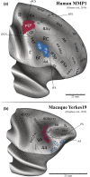
FIGURE 1
FIGURE 1
Putative homologies between the human and the macaque PFC displayed on the human MMP1 (a) and the macaque Yerkes19 atlases (b). The homologies between the human and the macaque PFC are, due to the lack of similar sulcal morphology (because the macaque brain has a single principal sulcus, in contrast to the human brain, which has two major sulci, the SFS and the IFS), best inferred based on structural and/or functional criteria, rather than morphological information. In the MMP1, the typical localization of the IFJ relative to the sulci likely corresponds to the IFJp (a) (see Section 2.2 for a detailed discussion). According to the study by Donahue et al. (2018), the human FEF would be the homologue of area 45b in the macaque (Schall et al., 1995), but it may also overlap with other brain regions (viz., 8a and 6DC(F2); see, e.g., fig. 1 in Schall, 2015). In the studies by Bichot et al. (2015, 2019), the authors identified a ventral prearcuate region (VPA) that was proposed as the human IFJ homologue and that overlapped with areas 46v, 45A and 12. Based on their injection sites, three regions that may correspond to the VPA were highlighted on the Yerkes19 atlas (b) (the regions 45A, 9/46v and 12r′ from the composite PFC parcellation by Donahue et al., ; PS, principal sulcus; AS, arcuate sulcus; both (a) and (b) were adapted from the datasets available in BALSA at https://balsa.wustl.edu/; Van Essen et al., 2017). At present, it is however unclear whether cytoarchitectonic maps in the macaque are appropriate for inferring homologies with the human PFC (see Section 2.1 for a discussion). In Section 5, we highlight some of the most intriguing proposals that aim at resolving these comparative neuroanatomical issues
FIGURE 2
FIGURE 2
Paradigms that allow disentangling spatial and non‐spatial attentional and working memory mechanisms. Red outline: Paradigms that involve a strong spatial component. The fundamental basis of all the tasks shown in this group is represented by the spatial cueing paradigm (a) (adapted from Carrasco, 2011). (b) The tasks from the study by Srimal and Curtis (2008), consisting of a memory‐guided saccade (top) and a spatial item recognition task (bottom). (c) The saccadic eye‐movement paradigm from the study by Amiez et al. (; adapted from Amiez & Petrides, 2009). Top row: Control task, in which subjects keep fixation. Bottom row: Experimental task, in which subjects need to perform a sequence of visually guided saccades. This paradigm represents the classical FEF fMRI localizer. (d) The memory‐guided saccade task from the study by Kastner et al. (2007). This study provided compelling evidence of the presence of topographic maps in the PFC (and crucially, in the sPCS/SFS, encompassing the FEF). Blue outline: Paradigms that involve purely non‐spatial mechanisms. (e and f) The feature‐based attention tasks from the studies by Zhang et al. (2018) and Liu et al. (2011). In these tasks, the subjects are asked to hold fixation and are instructed by an endogenous cue to pay attention to the features of a stimulus (either colour or motion) and to detect a sudden change in luminance of a dot or an increase in its speed of motion. Because this imperative event can happen randomly in each portion of the cloud‐like stimuli, spatial information is rendered ineffective for solving the task at hand (in the study by Zhang and colleagues, half of the dots were replaced and reappeared at new locations each 100 ms to discourage even more the use of spatial strategies; the attended and ignored sides were blocked). (g) The purely object‐based attention task devised by Liu (2016). The colour, orientation and spatial frequency of the two superimposed Gabor patches changed simultaneously over the trial, so the subjects needed to pay attention to their identity to effectively perform the task. (h and i) The working memory task used by Zanto et al. (2010) and the object‐based attention task from the study by Baldauf and Desimone (2014). All these feature‐ and object‐based attention and WM tasks engage the IFJ, which is responsible for modulating activity in downstream visual areas (e.g., MT+, V4, PPA and FFA) to enhance behavioural performance (Baldauf & Desimone, ; Zhang et al., 2018)
FIGURE 3
FIGURE 3
Structural connectivity and resting‐state functional connectivity of the posterior lateral prefrontal cortex. (a) depicts the comparison between the organization of the three branches of the superior longitudinal fasciculus (SLF) in macaques and humans. This frontoparietal bundle was delineated using a virtual dissection protocol in 20 adult participants (adapted from de Schotten et al., 2011). (b) presents a summary of the findings from the study by Parlatini et al. (2017). The activation maps resulting from an fMRI meta‐analysis of 14 distinct cognitive functions were clustered using a principal component analysis in a ‘spatial/motor’ and a ‘non‐spatial/motor’ component (b, right side). These maps' z scores were related to the cortical terminations of the three branches of the SLF obtained from a large sample of participants (n = 129), suggesting that the SLF1 could preferentially mediate ‘spatial’ processes, whereas the SLF3 could preferentially mediate ‘non‐spatial’ processes. The SLF2 overlapped with both activation components, therefore possibly underlying their interactions (b, left side). (c) The dorsal and the ventral attention networks identified for the first time during resting‐state fMRI in the study by Fox et al. (2006). These spontaneous activity patterns show high resemblance to the two attentional systems proposed by Corbetta and Shulman (2002) and highlight the potential of resting‐state data to reveal stable modes of organization of the attention networks in the absence of task demands (see also De Pasquale et al., , for converging evidence on the organization of the DAN using MEG). (d) Seed‐based analysis of resting‐state fMRI functional connectivity of the FEF and the ventro‐caudal prefrontal region (PrCv) from the study by Yeo et al. (2011). The authors defined the former nodes by performing an fMRI meta‐analysis and using a separate discovery sample, respectively, and employed a data‐driven approach to probe their connectivity patterns. In a subsequent stage, they tested them in an independent replication sample in a confirmatory fashion. They found that within the posterior parietal cortex, the medio‐caudal SPL showed increased functional coupling with FEF, whereas the lateral IPS complex showed increased functional coupling with PrCv in their replication sample. These results suggest segregated parallel pathways from the plPFC to the SPL and the IPS complex

References

    1. Abdollahi, R. O. , Kolster, H. , Glasser, M. F. , Robinson, E. C. , Coalson, T. S. , Dierker, D. , Jenkinson, M. , Van Essen, D. C. , & Orban, G. A. (2014). Correspondences between retinotopic areas and myelin maps in human visual cortex. NeuroImage, 99, 509–524. 10.1016/j.neuroimage.2014.06.042 - DOI - PMC - PubMed
    1. Amiez, C. , Kostopoulos, P. , Champod, A. S. , & Petrides, M. (2006). Local morphology predicts functional organization of the dorsal premotor region in the human brain. Journal of Neuroscience, 26(10), 2724–2731. 10.1523/JNEUROSCI.4739-05.2006 - DOI - PMC - PubMed
    1. Amiez, C. , & Petrides, M. (2009). Anatomical organization of the eye fields in the human and non‐human primate frontal cortex. Progress in Neurobiology, 89(2), 220–230. 10.1016/j.pneurobio.2009.07.010 - DOI - PubMed
    1. Amunts, K. , Lenzen, M. , Friederici, A. D. , Schleicher, A. , Morosan, P. , Palomero‐Gallagher, N. , & Zilles, K. (2010). Broca's region: Novel organizational principles and Multiple Receptor Mapping. PLoS Biology, 8(9), e1000489. 10.1371/journal.pbio.1000489 - DOI - PMC - PubMed
    1. Amunts, K. , Mohlberg, H. , Bludau, S. , & Zilles, K. (2020). Julich‐Brain: A 3D probabilistic atlas of the human brain's cytoarchitecture. Science, 369(6506), 988–992. 10.1126/science.abb4588 - DOI - PubMed

Publication types