Review
. 2021 Dec 20;224(12 Suppl 2):S856-S863.
doi: 10.1093/infdis/jiab372.

Intestinal Biopsies for the Evaluation of Environmental Enteropathy and Environmental Enteric Dysfunction

Affiliations
Review

Intestinal Biopsies for the Evaluation of Environmental Enteropathy and Environmental Enteric Dysfunction

Phoebe Hodges et al. J Infect Dis. .

Abstract

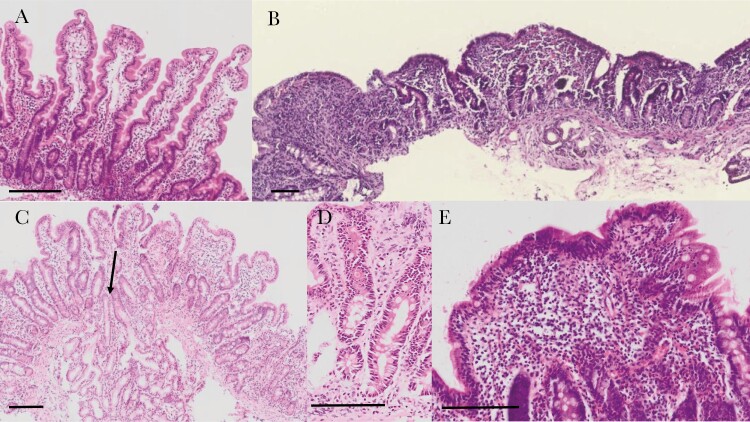
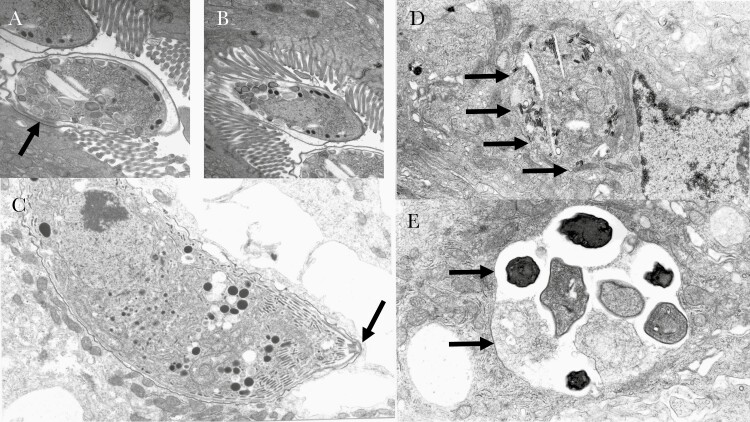
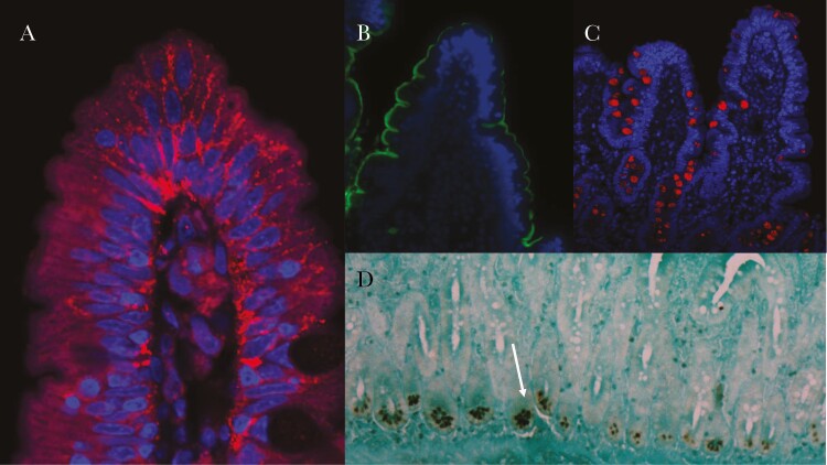
Environmental enteric dysfunction (EED) is a syndrome characterized by impairments of digestion and absorption and intestinal barrier failure in people living in insanitary or tropical environments. There is substantial evidence that it contributes to impaired linear growth of millions of children in low- and middle-income countries, to slowed neurocognitive development, and to diminished responses to oral vaccines. It represents the functional consequences of environmental enteropathy, an asymptomatic inflammatory disorder of the mucosa, and there is considerable overlap with the enteropathy observed in severe clinical malnutrition. The majority of studies of EED have employed functional tests based on lactulose permeation to define the presence of abnormal leak in the gut. However, where intestinal biopsies can safely be collected the opportunity then arises to study the underlying enteropathy in cellular and molecular detail, as well as to measure important functional elements such as enzyme expression. The purpose of this narrative review is to summarize the current understanding of environmental enteropathy obtained from small intestinal biopsies, and prospects for future work. We review histology, electron microscopy, transcription and protein expression, physiological measures, and the microbiome. We conclude that while noninvasive biomarkers of enteropathy and intestinal dysfunction permit large-scale studies of unquestionable value, intestinal biopsies are still required to investigate pathophysiology in depth.

Keywords: environmental enteric dysfunction; environmental enteropathy.


Figures

Figure 1.
Figure 1.
Histological sections of intestinal biopsies stained with hematoxylin and eosin. A, Long, slender villi in a well-orientated view as demonstrated by the crypts seen in longitudinal section. B, Severe enteropathy in a biopsy from a child with severe acute malnutrition complicated by human immunodeficiency virus infection and persistent diarrhea; there are no villi and severe inflammation of the lamina propria. C, Moderately inflamed section with shortened and widened villi visible, and with penetration of a Brunner gland into the mucosa (arrow). D, Section showing very long crypts and increased extracellular eosinophilic material from a biopsy showing subtotal villus atrophy. E, Lymphoid aggregate with marked intraepithelial lymphocytosis. Scale bars are 100 μm.
Figure 2.
Figure 2.
Electron microscopy of intestinal pathogens in small biopsies from AIDS patients in Zambia in the era before antiretroviral therapy. (A) and (B) fertilised macroamonts of Cryptosporidium spp which occupy an intracellular but extracytoplasmic niche, enclosed within plasma membrane and divided from the cytoplasm by the feeder organelle, an osmophilic band seen just beneath the parasite in (A) and indicated by an arrow. Obliteration of the brush border is evident across the area of attachment. (C) a trophozoite of Cystisospora belli (arrow) in a fully intracytoplasmic niche in an enterocyte, also from a Zambian AIDS patient. (D) Enterocytozoon bieneusi, and (E) Encephalitozoon intestinalis, both microsporidia which are now classified as fungi. Arrows indicate the boundary of the parasite.
Figure 3.
Figure 3.
Examples of immunostaining of small intestinal biopsies from Zambian adult volunteers to show (A) claudin 4 disorganisation at the tip of a villus; claudin 4 is a tight junction protein, here stained in red, showing departure from the usual pattern of small foci of localisation near the brush border along the intercellular junction. (B) sucrase-isomaltase staining (in green) of the brush border around a villus. (C) staining of trefoil factor 3 (in red) which highlights goblet cells. (D) human defensin 5 staining of Paneth cells shown (arrow) using peroxidase immunohistochemistry. In A-C nuclei are stained in blue.

References

    1. de Onis M, Branca F. Childhood stunting: a global perspective. Matern Child Nutr 2016; 12(Suppl 1):12–26. - PMC - PubMed
    1. Black RE, Victora CG, Walker SP, et al. Maternal and Child Nutrition Study Group. Maternal and child undernutrition and overweight in low-income and middle-income countries. Lancet 2013; 382:427–51. - PubMed
    1. Central Statistical Office of Zambia, Ministry of Health of Zambia, and ICF. Zambia demographic and health survey 2018: key indicators. Rockville, MD: Central Statistical Office, Ministry of Health, and ICF,2019.
    1. Olofin I, McDonald CM, Ezzati M, et al. Nutrition Impact Model Study. Associations of suboptimal growth with all-cause and cause-specific mortality in children under five years: a pooled analysis of ten prospective studies. PLoS One 2013; 8:e64636. - PMC - PubMed
    1. Prendergast AJ, Humphrey JH. The stunting syndrome in developing countries. Paediatr Int Child Health 2014; 34:250–65. - PMC - PubMed

Publication types