

Deep-LIBRA: An artificial-intelligence method for robust quantification of breast density with independent validation in breast cancer risk assessment
- PMID: 34274690
- PMCID: PMC8453099
- DOI: 10.1016/j.media.2021.102138
Deep-LIBRA: An artificial-intelligence method for robust quantification of breast density with independent validation in breast cancer risk assessment
Abstract
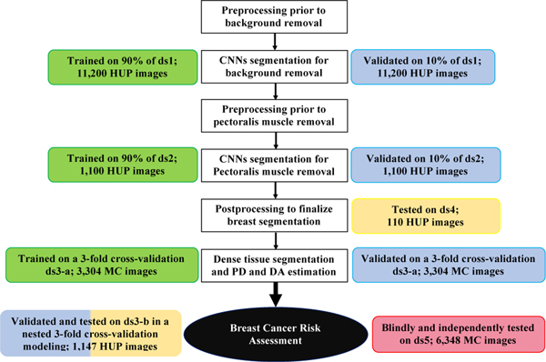
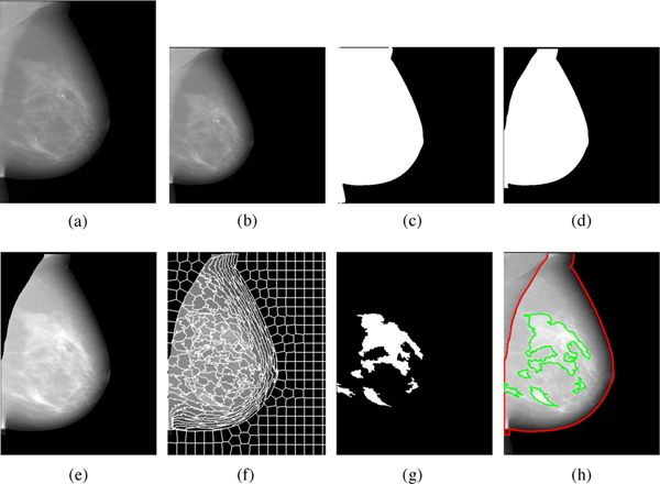
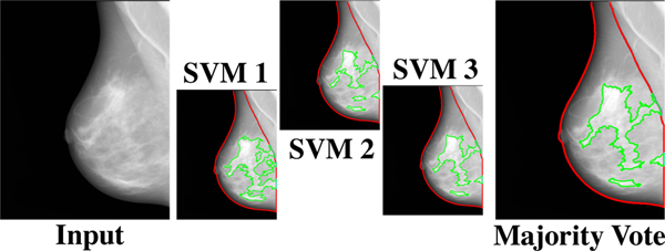
Breast density is an important risk factor for breast cancer that also affects the specificity and sensitivity of screening mammography. Current federal legislation mandates reporting of breast density for all women undergoing breast cancer screening. Clinically, breast density is assessed visually using the American College of Radiology Breast Imaging Reporting And Data System (BI-RADS) scale. Here, we introduce an artificial intelligence (AI) method to estimate breast density from digital mammograms. Our method leverages deep learning using two convolutional neural network architectures to accurately segment the breast area. An AI algorithm combining superpixel generation and radiomic machine learning is then applied to differentiate dense from non-dense tissue regions within the breast, from which breast density is estimated. Our method was trained and validated on a multi-racial, multi-institutional dataset of 15,661 images (4,437 women), and then tested on an independent matched case-control dataset of 6368 digital mammograms (414 cases; 1178 controls) for both breast density estimation and case-control discrimination. On the independent dataset, breast percent density (PD) estimates from Deep-LIBRA and an expert reader were strongly correlated (Spearman correlation coefficient = 0.90). Moreover, in a model adjusted for age and BMI, Deep-LIBRA yielded a higher case-control discrimination performance (area under the ROC curve, AUC = 0.612 [95% confidence interval (CI): 0.584, 0.640]) compared to four other widely-used research and commercial breast density assessment methods (AUCs = 0.528 to 0.599). Our results suggest a strong agreement of breast density estimates between Deep-LIBRA and gold-standard assessment by an expert reader, as well as improved performance in breast cancer risk assessment over state-of-the-art open-source and commercial methods.
Keywords: Artificial intelligence; Breast cancer risk; Breast density; Deep learning; Digital mammography.
Copyright © 2021 The Authors. Published by Elsevier B.V. All rights reserved.
Conflict of interest statement
Declaration of Competing Interest The authors declare the following financial interests/personal relationships which may be considered as potential competing interests Dr. Emily Conant reports research grants and membership on the Scientific Advisory Boards of Hologic, Inc., and iCAD, Inc. The other nine authors have no conflict of interests.
Figures
References
-
- Achanta R, Shaji A, Smith K, Lucchi A, Fua P, Süsstrunk S, 2012. Slic superpixels compared to state-of-the-art superpixel methods. IEEE transactions on pattern analysis and machine intelligence 34, 2274–2282. - PubMed
-
- Anitha J, Peter JD, Pandian SIA, 2017. A dual stage adaptive thresholding (dusat) for automatic mass detection in mammograms. Computer methods and programs in biomedicine 138, 93–104. - PubMed
-
- Are-You-Dense-Advocacy, 2019. D.E.N.S.E. State Efforts. http://areyoudenseadvocacy.org/. [Online; accessed 1-April-2021].
-
- Becker AS, Marcon M, Ghafoor S, Wurnig MC, Frauenfelder T, Boss A, 2017. Deep learning in mammography: diagnostic accuracy of a multipurpose image analysis software in the detection of breast cancer. Investigative radiology 52, 434–440. - PubMed
-
- Boyd NF, Guo H, Martin LJ, Sun L, Stone J, Fishell E, Jong RA, Hislop G, Chiarelli A, Minkin S, et al., 2007. Mammographic density and the risk and detection of breast cancer. New England journal of medicine 356, 227–236. - PubMed
Publication types
MeSH terms
Grants and funding
LinkOut - more resources
Full Text Sources
Medical
