. 2021:31:102755.
doi: 10.1016/j.nicl.2021.102755. Epub 2021 Jul 7.

Resting functional connectivity in the semantic appraisal network predicts accuracy of emotion identification

Affiliations

Resting functional connectivity in the semantic appraisal network predicts accuracy of emotion identification

Winson F Z Yang et al. Neuroimage Clin. 2021.

Abstract

Objective: Structural and task-based functional studies associate emotion reading with frontotemporal brain networks, though it remains unclear whether functional connectivity (FC) alone predicts emotion reading ability. The predominantly frontotemporal salience and semantic appraisal (SAN) networks are selectively impacted in neurodegenerative disease syndromes like behavioral-variant frontotemporal dementia (bvFTD) and semantic-variant primary progressive aphasia (svPPA). Accurate emotion identification diminishes in some of these patients, but studies investigating the source of this symptom in patients have predominantly examined structural rather than functional brain changes. Thus, we investigated the impact of altered connectivity on their emotion reading.

Methods: One-hundred-eighty-five participants (26 bvFTD, 21 svPPA, 24 non-fluent variant PPA, 24 progressive supranuclear palsy, 49 Alzheimer's disease, 41 neurologically healthy older controls) underwent task-free fMRI, and completed the Emotion Evaluation subtest of The Awareness of Social Inference Test (TASIT-EET), watching videos and selecting labels for actors' emotions.

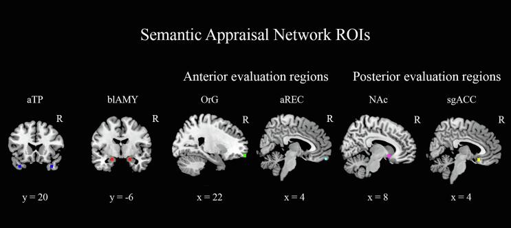
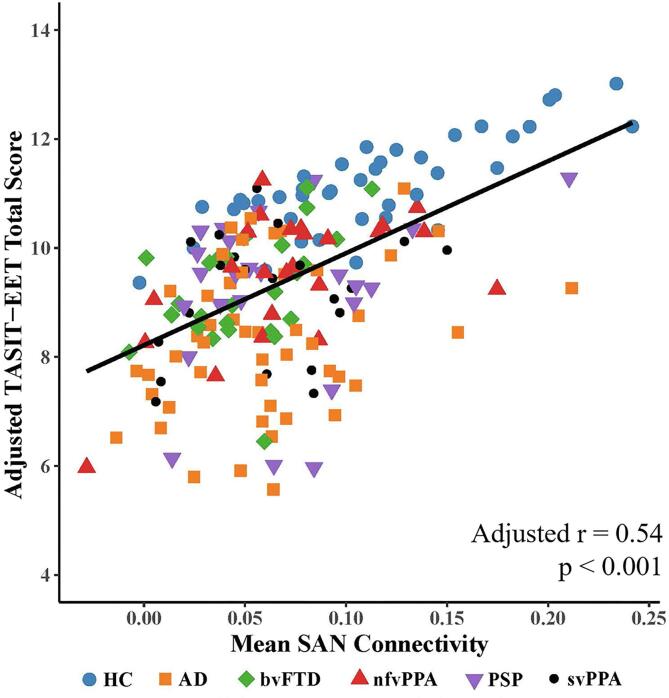
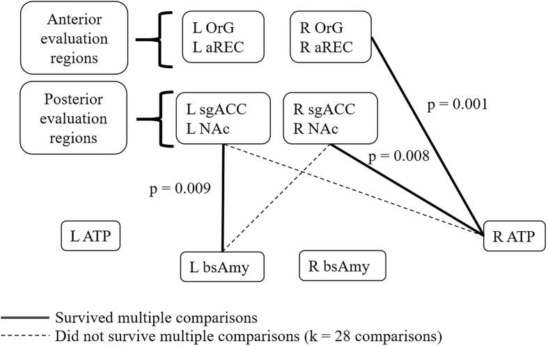
Results: As expected, patients averaged significantly worse on emotion reading, but with wide inter-individual variability. Across all groups, lower mean FC in the SAN, but not other ICNs, predicted worse TASIT-EET performance. Node-pair analysis revealed that emotion identification was predicted by FC between 1) right anterior temporal lobe (RaTL) and right anterior orbitofrontal (OFC), 2) RaTL and right posterior OFC, and 3) left basolateral amygdala and left posterior OFC.

Conclusion: Emotion reading test performance predicts FC in specific SAN regions mediating socioemotional semantics, personalized evaluations, and salience-driven attention, highlighting the value of emotion testing in clinical and research settings to index neural circuit dysfunction in patients with neurodegeneration and other neurologic disorders.

Keywords: Emotion reading; Frontotemporal dementia; Functional connectivity; Neurodegeneration; Right anterior temporal lobe; Semantic appraisal network.


Conflict of interest statement

The authors declare that they have no known competing financial interests or personal relationships that could have appeared to influence the work reported in this paper.

Figures

Fig. 1
Fig. 1
shows the regions of interest used for the node pair connectivity analyses in the semantic appraisal network. Sagittal slices shown are in the right hemisphere. aTP = anterior temporal pole, blAMY = basolateral amygdala, OrG = orbital gyrus, aREC = anterior gyrus rectus, NAc = nucleus accumbens, sgACC = subgenual anterior cingulate cortex. Blue = aTP [±36, 22, −34], red = blAMY [±20, −6, −18], green = OrG [±20, 59, −15], cyan = aREC [±4, 58, −20], violet = NAc [±10, 11, −9], yellow = sgACC [±4, 20, −15]. Coordinates shown are the Montreal Neurological Institute (MNI) coordinates. (For interpretation of the references to colour in this figure legend, the reader is referred to the web version of this article.)
Fig. 2
Fig. 2
displays the relationship between mean connectivity in the semantic appraisal network (SAN) and the TASIT-EET in the full sample of healthy older controls and patients with neurodegenerative diseases (n = 185), after adjusting for age, sex, MMSE, and mean salience network connectivity. Mean SAN connectivity significantly predicted TASIT-EET score after adjusting for age, sex, MMSE, and after including the salience network in the model (r = 0.54, p < 0.01). Higher mean SAN connectivity was associated with higher TASIT-EET score. Mean connectivity values were calculated as each participant’s mean beta value across all voxels within their given ICN map, masked at the ICN as defined in an independent sample of healthy older participants (see methods). HC = healthy control, AD = Alzheimer’s disease, bvFTD = behavioral variant frontotemporal dementia, nfvPPA = nonfluent variant primary progressive aphasia, PSP = progressive supranuclear palsy, svPPA = semantic variant primary progressive aphasia, TASIT-EET = The Emotional Evaluation Task of the Awareness of Social Interference Test.
Fig. 3
Fig. 3
shows the functional connectivity between nodes of the semantic appraisal network (SAN). Several regression models were conducted to investigate the functional connectivity between node-pairs of the SAN, after controlling for age, sex, and MMSE. A Benjamini-Yekutieli correction for k = 28 multiple comparisons was applied to the primary analysis, yielding a corrected significance threshold of p < 0.01273. We found that the functional connectivity between (1) the RaTP and the right posterior OFC evaluation node, (2) the RaTP and the right anterior OFC evaluation node, and (3) the left basolateral amygdala and the left posterior OFC evaluation node predicted TASIT-EET total score. All of these relationships remained significant after atrophy correction, and (1) and (2) remained significant in a rigorous error check for diagnostic confounding. OrG = orbital gyrus, aREC = anterior gyrus rectus, sgACC = subgenual anterior cingulate cortex, NAc = nucleus accumbens, aTP = anterior temporal pole, blAMY = basolateral amygdala, R = Right, L = Left.

References

    1. Ambroggi F., Ishikawa A., Fields H.L., Nicola S.M. Basolateral amygdala neurons facilitate reward-seeking behaviour by exciting nucelus accumbens neurons. Neuron. 2009;59:648–661. - PMC - PubMed
    1. Andrews-Hanna J.R., Reidler J.S., Sepulcre J., Poulin R., Buckner R.L. Functional-anatomic fractionation of the brain's default network. Neuron. 2010;65:550–562. - PMC - PubMed
    1. Andrews-Hanna J.R., Smallwood J., Spreng R.N. The default network and self-generated thought: component processes, dynamic control, and clinical relevance. Ann. N. Y. Acad. Sci. 2014;1316:29–52. - PMC - PubMed
    1. Ashburner, J., Barnes, G., Chen, C.-C., Daunizeau, J., Flandin, G., Friston, K., Jafarian, A., Kiebel, S., Kilner, J., Litvak, V., Moran, R., Penny, W., Razi, A., Stephan, K., Tak, S., Zeidman, P., Gitelman, D., Henson, R., Hutton, C., Glauche, V., Mattout, J., Phillips, C., 2020. SPM12 Manual.
    1. Báez-Mendoza R., Schultz W. The role of the striatum in social behavior. Front. Neurosci. 2013;7:1–14. - PMC - PubMed

Publication types