Review
. 2021 Jul 19;15(1):351.
doi: 10.1186/s13256-021-02927-x.

Diagnostic dilemma of Hodgkin's lymphoma versus tuberculosis: a case report and review of the literature

Affiliations
Review

Diagnostic dilemma of Hodgkin's lymphoma versus tuberculosis: a case report and review of the literature

Anamika Banerjee et al. J Med Case Rep. .

Abstract

Background: Hodgkin's Lymphoma (HL) is a rare malignancy characterised histologically by the presence of Reed-Sternberg cells. Diagnosis of lymphomas can be difficult due to broad, non-specific presentations of disease, which can be similar to several other conditions ranging from infective, inflammatory or malignant causes, with one of the most common differentials being tuberculosis (TB). We aim to highlight the diagnostic dilemma of TB versus lymphoma with an atypical presentation of HL and explored areas for further research and improvement with a non-systematic literature review using MEDLINE database and Google Scholar. Written consent was obtained from the patient in compliance with ethical guidelines.

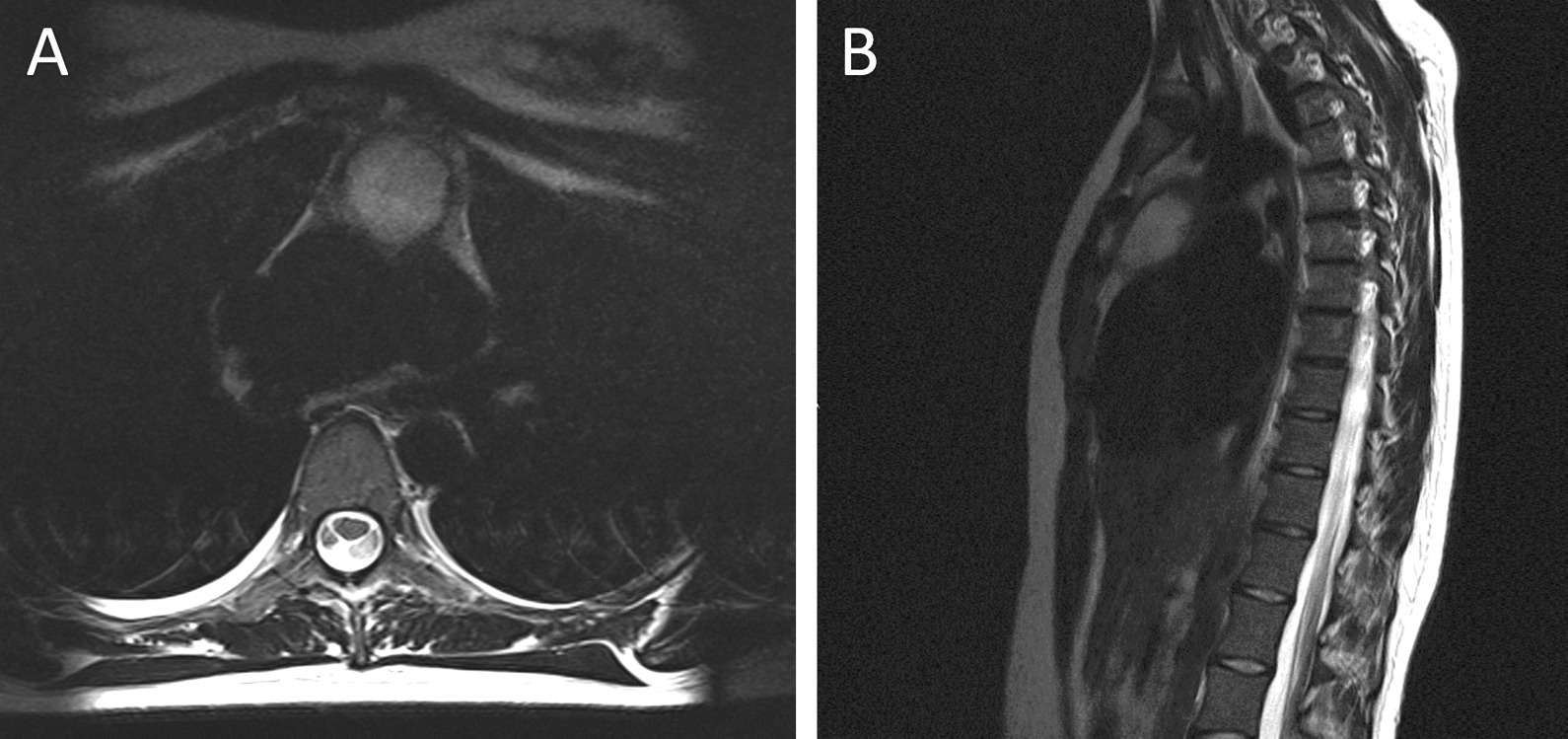
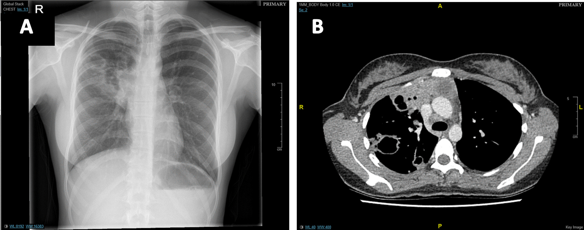
Case presentation: A 23-year-old Asian female initially presented to rheumatology with over a one-year history of neuropathic pain, alongside abnormal white cell count and inflammatory markers. This was investigated with magnetic resonance imaging resulting in an incidental finding of mediastinal mass and pulmonary infiltrates. An initial diagnosis of TB was made despite testing negative for acid-fast bacilli and anti-tubercular treatment was commenced. Four months later, following clinical deterioration and further investigations, a mediastinal biopsy assisted in diagnosing Stage IV HL.

Conclusions: Lymphoma is often misdiagnosed as TB, prolonging time to treatment and may adversely impact patient prognosis due to disease progression. Existing TB guidelines for smear-negative cases are not clear when to consider alternative diagnoses. In smear-negative TB, lymphoma should be considered as a differential and definitive diagnostic tests such as molecular testing and histological examination of biopsies should be considered earlier in the diagnostic work-up to prevent diagnostic delay.

Keywords: Case report; Diagnostic delay; Guidelines; Hodgkin’s lymphoma; Literature review; Tuberculosis.


Conflict of interest statement

The authors declare no competing of interests.

Figures

Fig. 1
Fig. 1
Magnetic resonance imaging (MRI) demonstrating anterior mediastinal mass (A) Transverse T2 image showing a heterogeneous anterior mediastinal mass. B Sagittal fat suppressed image demonstrating anterior mediastinal mass
Fig. 2
Fig. 2
Sagittal plane of CT. This image highlights the presence of a 3.3cm × 3.8 cm cystic mass in the anterior mediastinum
Fig. 3
Fig. 3
Imaging demonstrating cavitating lung lesions. A Chest X-ray demonstrating cavitating lesions in the right upper lobe. B Contrast-enhanced Chest CT scan showing cavitating lesions in the right lung with multiple nodular lesions bilaterally, along with an anterior mediastinal mass that is continuous with the posterior sternum
Fig. 4
Fig. 4
FDG-PET-CT scan images pre-and post-chemotherapy. A Pre-treatment axial section of FDG-PET-CT at mid-thoracic level showing metabolically active necrotic mediastinal lymph nodes and parenchymal lesions in the lungs. B Pre-treatment coronal section demonstrating metabolically active necrotic mediastinal and lung lesions. C Post-treatment axial images of FDG-PET-CT at mid-thoracic level showing complete resolution of the previously noted lung lesions with no significant residual activity. Background uptake seen on these images is due to physiological brown fat activity. D Post-treatment coronal section of FDG-PET-CT with no residual metabolically active disease. Supraclavicular fossa shows marked background of physiological brown fat activity
Fig. 5
Fig. 5
Timeline of case report. This timeline summarises the historical information and clinical interventions and management of this case

References

    1. Agostinelli C, Pileri S. Pathobiology of hodgkin lymphoma. Mediterranean J Hematol Infect Dis. 2014;6(1):2014040. doi: 10.4084/MJHID.2014.040. - DOI - PMC - PubMed
    1. Zhou L, Deng Y, Li N, et al. Global, regional, and national burden of Hodgkin lymphoma from 1990 to 2017: estimates from the 2017 Global Burden of Disease study. J Hematol Oncol. 2019 doi: 10.1186/s13045-019-0799-1. - DOI - PMC - PubMed
    1. Eichenauer DA, Aleman BMP, Andre M, Federico M, Hutchings M, Illidge T, Ladetto M, ESMO Guidelines Committee Hodgkin lymphoma: ESMO Clinical Practice Guidelines for diagnosis treatment and follow-up. HaematolMalig. 2018 doi: 10.1093/annonc/mdy080. - DOI - PubMed
    1. Howell DA, Smith AG, Jack A, Patmore R, Macleod U, Mironska E, Roman E. Time-to-diagnosis and symptoms of myeloma, lymphomas and leukaemias: a report from the Haematological Malignancy Research Network. BMC Hematol. 2013 doi: 10.1186/2052-1839-13-9. - DOI - PMC - PubMed
    1. Puvaneswaran B, Shoba B. Misdiagnosis of tuberculosis in patients with lymphoma. South African Med J. 2012;103(1):32–33. doi: 10.7196/SAMJ.6093. - DOI - PubMed