Review
. 2021 Jul 1:15:703944.
doi: 10.3389/fncel.2021.703944. eCollection 2021.

Living on the Edge of the CNS: Meninges Cell Diversity in Health and Disease

Affiliations
Review

Living on the Edge of the CNS: Meninges Cell Diversity in Health and Disease

Julia Derk et al. Front Cell Neurosci. .

Erratum in

Abstract

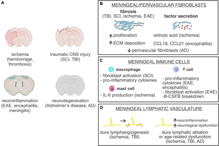
The meninges are the fibrous covering of the central nervous system (CNS) which contain vastly heterogeneous cell types within its three layers (dura, arachnoid, and pia). The dural compartment of the meninges, closest to the skull, is predominantly composed of fibroblasts, but also includes fenestrated blood vasculature, an elaborate lymphatic system, as well as immune cells which are distinct from the CNS. Segregating the outer and inner meningeal compartments is the epithelial-like arachnoid barrier cells, connected by tight and adherens junctions, which regulate the movement of pathogens, molecules, and cells into and out of the cerebral spinal fluid (CSF) and brain parenchyma. Most proximate to the brain is the collagen and basement membrane-rich pia matter that abuts the glial limitans and has recently be shown to have regional heterogeneity within the developing mouse brain. While the meninges were historically seen as a purely structural support for the CNS and protection from trauma, the emerging view of the meninges is as an essential interface between the CNS and the periphery, critical to brain development, required for brain homeostasis, and involved in a variety of diseases. In this review, we will summarize what is known regarding the development, specification, and maturation of the meninges during homeostatic conditions and discuss the rapidly emerging evidence that specific meningeal cell compartments play differential and important roles in the pathophysiology of a myriad of diseases including: multiple sclerosis, dementia, stroke, viral/bacterial meningitis, traumatic brain injury, and cancer. We will conclude with a list of major questions and mechanisms that remain unknown, the study of which represent new, future directions for the field of meninges biology.

Keywords: arachnoid barrier; blood-CSF barrier; border-associated macrophages; fibroblast; meningeal lymphatic system; meninges.


Conflict of interest statement

The authors declare that the research was conducted in the absence of any commercial or financial relationships that could be construed as a potential conflict of interest.

Figures

FIGURE 1
FIGURE 1
Meninges structure and cellular heterogeneity (A) Schematic of cellular make up and structure of the mouse meninges and contiguous perivascular space. (B) Electron microscopy image of a human arachnoid trabecula, the collagen fibril structures that span the wide of the sub-arachnoid space. A cell, potentially an arachnoid, is seen associated with the collagen fibril. Image reproduced with permission from Alcolado et al. (1988). (C) Electron microscopy image of the mouse leptomeninges, a “pia-arachnoid” cell process (PA) spans the subarachnoid space (SA) containing blood vessels (BV). Cells of the inner arachnoid are immediately adjacent to the arachnoid barrier cell layer, which contains microfibrils (f) of extracellular matrix material. Image reproduced with permission from McLone and Bondareff (1975). (D) Immunofluorescence image of human fetal leptomeninges in the Sylvian sulcus labeled with CRABP2 (magenta) and DAPI (cyan). CRABP2 immunoreactivity in the meninges is limited to the arachnoid layer and cells associated with the arachnoid trabecula (AT) in the subarachnoid space (SA). Image reproduced with permission from DeSisto et al. (2020). BV, blood vessels. (E) Immunofluorescence image of mouse leptomeninges at postnatal day 14 labeled with CRABP2 (magenta) and DAPI (cyan). CRABP2 immunoreactivity is seen in cells of the arachnoid layer but not in pia-located cells. Image reproduced with permission from DeSisto et al. (2020).
FIGURE 2
FIGURE 2
Function and structure of the arachnoid barrier layer in the meninges. (A) Graphical depiction of proposed functions of the arachnoid barrier layer of the meninges including (1) as a physical barrier preventing free movement of molecules and cells into the subarachnoid space by virtue of its tight junctions, (2) enriched expression of efflux (P-glycoprotein, BCRP) and solute transporters (SLC) by arachnoid barrier cells support regulated movement of molecules across this barrier layer. (B) Electron microscopy image of the mouse arachnoid-dura interface, reproduced with permission from Nabeshima et al. (1975). Pseudo-coloring of the cell bodies highlights close interface between cells of the arachnoid (A) (inner arachnoid fibroblasts and arachnoid barrier cells connected by electron-dense tight junctions) and cells of the dura, [dura border cells (Db) and dural fibroblasts within collagen fibril dense dura layer]. (C) Immunofluorescence image of mouse leptomeninges from a postnatal day 14 Col1a1-GFP mouse brain with E-cadherin (magenta) and DAPI (cyan). E-cadherin is expressed by arachnoid barrier cells (representing the outer part of the arachnoid layer) but not by inner Col1a1-GFP+ fibroblasts, representing pial and inner arachnoid fibroblasts. Image reproduced with permission from DeSisto et al. (2020).
FIGURE 3
FIGURE 3
Meningeal cells or structures in CNS injury and disease. (A) Depiction of CNS injury or diseases in which meningeal cells, meninges located cell types or parenchyma located perivascular fibroblasts are part of the pathology. SCI, spinal cord injury; TBI, traumatic brain injury; EAE, experimental autoimmune encephalomyelitis. (B) Graphical depiction of fibrosis, caused by increased meningeal/perivascular fibroblast proliferation and Extracellular matrix (ECM) deposition and summary of known CNS fibroblast-derived factors identified in specific CNS disease states. AD, Alzheimer’s disease. (C) Summary of functional roles of meningeal-located immune cells in specific CNS disease states. B-CSFB, blood-CSF barrier. (D) Summary of cellular changes in meninges located lymphatic vasculature in response to different CNS injuries and disease states.

References

    1. Absinta M., Ha S.-K., Nair G., Sati P., Luciano N. J., Palisoc M., et al. (2017). Human and nonhuman primate meninges harbor lymphatic vessels that can be visualized noninvasively by MRI. eLife 6:e29738. 10.7554/eLife.29738 - DOI - PMC - PubMed
    1. Ahn J. H., Cho H., Kim J.-H., Kim S. H., Ham J.-S., Park I., et al. (2019). Meningeal lymphatic vessels at the skull base drain cerebrospinal fluid. Nature 572 62–66. 10.1038/s41586-019-1419-5 - DOI - PubMed
    1. Alcolado R., Weller R. O., Parrish E. P., Garrod D. (1988). The cranial arachnoid and pia mater in man: anatomical and ultrastructural observations. Neuropathol. Appl. Neurobiol. 14 1–17. 10.1111/j.1365-2990.1988.tb00862.x - DOI - PubMed
    1. Alkuwaity K., Taylor A., Heckels J. E., Doran K. S., Christodoulides M. (2012). Group B streptococcus interactions with human meningeal cells and astrocytes in vitro. PLoS One 7:e42660. 10.1371/journal.pone.0042660 - DOI - PMC - PubMed
    1. Alves de Lima K., Rustenhoven J., Kipnis J. (2020). Meningeal immunity and its function in maintenance of the central nervous system in health and disease. Annu. Rev. Immunol. 38 597–620. 10.1146/annurev-immunol-102319-103410 - DOI - PubMed

LinkOut - more resources