Review
. 2021 Jun 30:12:689747.
doi: 10.3389/fphys.2021.689747. eCollection 2021.

Lactate Fluxes and Plasticity of Adipose Tissues: A Redox Perspective

Affiliations
Review

Lactate Fluxes and Plasticity of Adipose Tissues: A Redox Perspective

Damien Lagarde et al. Front Physiol. .

Abstract

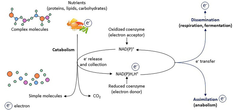
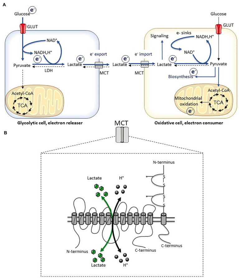
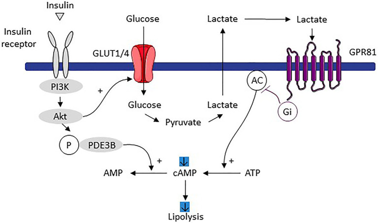
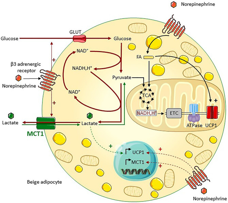
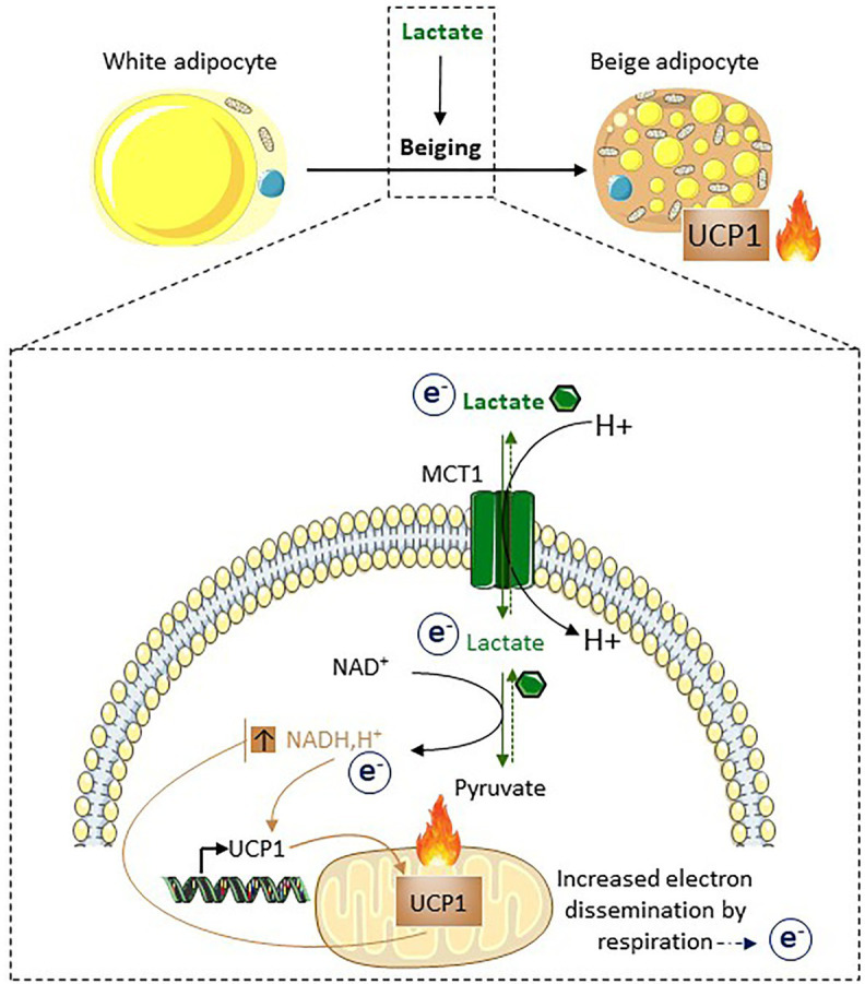
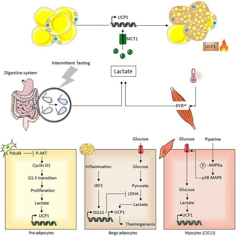
Lactate, a metabolite produced when the glycolytic flux exceeds mitochondrial oxidative capacities, is now viewed as a critical regulator of metabolism by acting as both a carbon and electron carrier and a signaling molecule between cells and tissues. In recent years, increasing evidence report its key role in white, beige, and brown adipose tissue biology, and highlights new mechanisms by which lactate participates in the maintenance of whole-body energy homeostasis. Lactate displays a wide range of biological effects in adipose cells not only through its binding to the membrane receptor but also through its transport and the subsequent effect on intracellular metabolism notably on redox balance. This study explores how lactate regulates adipocyte metabolism and plasticity by balancing intracellular redox state and by regulating specific signaling pathways. We also emphasized the contribution of adipose tissues to the regulation of systemic lactate metabolism, their roles in redox homeostasis, and related putative physiopathological repercussions associated with their decline in metabolic diseases and aging.

Keywords: adipose tissues; beige adipocytes; brown adipocytes; lactate; metabolic dialogs; redox metabolism; white adipocytes.


Conflict of interest statement

The authors declare that the research was conducted in the absence of any commercial or financial relationships that could be construed as a potential conflict of interest.

Figures

Figure 1
Figure 1
Electrons as primary source of energy for cellular activity. Electrons represent the primary source of energy in cells. During catabolic processes, electrons are released from nutrients (which are oxidized) and are accepted by co-enzymes (which become reduced). Two processes are key for regenerating oxidized forms of co-enzymes and thus for redox homeostasis: the assimilation and dissemination processes.
Figure 2
Figure 2
Lactate transport and redox balance. (A) Inter-cellular lactate exchange is associated with inter-cellular electron fluxes. (B) Schematic representation of monocarboxylate transporters (MCT) which are constituted by 12 transmembrane domains, and their chaperone proteins (basigin and embigin). The transport of monocarboxylates (lactate, pyruvate, and ketone bodies) is associated with a proton transport (symport) and occurs bidirectionally depending on the electrochemical gradient. Adapted from Carriere et al. (2020). TCA, tricarboxylic acid cycle.
Figure 3
Figure 3
Mechanism of lipolysis inhibition induced by adipocyte dependent lactate production in response to increased glucose utilization stimulated by insulin. Imported glucose following insulin signaling is converted to lactate and then exported. By an autocrine/paracrine action, lactate activates the GPR81 receptor which, through its effects on adenylate cyclase, decreases the level of cAMP and therefore lipolysis. Adapted from Ahmed et al. (2010). Pl3K, phosphoinositide 3-kinase; POE3B, phosphodiesterase 3B; AMPc, cyclic adenosine monophosphate; ATP, adenosine triphosphate; AC, adenylate cyclase; and GPR81, G protein-coupled receptors associated with inhibitory regulative G-protein (Gi).
Figure 4
Figure 4
MCT1 is a key regulator of lactate bidirectional fluxes in beige adipocytes. MCT1, which is expressed at the cell surface of beige adipocytes, sustains bidirectional lactate fluxes. Lactate export, which is predominant under noradrenergic stimulation such as cold exposure, supports glycolysis through NAD+ regeneration (red arrows). Concomitant lactate import occurs (green arrows), which fuels the oxidative metabolism and promotes the induction of UCPl, thereby increasing the oxidative capacity of beige adipocytes (adapted from Lagarde et al., 2020). ETC, electron transport chain; ATPase, ATP synthase; and FA, fatty acids.
Figure 5
Figure 5
Lactate induces beiging as a way to dissipate redox pressure. Following its import through MCT1, lactate is converted into pyruvate. This conversion is associated with the reduction of NAD+ into NADH,H+. The increased NADH, H+/NAD+ ratio triggers UCP1 expression. Due to the properties of UCP1 and its effects on the respiratory chain, UCP1-dependent uncoupling accelerates oxidation of NADH,H+ into NAD+. Thus, UCP1-dependent uncoupling, in addition to its involvement in non-shivering thermogenesis, may also play an active role in redox homeostasis. Of note, MCT1 is expressed at the cell surface of the subpopulation of adipocytes that will express UCP1 after cold exposure, highlighting it as a marker of inducible beige adipocytes (Carriere et al., 2014; Jeanson et al., 2015; Lagarde et al., 2020).
Figure 6
Figure 6
Different contexts associated with lactate-induced beiging. In vivo, reprogramming of gut microbiota following intermittent fasting is associated with increased lactate production, which may contribute to beiging that improved systemic metabolic parameters (Li et al., 2017). Increased muscle lactate production caused by a calcium channel mutation is associated with beiging (Wang et al., 2020a). Intracellular production of lactate induced by pdcd4 and piperine in preadipocytes and C2C12 cells respectively, upregulates UCPl expression (Bai et al., 2016; Kim et al., 2017). IRF3/ISG15-mediated inhibition of lactate dehydrogenase decreased lactate-induced UCPl expression thus impairing thermogenesis during inflammation (Yan et al., 2021).

References

    1. Adeva-Andany M., Lopez-Ojen M., Funcasta-Calderon R., Ameneiros-Rodriguez E., Donapetry-Garcia C., Vila-Altesor M., et al. . (2014). Comprehensive review on lactate metabolism in human health. Mitochondrion 17, 76–100. 10.1016/j.mito.2014.05.007, PMID: - DOI - PubMed
    1. Ahmed K., Tunaru S., Tang C., Muller M., Gille A., Sassmann A., et al. . (2010). An autocrine lactate loop mediates insulin-dependent inhibition of lipolysis through GPR81. Cell Metab. 11, 311–319. 10.1016/j.cmet.2010.02.012, PMID: - DOI - PubMed
    1. Bai Y., Shang Q., Zhao H., Pan Z., Guo C., Zhang L., et al. . (2016). Pdcd4 restrains the self-renewal and white-to-beige transdifferentiation of adipose-derived stem cells. Cell Death Dis. 7:e2169. 10.1038/cddis.2016.75, PMID: - DOI - PMC - PubMed
    1. Barbatelli G., Murano I., Madsen L., Hao Q., Jimenez M., Kristiansen K., et al. . (2010). The emergence of cold-induced brown adipocytes in mouse white fat depots is determined predominantly by white to brown adipocyte transdifferentiation. Am. J. Physiol. Endocrinol. Metab. 298, E1244–E1253. 10.1152/ajpendo.00600.2009, PMID: - DOI - PubMed
    1. Barreau C., Labit E., Guissard C., Rouquette J., Boizeau M. L., Gani Koumassi S., et al. . (2016). Regionalization of browning revealed by whole subcutaneous adipose tissue imaging. Obesity 24, 1081–1089. 10.1002/oby.21455, PMID: - DOI - PubMed

LinkOut - more resources