. 2021 Jul 1:12:696125.
doi: 10.3389/fmicb.2021.696125. eCollection 2021.

Discovery and Characterization of a Novel Bipartite Botrexvirus From the Phytopathogenic Fungus Botryosphaeria dothidea

Affiliations

Discovery and Characterization of a Novel Bipartite Botrexvirus From the Phytopathogenic Fungus Botryosphaeria dothidea

Mengmeng Yang et al. Front Microbiol. .

Abstract

In this study, we describe a novel positive, single-stranded (+ss) RNA mycovirus, named Botryosphaeria dothidea botrexvirus 1 (BdBV1), from a phytopathogenic fungus Botryosphaeria dothidea showing abnormal morphology and attenuated virulence. BdBV1 is phylogenetically related to Botrytis virus X (BotVX) and is the second potential member of the proposed genus Botrexvirus in the family Alphaflexiviridae. However, it differs from the monopartite BotVX in that BdBV1 possesses a bipartite genome comprised of two ssRNA segments (RNA1 and RNA2 with lengths of 5,035 and 1,063 nt, respectively). BdBV1 RNA1 and RNA2 encode putative RNA-dependent RNA polymerase (RdRp) and coat protein (CP) genes, which share significant identity with corresponding genes in both fungal and plant viruses. Moreover, open reading frames (ORFs) 2-4 of BdBV1 RNA1 shared no detectable identity with any known viral proteins. Immunosorbent electron microscopy (ISEM) analysis using an antibody against the virus CP generated in vitro revealed that BdBV1 is encapsidated in filamentous particles. A comparison of the biological effects of BdBV1 infection on symptoms and growth in isogenic lines of virus-free and virus-infected B. dothidea revealed that BdBV1 is probably involved in reduced growth and virulence of the host fungus. This study describes and characterizes a novel bipartite botrexvirus, which is closely related to uni- and multi-partite fungal and plant viruses and contributes useful information to a better understanding of virus evolution.

Keywords: Alphaflexiviridae; Botryosphaeria dothidea; Botryosphaeria dothidea botrexvirus 1; bipartite botrexvirus; filamentous particles; genome.


Conflict of interest statement

The authors declare that the research was conducted in the absence of any commercial or financial relationships that could be construed as a potential conflict of interest.

Figures

FIGURE 1
FIGURE 1
Genomic properties of Botryosphaeria dothidea botrexvirus 1 (BdBV1). (A) Schematic representation of the genomic organization of BdBV1. Different proteins are represented by different colored boxes, three conserved domains in ORF1 are indicated by darker areas. (B) Multiple alignment of the region corresponding to RdRp domain of BdBV1 and other selected alphaflexiviruses. Eight core RdRp motifs are indicated with lines above. The starting amino acid (aa) position and spacing between motifs are indicated. (C) Multiple alignment of the CP aa sequences of BdBV1 and other flexuous rod-shaped viruses potentially involved in salt-bridge formation. The conserved positively (Arg) and negatively (Asp) charged residues are indicated by yellow boxes.
FIGURE 2
FIGURE 2
Phylogenetic relationships of BdBV1 with other related viruses. (A) Tree created based on the replicase sequences. (B) Tree created based on the CP sequences. BdBV1 is indicated by a black circle, genera within the Alphaflexiviridae are indicated in different colors.
FIGURE 3
FIGURE 3
Colony and hyphal characteristics of B. dothidea strains. (A) Colonial morphology of strains L153, L153-29, and JNT1111 growing on potato dextrose agar (PDA) at 25°C for 3, 6, and 9 days. The front view and rear view were indicated by left and right sides, respectively. (B) Hyphal tips of B. dothidea strains. Scale bar, 100 μm.
FIGURE 4
FIGURE 4
Effects of BdBV1 infection on fungal growth and virulence. (A) Growth rates of strains L153, L153-29, and JNT1111 on PDA 25°C. (B) Pear fruits (P. Brettschneider cv. Huang guan) wound-inoculated with colonized plugs of fungal strains. (C) The average diameter of lesions induced on pear fruits. The presence of BdBV1, BdNV2–4, and BdPV1 in these isolates is indicated below the histograms. Symbols “+” and “−” represent the presence and absence of these viruses based on the results of RT-PCR amplification and dsRNA electrophoresis.
FIGURE 5
FIGURE 5
Transmission electron microscopy (TEM) analysis of virus particles and SDS-PAGE analysis of viral and prokaryotic expressed fusion proteins using 12% gel. (A) TEM images of virus particles purified from strain L153. Two types of isometric and filamentous particles were observed and indicated by red and black arrows, respectively. (B) SDS-PAGE analysis of virus preparations in different sucrose gradients of strains L153-29 and L153. (C) SDS-PAGE analysis of expression of fusion proteins in E. coli was induced at 28°C overnight. Lines 1 and 2, uninduced and induced E. coli transformed with plasmid pGEX-BdBV1-ORF5. Lanes 3 and 4, uninduced and induced E. coli transformed with plasmid plasmid pGEX-KG. Lane 4, induced E. coli transformed with plasmid pGEX-KG. Lanes 5 and 6, supernatant and precipitated proteins of induced E. coli transformed with plasmid pGEX-BdBV1-ORF5 after sonication. Target protein band was indicated by black arrow.
FIGURE 6
FIGURE 6
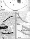
Immunosorbent electron microscopy (ISEM) analysis using PAb-P5 in 4,000- (A,C) or 2,000-fold (B) dilution. The areas marked by arrow were enlarged in black frame.

References

    1. Agranovsky A. A., Lesemann D. E., Maiss E., Hull R., Atabekov J. G. (1995). “Rattlesnake” structure of a fiamentous plant RNA virus built of two capsid proteins. Proc. Natl Acad. Sci. USA 92 2470–2473. 10.1073/pnas.92.7.2470 - DOI - PMC - PubMed
    1. Ayllón M., Turina M., Xie J., Nerva L., Marzano S. Y., Donaire L., et al. (2020). ICTV virus taxonomy profile: botourmiaviridae. J. Gen. Virol. 101 454–455. 10.1099/jgv.0.001409 - DOI - PMC - PubMed
    1. Beever R. E., Howitt R. L. J., Forster R. L. S., Pearson M. N. (2001). Genome characterization of Botrytis virus F, a flexuous rod-shaped mycovirus resembling plant ‘potex-like’ viruses. J. Gen. Virol. 82 67–78. 10.1099/0022-1317-82-1-67 - DOI - PubMed
    1. Burland T. G. (2000). DNASTAR’s Lasergene sequence analysis software. Methods Mol. Biol. 132 71–91. 10.1385/1-59259-192-2:71 - DOI - PubMed
    1. Chare E. R., Holmes E. C. (2006). A phylogenetic survey of recombination frequency in plant RNA viruses. Arch. Virol. 151 933–946. 10.1007/s00705-005-0675-x - DOI - PubMed