Review
. 2021 Jul 1:12:694055.
doi: 10.3389/fimmu.2021.694055. eCollection 2021.

Detection of Immune Checkpoint Receptors - A Current Challenge in Clinical Flow Cytometry

Affiliations
Review

Detection of Immune Checkpoint Receptors - A Current Challenge in Clinical Flow Cytometry

Benjamin Shibru et al. Front Immunol. .

Abstract

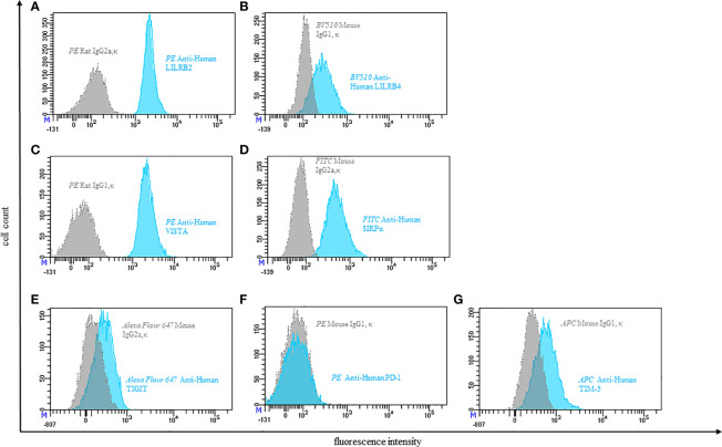
Immunological therapy principles are increasingly determining modern medicine. They are used to treat diseases of the immune system, for tumors, but also for infections, neurological diseases, and many others. Most of these therapies base on antibodies, but small molecules, soluble receptors or cells and modified cells are also used. The development of immune checkpoint inhibitors is amazingly fast. T-cell directed antibody therapies against PD-1 or CTLA-4 are already firmly established in the clinic. Further targets are constantly being added and it is becoming increasingly clear that their expression is not only relevant on T cells. Furthermore, we do not yet have any experience with the long-term systemic effects of the treatment. Flow cytometry can be used for diagnosis, monitoring, and detection of side effects. In this review, we focus on checkpoint molecules as target molecules and functional markers of cells of the innate and acquired immune system. However, for most of the interesting and potentially relevant parameters, there are still no test kits suitable for routine use. Here we give an overview of the detection of checkpoint molecules on immune cells in the peripheral blood and show examples of a possible design of antibody panels.

Keywords: autoimmunity; checkpoint receptors; flow cytometry; immune diagnostics; immune oncology; immunity; infection; laboratory diagnose.


Conflict of interest statement

The authors declare that the research was conducted in the absence of any commercial or financial relationships that could be construed as a potential conflict of interest.

Figures

Figure 1
Figure 1
Gating strategy. The basis of all measurements in this publication is the gating strategy shown here. After exclusion of doublets (A), Neutrophils, Monocytes and Lymphocytes were identified based on the expression of CD45 and granularity (SSC) (B). Neutrophils are also defined by high CD16 and low CD14 expression (CD14-CD16+) (C). Monocytes can be categorized into 3 subpopulations, based on their expression pattern of CD14 and CD16: i) “classical” CD14+CD16-, ii) “intermediate” CD14+CD16+ and iii) “non-classical” CD14-CD16+ (D). T cells were defined as Lymphocytes expressing CD3 (E). By confronting CD4 and CD8 we then identified cytotoxic T cells (CD4- CD8+) and T helper cells (CD4+ CD8-) (F). Among Lymphocytes, those cells that express CD56 but not CD3 were defined as NK cells (G). They were further divided into a CD56dim (CD56+) and a CD56bright (CD56++) subset (H). Antibody panels used can be found in Table 1 .
Figure 2
Figure 2
Representative flow cytometric analysis of the expression of immune checkpoints (green): LAG-3 (A), TIM-3 (B), Siglec-7 (C) and TIGIT (D) on resting NK cells of a healthy donor (male, 23 years old) compared with isotype control (grey).
Figure 3
Figure 3
Representative flow cytometric analysis of the expression of immune checkpoints (blue): LILRB2 (A), LILRB4 (B), VISTA (C), SIRPα (D), TIGIT (E), PD-1 (F), TIM-3 (G) on resting peripheral blood monocytes of a healthy donor compared with isotype control (grey).
Figure 4
Figure 4
Representative flow cytometric analysis of the expression of immune checkpoints (orange): PD-1 (A), VISTA (B), TIM-3 (C), SIRPα (D), LILRB2 (E), TIGIT (F) on resting neutrophils of a healthy donor compared with isotype control (grey).
Figure 5
Figure 5
Schematic overview of immune checkpoints expressed on innate and adaptive immune cells. Only immune checkpoints included in our panels ( Table 1 ) are shown. This selection is by no means a complete representation of all immune checkpoints.
Figure 6
Figure 6
Representative flow cytometric analysis of TIM-3 expression on T helper cells (CD4+) (A) and cytotoxic T cells (CD8+) (B). Comparison of unstimulated (left) and CD3/28 stimulated results after 24h (right) (healthy donor, male, 23 years old).
Figure 7
Figure 7
Representative flow cytometric analysis of the expression of the immune checkpoint TIM-3 on resting NK cells of a healthy donor (male, 23 years old) compared with isotype control.
Figure 8
Figure 8
Representative flow cytometric analysis of LAG-3 expression on T helper cells (CD4+) (A) and cytotoxic T cells (CD8+) (B). Comparison of unstimulated (left) and CD3/28 stimulated results after 24h (right) (healthy donor, male, 23 years old).
Figure 9
Figure 9
Representative flow cytometric analysis of LAG-3 Expression on NK cells. Comparison of unstimulated NK cells after 48h of co-incubation with complete medium (left) and stimulated NK cells after 48h of co-incubation with 10ng/ml IL-15 (right). (healthy donor, female, 65 years old).
Figure 10
Figure 10
Representative flow cytometric analysis of the expression of immune checkpoints TIGIT and PD-1 on unstimulated whole blood T lymphocytes of a healthy 41-year-old female.
Figure 11
Figure 11
Representative flow cytometric analysis of the expression of the immune checkpoint TIGIT on resting NK cells of a healthy donor (male, 23 years old) compared with isotype control.
Figure 12
Figure 12
Representative flow cytometric analysis of the expression of the immune checkpoint Siglec-7 on resting NK cells of a healthy donor (male, 23 years old) compared with isotype control.
Figure 13
Figure 13
Immune checkpoints observed on different immune cells. Inhibitory receptors expressed on different immune cells are illustrated as blue rods, and ligands for these receptors are illustrated as green rods. FDA approved monoclonal antibodies that block receptor-ligand interaction are shown within the outlined boxes. Checkpoint inhibitors targeting the receptor are marked in blue, checkpoint inhibitors targeting ligands are marked in green. Immune cell populations printed in bold signalize that the respective immune checkpoint was included in our own antibody-panel (provided in Table 1 ) and that we were able to detect expression.

References

    1. Regulation (EU) 2017/746 of the European Parliament and of the Council of 5 April 2017 on In Vitro Diagnostic Medical Devices and Repealing Directive 98/79/EC and Commission Decision 2010/227/Eu (2017).
    1. Laskowski TJ, Hazen AL, Collazo RS, Haviland D. Rigor and Reproducibility of Cytometry Practices for Immuno-Oncology: A Multifaceted Challenge. Cytometry A (2020) 97(2):116–25. 10.1002/cyto.a.23882 - DOI - PubMed
    1. Lambert C, Yanikkaya Demirel G, Keller T, Preijers F, Psarra K, Schiemann M, et al. . Flow Cytometric Analyses of Lymphocyte Markers in Immune Oncology: A Comprehensive Guidance for Validation Practice According to Laws and Standards. Front Immunol (2020) 11:2169. 10.3389/fimmu.2020.02169 - DOI - PMC - PubMed
    1. Özcürümez MK, Haeckel R, Gurr E, Streichert T, Sack U. Determination and Verification of Reference Interval Limits in Clinical Chemistry. Recommendations for Laboratories on Behalf of the Working Group Guide Limits of the DGKL With Respect to ISO Standard 15189 and the Guideline of the German Medical Association on Quality Assurance in Medical Laboratory Examinations (Rili-Baek). J Lab Med (2019) 43:127–33. 10.1515/labmed-2018-0500 - DOI
    1. ISO . Iso 15189-2012 Medical Laboratories - Requirements for Quality and Competence. Geneva: ISO; (2012).

Publication types

MeSH terms

Substances