. 2021 Jun 3;125(21):11782-11790.
doi: 10.1021/acs.jpcc.1c03278. Epub 2021 May 19.

EPR of Photoexcited Triplet-State Acceptor Porphyrins

Affiliations

EPR of Photoexcited Triplet-State Acceptor Porphyrins

Ashley J Redman et al. J Phys Chem C Nanomater Interfaces. .

Abstract

The photoexcited triplet states of porphyrin architectures are of significant interest in a wide range of fields including molecular wires, nonlinear optics, and molecular spintronics. Electron paramagnetic resonance (EPR) is a key spectroscopic tool in the characterization of these transient paramagnetic states singularly well suited to quantify spin delocalization. Previous work proposed a means of extracting the absolute signs of the zero-field splitting (ZFS) parameters, D and E, and triplet sublevel populations by transient continuous wave, hyperfine measurements, and magnetophotoselection. Here, we present challenges of this methodology for a series of meso-perfluoroalkyl-substituted zinc porphyrin monomers with orthorhombic symmetries, where interpretation of experimental data must proceed with caution and the validity of the assumptions used in the analysis must be scrutinized. The EPR data are discussed alongside quantum chemical calculations, employing both DFT and CASSCF methodologies. Despite some success of the latter in quantifying the magnitude of the ZFS interaction, the results clearly provide motivation to develop improved methods for ZFS calculations of highly delocalized organic triplet states.


Conflict of interest statement

The authors declare no competing financial interest.

Figures
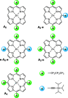
Figure 1
Figure 1
Chemical structures of the acceptor-substituted porphyrins (denoted A2, A2-e, e-A2-e, A3-e, and A4) investigated in this work.
Figure 2
Figure 2
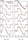
Experimental frozen-solution trEPR spectra of A2, A2-e, e-A2-e, A3-e, and A4 (black) recorded at 20 K at X- and W-band microwave frequencies after photoexcitation at 532 nm. The spectral simulations obtained using the parameters in Table 1 are shown in red. Note: the experimental spectra are the average obtained over a 0.8 μs time window following the laser pulse.
Figure 3
Figure 3
Experimental and simulated MPS spectra recorded at the X-band for A2 (top) and A2-e (bottom). For the MPS spectra presented, excitation was performed with linearly polarized laser light, aligned either parallel (∥) or perpendicular (⊥) to the applied magnetic field direction. The laser wavelengths specified in each panel correspond to absorptions in the optical Q-band region of these porphyrins (see SI). The α and β angles define the relative orientations of the principal axes of the ZFS tensor (X, Y, and Z) and the optical transition dipole moments (Qx and Qy), depicted in the diagrams adjacent to the spectra and were obtained by least-squares fitting. The EPR fitting parameters are given in Table 1. The simulations for D-values of opposite signs as well as a more in-depth discussion of the fitting procedures and interpretation are presented in the SI.
Figure 4
Figure 4
Visualization of the spin-density distributions of the photoexcited triplet states for the five perfluoroalkyl-substituted porphyrins calculated at the B3LYP/EPR-II level.
Figure 5
Figure 5
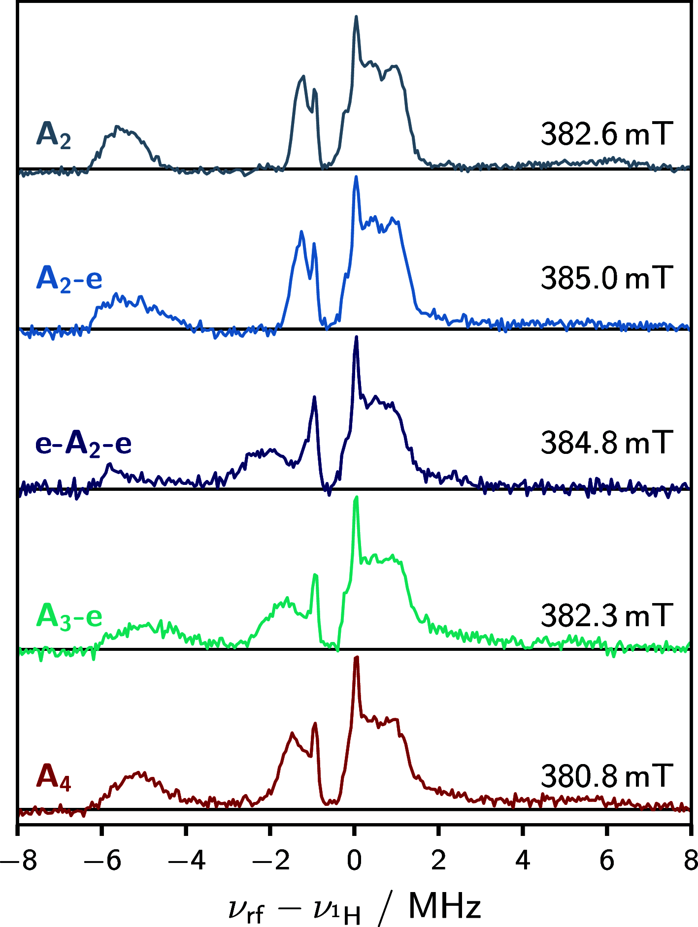
Proton Mims ENDOR spectra for all compounds recorded at the specified magnetic field position. The resonance fields correspond to both the X and Z canonical orientations of the D-tensor. The extent to which the X orientation contributes to the spectrum depends on the orthorhombicity of the D-tensor. The narrow and intense lines located at ≈−1 and 0 MHz, present in all spectra, correspond to the 19F and 1H Larmor frequencies, respectively.

References

    1. Anderson H. L. Building Molecular Wires from the Colours of Life: Conjugated Porphyrin Oligomers. Chem. Commun. 1999, 2323–2330. 10.1039/a904209a. - DOI
    1. Wolf S.; Awschalom D.; Buhrman R.; Daughton J.; Von Molnar S.; Roukes M.; Chtchelkanova A. Y.; Treger D. Spintronics: A Spin-Based Electronics Vision for the Future. Science 2001, 294, 1488–1495. 10.1126/science.1065389. - DOI - PubMed
    1. Ratner M. A Brief History of Molecular Electronics. Nat. Nanotechnol. 2013, 8, 378.10.1038/nnano.2013.110. - DOI - PubMed
    1. Bakulin A. A.; Rao A.; Pavelyev V. G.; van Loosdrecht P. H.; Pshenichnikov M. S.; Niedzialek D.; Cornil J.; Beljonne D.; Friend R. H. The Role of Driving Energy and Delocalized States for Charge Separation in Organic Semiconductors. Science 2012, 335, 1340–1344. 10.1126/science.1217745. - DOI - PubMed
    1. Bai Y.; Olivier J.-H.; Yoo H.; Polizzi N. F.; Park J.; Rawson J.; Therien M. J. Molecular Road Map to Tuning Ground State Absorption and Excited State Dynamics of Long-Wavelength Absorbers. J. Am. Chem. Soc. 2017, 139, 16946–16958. 10.1021/jacs.7b09982. - DOI - PubMed