Review
. 2021 Jun 16;21(3):e20.
doi: 10.4110/in.2021.21.e20. eCollection 2021 Jun.

Gut-Brain Connection: Microbiome, Gut Barrier, and Environmental Sensors

Affiliations
Review

Gut-Brain Connection: Microbiome, Gut Barrier, and Environmental Sensors

Min-Gyu Gwak et al. Immune Netw. .

Abstract

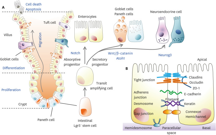
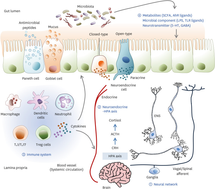
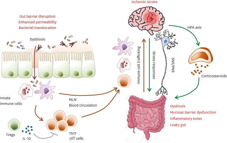
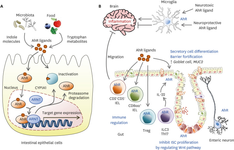
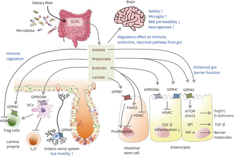
The gut is an important organ with digestive and immune regulatory function which consistently harbors microbiome ecosystem. The gut microbiome cooperates with the host to regulate the development and function of the immune, metabolic, and nervous systems. It can influence disease processes in the gut as well as extra-intestinal organs, including the brain. The gut closely connects with the central nervous system through dynamic bidirectional communication along the gut-brain axis. The connection between gut environment and brain may affect host mood and behaviors. Disruptions in microbial communities have been implicated in several neurological disorders. A link between the gut microbiota and the brain has long been described, but recent studies have started to reveal the underlying mechanism of the impact of the gut microbiota and gut barrier integrity on the brain and behavior. Here, we summarized the gut barrier environment and the 4 main gut-brain axis pathways. We focused on the important function of gut barrier on neurological diseases such as stress responses and ischemic stroke. Finally, we described the impact of representative environmental sensors generated by gut bacteria on acute neurological disease via the gut-brain axis.

Keywords: Aryl hydrocarbon receptor; Brain; Intestine; Microbiome; Short-chain fatty acid; Stroke.


Conflict of interest statement

Conflict of Interest: The authors declare no potential conflicts of interest.

Figures

Figure 1
Figure 1. Dynamic homeostasis of the gut epithelium and gut barrier integrity. (A) Gross features of the small intestine. Proliferation and differentiation of Lgr5+ ISCs into TA cells occur at the intestinal crypt. TA cells are differentiated into secretory and absorptive epithelial lineages during migration from the crypt to the villi. Secretory lineage cells are more differentiated into mucin/antimicrobial peptide-secreting goblet cells and Paneth cells or neuroendocrine cells producing hormones. When these fully differentiated cells reach the villi tips, apoptotic cell death occurs via cell signaling. (B) The intestinal epithelial barrier is constructed of cellular junctions in the paracellular spaces between adjacent cells. Tight junctions are composed of claudin, occludin, and ZO-1, and adherens junctions are composed of E-cadherin. There are also desmosomes, gap junctions, and hemidesmosomes in the paracellular space, which are involved in transporting nutrients and forming the physical intestinal barrier.
TA, transit-amplifying.
Figure 2
Figure 2. The dynamic communication of the gut microbiota-brain axis. Bidirectional communication between the gut and brain can be mediated by several direct and indirect pathways. The communication routes involve ① the nervous system including the ENS and the VN, ② the neuroendocrine and HPA axis, ③ the immune system, and ④ microbiota-derived neuroactive compounds. The gut microbiota can produce neurotransmitters such as GABA, dopamine, and serotonin; amino acids such as tyramine and tryptophan; and microbial metabolites such as SCFAs and AhR ligands. The gut epithelium consists of various kinds of hormone-secreting specialized neuroendocrine cells. These hormone and neuroactive compounds influence the local gut physiology and can travel through the blood circulation to interact with the host immune system and metabolism that directly signals the brain. The gut microbiota can influence epithelial barrier integrity, controlling the transit of signaling molecules from the gut lumen to the lamina propria, which contains various immune cells and neurons, or the blood circulation. Some neuropsychiatric conditions can disrupt gut barrier integrity. Stress can activate an HPA axis response that involves CRH, ACTH, and cortisol sequentially. Cortisol regulates neuro-immune signaling and affects intestinal barrier integrity. Therefore, stress hormones, immune mediators, and CNS neurotransmitters can change the gut environment and alter microbiota composition.
5-HT, 5-hydroxytryptamine.
Figure 3
Figure 3. Gut-brain system crosstalk via immunity and HPA axis after stroke. Stroke induces dysbiosis, mucosal barrier dysfunction, an increase in gut permeability, and bacteria translocation, leading to post-stroke infection. In the gut, dysbiosis and bacteria translocation induce a pro-inflammatory T cell response via innate immune cells. Immune cells, especially IL-17+ γδ T cells, macrophages, and DCs migrate from the gut to the meninges and the brain after stroke, which leads to post-stroke inflammation and exacerbated brain tissue damage.
SNS, sympathetic nervous system; PNS, parasympathetic nervous system; MLN, mesenteric lymph node.
Figure 4
Figure 4. The role of AhR signals in the gut and brain. (A) Upon agonist binding, AhR and some components of the chaperone complex translocate to the nucleus, where AhR binds DNA-responsive elements to control target gene expression such as the CYP1A1. CYP1A1 inactivates AhR ligands in the cytosol. (B) In AhR activation in T cells, AhR induces Th17 differentiation and stabilizes Treg and Tr1 cells. In IELs, AhR leads to differentiation into immunoregulatory TCRαβ+CD8αα+ IELs. AhR activation in CD2+CD5+ IELs specific to myelin induces migration into the CNS and limits inflammation. AhR functions in the homeostasis of TCRγδ+CD8 αα+ IELs. AhR mediates the homeostasis of and IL-22 secretion from ILC3s. AhR controls IEC regeneration, preventing malignant outgrowth. AhR agonists derived from the diet, gut flora, and host metabolism may affect brain tissues crossing the BBB. In astrocytes and microglia, AhR suppresses or enhances inflammatory tones in the CNS in a ligand-specific manner.
CYP1A1, cytochrome P450 family 1 subfamily A member 1; ARNT, aryl hydrocarbon receptor nuclear translocator.
Figure 5
Figure 5. Potential gut-brain pathways through SCFAs modulating brain function. Dietary fiber-derived SCFAs fermented by gut microbiota influence gut-brain communication. SCFAs locally interact with IECs and immune cells by their receptors, GPRs, or by inhibiting histone deacetylases, which influence intestinal mucosal immunity and barrier function. In IEC, SCFAs can enhance barrier integrity by upregulating the expression of tight-junction proteins and RegIIIγ. Butyrate suppresses the proliferation of IEC, but lactate enhances it. SCFAs induce Treg expansion but inhibit Th17 generation via lamina propria DCs. SCFA promotes the secretion of gut hormones from the enteroendocrine cells and indirectly affects the brain via the systemic circulation or vagal pathways. In the brain, SCFAs also increased satiety, microglia, and neurogenesis, and decreased BBB permeability. After a stroke, SCFAs have a critical role in balancing γδT-IL-17+ cells and Tregs and decrease in microglial activation, associated with better recovery after a stroke.

References

    1. Gribble FM, Reimann F. Enteroendocrine cells: chemosensors in the intestinal epithelium. Annu Rev Physiol. 2016;78:277–299. - PubMed
    1. Kim SH, Jang YS. Recent insights into cellular crosstalk in respiratory and gastrointestinal mucosal immune systems. Immune Netw. 2020;20:e44. - PMC - PubMed
    1. Seo K, Seo J, Yeun J, Choi H, Kim YI, Chang SY. The role of mucosal barriers in human gut health. Arch Pharm Res. 2021;44:325–341. - PubMed
    1. Chelakkot C, Ghim J, Ryu SH. Mechanisms regulating intestinal barrier integrity and its pathological implications. Exp Mol Med. 2018;50:1–9. - PMC - PubMed
    1. Szakál DN, Gyorffy H, Arató A, Cseh A, Molnár K, Papp M, Dezsofi A, Veres G. Mucosal expression of claudins 2, 3 and 4 in proximal and distal part of duodenum in children with coeliac disease. Virchows Arch. 2010;456:245–250. - PubMed