Review
. 2021 Sep;59(3):68.
doi: 10.3892/ijo.2021.5248. Epub 2021 Jul 19.

MUC1 is an oncoprotein with a significant role in apoptosis (Review)

Affiliations
Review

MUC1 is an oncoprotein with a significant role in apoptosis (Review)

Katarzyna Supruniuk et al. Int J Oncol. 2021 Sep.

Abstract

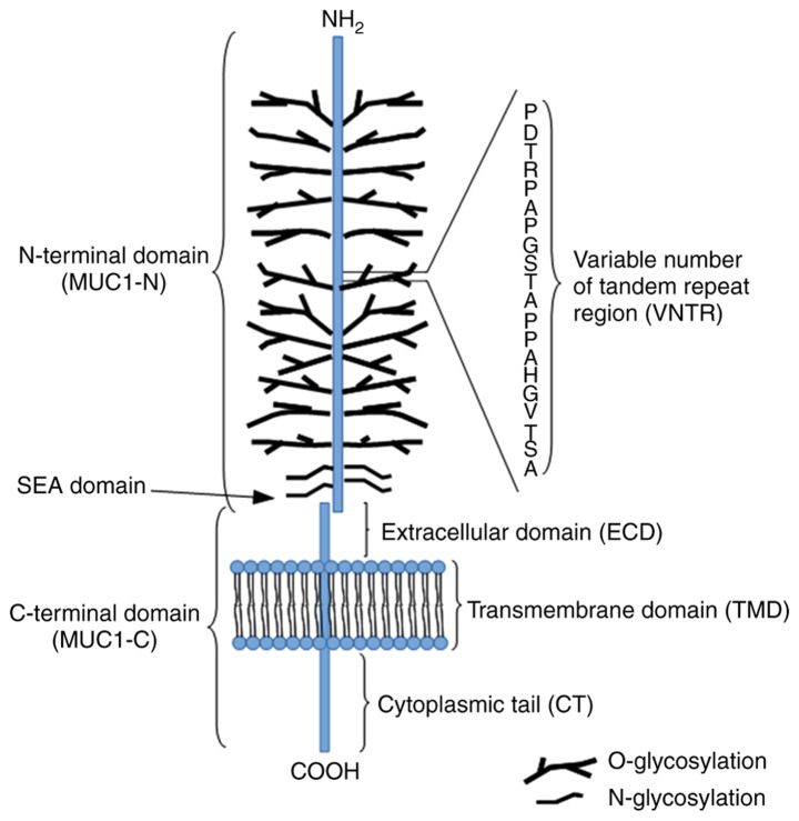
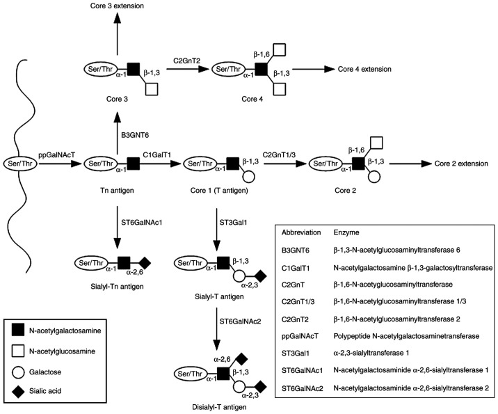
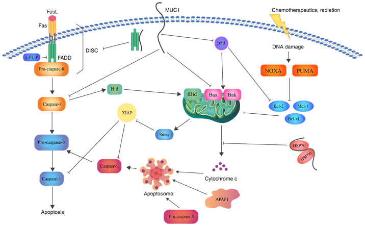
Mucin 1 (MUC1) is a membrane‑bound, highly glycosylated protein that is overexpressed in all stages of malignant transformation. Overexpression of MUC1 together with loss of polarization and hypoglycosylation are associated with resistance to apoptosis, which is the process that results in efficient removal of damaged cells. Inhibition of the apoptotic process is responsible for tumor development, tumor progression and drug resistance. MUC1 is considered as an oncogenic molecule that is involved in various signaling pathways responsible for the regulation of apoptosis. Based on this, the aim of the present study was to discuss the involvement of MUC1 in the divergent mechanisms regulating programmed cell death.

Keywords: MUC1; anoikis; apoptosis; cancer; glycosylation.


Conflict of interest statement

The authors declare that they have no competing interests.

Figures

Figure 1
Figure 1
Structure of MUC1. MUC1, mucin 1.
Figure 2
Figure 2
Mucin-type O-glycan synthesis. O-glycosylation is initiated by attachment of GalNAc to the hydroxyl groups of the Ser/Thr of the protein chain. The reaction is catalyzed by ppGalNAcT enzymes and results in the formation of the Tn antigen (GalNAcα1-O- Ser/Thr). The four basic core structures are generated in the Golgi apparatus by specific glycosyltransferases. The Tn antigen can be elongated through galactose addition catalyzed by C1GalT1 or core 1 synthase, which results in the synthesis of the T antigen or core 1. The antigen can also be elongated by B3GNT6, which transfers GlcNAc to the GalNAc-Ser/Thr structure to form core 3. Subsequent GlcNAc addition to core 3 forms core 4. Core structures can be further elongated or terminated by attachment of fucose or sialic acid. GalNAc, N-acetylgalactosamine; Ser, serine; Thr, threonine; ppGalNAcT, N-acetylgalactosaminyltransferase; C1GalT1, N-acetylgalactosamine β-1,3-galactosyltransferase; Galβ1, galactosamine β1; T, Galβ1-3GalNAcα1-O-Ser/Thr; Tn, GalNAcα1-O-Ser/Thr.
Figure 3
Figure 3
Schematic representation of the role of MUC1 in apoptosis. MUC1 interacts with FADD DED, blocking the formation of DISC and suppressing the induction of the extrinsic apoptotic pathway. Direct association with caspase-8 inhibits its activation. In addition, MUC1-C suppresses Bax translocation to the MOM and cytochrome c release. Binding of MUC1 to the HSP90/HSP70 complex weakens the activation of the mitochondrial pathway. Moreover, direct binding of MUC1 to the p53 regulatory domain is associated with stimulation of growth-arresting gene transcription, thereby inhibiting apoptosis. Bcl, B-cell lymphoma; Bax, Bcl-2-associated X protein; Bcl-xL, Bcl-extra large; MUC1, mucin 1; FADD, FAS-associated with death domain; DED, death effector domain; DISC, death inducing signaling complex; MUC1-C, MUC1 C-terminal subunit; MOM, mitochondrial outer membrane; HSP, heat shock protein; FasL, fatty acid synthetase ligand; FLIP, FLICE (FADD-like IL-1β-converting enzyme)-inhibitory protein; Bid, BH3 interacting domain death agonist; Mcl-1, induced myeloid leukemia cell differentiation protein; NOXA, NADPH oxidase activator; PUMA, p53 upregulated modulator of apoptosis; Smac, second mitochondria-derived activator of caspase; XIAP, X-linked inhibitor of apoptosis protein.

References

    1. Behera SK, Praharaj AB, Dehury B, Negi S. Exploring the role and diversity of mucins in health and disease with special insight into non-communicable diseases. Glyconconj J. 2015;32:575–613. doi: 10.1007/s10719-015-9606-6. - DOI - PubMed
    1. Dhanisha SS, Guruvayoorappan C, Drishya S, Abeesh P. Mucins: Structural diversity, biosynthesis, its role in pathogenesis and as possible therapeutic targets. Crit Rev Oncol Hematol. 2018;122:98–122. doi: 10.1016/j.critrevonc.2017.12.006. - DOI - PubMed
    1. Cornick S, Tawiah A, Chadee K. Roles and regulation of the mucus barrier in the gut. Tissue Barriers. 2015;3:e982426. doi: 10.4161/21688370.2014.982426. - DOI - PMC - PubMed
    1. Moniaux N, Escande F, Porchet N, Aubert JP, Batra SK. Structural organization and classification of the human mucin genes. Front Biosci. 2001;6:D1192–D1206. doi: 10.2741/Moniaux. - DOI - PubMed
    1. Nath S, Mukherjee P. MUC1: A multifaceted oncoprotein with a key role in cancer progression. Trends Mol Med. 2014;20:332–342. doi: 10.1016/j.molmed.2014.02.007. - DOI - PMC - PubMed