. 2021 Sep;24(3):636.
doi: 10.3892/mmr.2021.12275. Epub 2021 Jul 19.

Interactions between the ERK1/2 signaling pathway and PCAF play a key role in PE‑induced cardiomyocyte hypertrophy

Affiliations

Interactions between the ERK1/2 signaling pathway and PCAF play a key role in PE‑induced cardiomyocyte hypertrophy

Qian Mao et al. Mol Med Rep. 2021 Sep.

Abstract

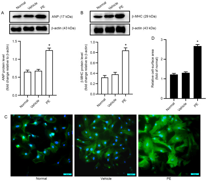
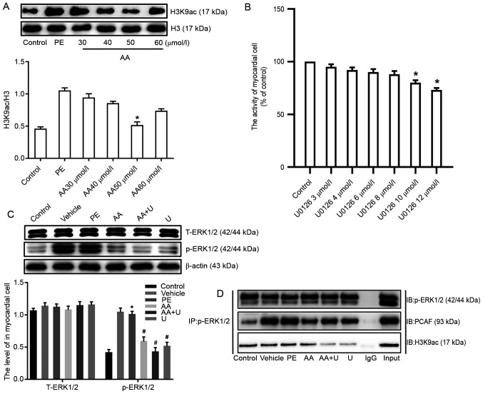
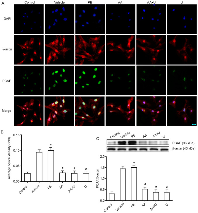
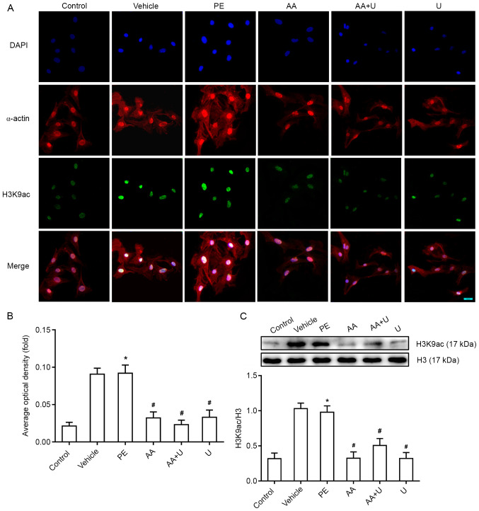
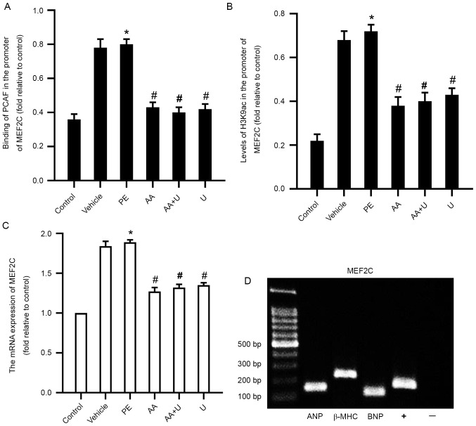
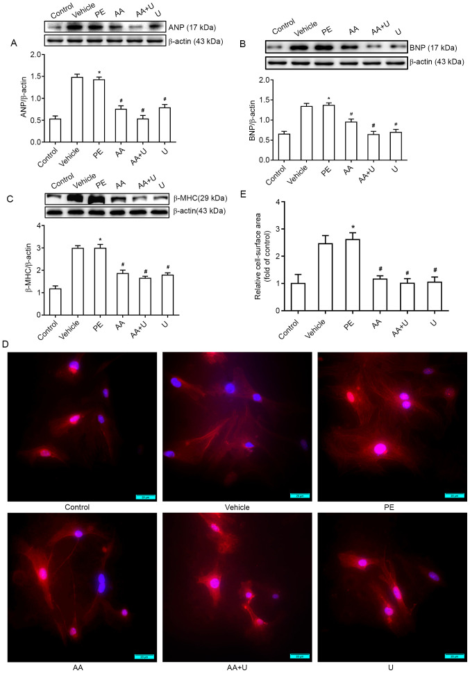
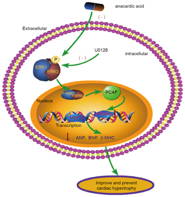
Cardiomyocyte hypertrophy is a compensatory phase of chronic heart failure that is induced by the activation of multiple signaling pathways. The extracellular signal‑regulated protein kinase (ERK) signaling pathway is an important regulator of cardiomyocyte hypertrophy. In our previous study, it was demonstrated that phenylephrine (PE)‑induced cardiomyocyte hypertrophy involves the hyperacetylation of histone H3K9ac by P300/CBP‑associated factor (PCAF). However, the upstream signaling pathway has yet to be fully identified. In the present study, the role of the extracellular signal‑regulated protein kinase (ERK)1/2 signaling pathway in PE‑induced cardiomyocyte hypertrophy was investigated. The mice cardiomyocyte hypertrophy model was successfully established by treating cells with PE in vitro. The results showed that phospho‑(p‑)ERK1/2 interacted with PCAF and modified the pattern of histone H3K9ac acetylation. An ERK inhibitor (U0126) and/or a histone acetylase inhibitor (anacardic acid; AA) attenuated the overexpression of phospho‑ERK1/2 and H3K9ac hyperacetylation by inhibiting the expression of PCAF in PE‑induced cardiomyocyte hypertrophy. Moreover, U0126 and/or AA could attenuate the overexpression of several biomarker genes related to cardiac hypertrophy (myocyte enhancer factor 2C, atrial natriuretic peptide, brain natriuretic peptide and β‑myosin heavy chain) and prevented cardiomyocyte hypertrophy. These results revealed a novel mechanism in that AA protects against PE‑induced cardiomyocyte hypertrophy in mice via the ERK1/2 signaling pathway, and by modifying the acetylation of H3K9ac. These findings may assist in the development of novel methods for preventing and treating hypertrophic cardiomyopathy.

Keywords: ERK‑signaling pathway; anacardic acid; cardiomyocyte hypertrophy; histone acetylation.


Conflict of interest statement

The authors declare that they have no competing interests.

Figures

Figure 1.
Figure 1.
Cardiomyocyte hypertrophy induced by PE. (A) Effects of PE on the expression levels of the myocardial hypertrophy biomarker ANP in primary cardiomyocytes from a neonatal mouse. (B) Effects of PE on the expression levels of the myocardial hypertrophy biomarker β-MHC in primary cardiomyocytes from a neonatal mouse. (C) Immunofluorescent staining analysis of the cell surface area in primary cardiomyocytes from a neonatal mouse. (D) Quantification of changes in the cell surface area in each group. Cells were treated with 100 µmol/l PE for 48 h. n=6. Scale bar, 20 µm. *P<0.05 vs. vehicle. PE, phenylephrine; ANP, atrial natriuretic peptide; β-MHC, β-myosin heavy chain; Vehicle, 100 µmol/l phenylephrine + equal volume of DMSO. for 48 h.
Figure 2.
Figure 2.
p-ERK1/2 interacted with PCAF and modified H3K9ac acetylation in hypertrophic cardiomyocytes induced by PE. (A) Different concentrations of AA (30, 40, 50 and 60 µmol/l) were used to identify the optimal concentration of AA; 50 µmol/l was selected for subsequent experiments, based on the levels of histone H3K9ac. (B) Effects of different concentrations of the ERK inhibitor U0126 (2, 4, 6, 8 and 10 µmol/l) on cell viability in neonatal mouse cardiomyocytes. (C) Expression of T-ERK1/2 and p-ERK1/2 in myocardial cells from neonatal mice. (D) Co-immunoprecipitation in cell lysates of mouse myocardial cells exposed to six different experimental conditions with anti-p-ERK1/2-protein G magnetic beads and IB with anti-PCAF, anti-H3K9ac or anti-p-ERK1/2 antibodies to evaluate protein expression. Input, positive control; IgG, negative control. n=6. *P<0.05 vs. control group; #P<0.05 vs. PE group. P-, phospho-; PCAF, P300/CBP-associated factor; PE, phenylephrine; H3K9ac, histone 3 acetylation K9; AA, anacardic acid; T-, total-; IB, immunoblotting; ERK, extracellular signal-regulated protein kinase; PE, 100 µmol/l phenylephrine for 48 h; Vehicle, 100 µmol/l phenylephrine + equal volume of DMSO for 48 h; AA, 50 µmol/l AA for 30 min + 100 µmol/l phenylephrine for 48 h; AA + U, 50 µmol/l AA + 10 µmol/l U0126 for 30 min + 100 µmol/l phenylephrine for 48 h; U, 10 µmol/l U0126 for 30 min + 100 µmol/l phenylephrine for 48 h; Control, no drug.
Figure 3.
Figure 3.
Effects of AA and U0126 on the expression of PCAF in PE-induced cardiomyocyte hypertrophy. (A) PCAF (green fluorescence) and α-actin (red fluorescence), in combination with DAPI staining (blue fluorescence), in cardiomyocytes exposed to six different conditions. Scale bar, 20 µm. (B) Mean optical density of PCAF immunofluorescence in the six groups. (C) Western blotting of PCAF expression, showed PCAF levels were significantly higher in the hypertrophic cardiomyocytes induced by PE, whereas AA and/or U0126 prevented this effect. n=6. *P<0.05 vs. control group; #P<0.05 vs. PE group. PCAF, P300/CBP-associated factor; PE, phenylephrine; AA, anacardic acid; PE, 100 µmol/l phenylephrine for 48 h; Vehicle, 100 µmol/l phenylephrine + equal volume of DMSO for 48 h; AA, 50 µmol/l AA for 30 min + 100 µmol/l phenylephrine for 48 h; AA + U, 50 µmol/l AA + 10 µmol/l U0126 for 30 min + 100 µmol/l phenylephrine for 48 h; U, 10 µmol/l U0126 for 30 min + 100 µmol/l phenylephrine for 48 h; Control, no drug.
Figure 4.
Figure 4.
Acetylation levels of histone H3K9ac in mouse myocardial cells. (A) H3K9ac (green fluorescence) and α-actin (red fluorescence), combined with DAPI staining (blue fluorescence), in cardiomyocytes exposed to six different conditions. Scale bar, 20 µm. (B) Mean optical density of H3K9ac immunofluorescence in the six groups. (C) Western blotting data showed that the levels of H3K9ac were significantly higher in the hypertrophic cardiomyocytes induced by PE, whereas AA and/or U0126 prevented this effect. n=6. *P<0.05 vs. control group; #P<0.05 vs. PE group. PCAF, P300/CBP-associated factor; PE, phenylephrine; AA, anacardic acid; PE, 100 µmol/l phenylephrine for 48 h; Vehicle, 100 µmol/l phenylephrine + equal volume of DMSO for 48 h; AA, 50 µmol/l AA for 30 min + 100 µmol/l phenylephrine for 48 h; AA + U, 50 µmol/l AA + 10 µmol/l U0126 for 30 min + 100 µmol/l phenylephrine for 48 h; U, 10 µmol/l U0126 for 30 min + 100 µmol/l phenylephrine for 48 h; Control, no drug; H3K9ac, histone 3 acetylation K9.
Figure 5.
Figure 5.
PCAF binding, acetylation levels of H3K9ac in the MEF2C promoter and the expression of MEF2C in myocardial cells. (A) Binding of PCAF to the promoter in MEF2C was assessed using ChIP-qPCR. (B) Acetylation levels of histone H3K9ac in the promoter of MEF2C were assessed using ChIP-qPCR. (C) mRNA expression of MEF2C in cardiomyocytes exposed to six different conditions. (D) ChIP-qPCR data showed that the cardiac nuclear transcription factor MEF2C could bind to the promoter of biomarker genes for cardiac hypertrophy (ANP, BNP and β-MHC). n=6. *P<0.05 vs. control group; #P<0.05 vs. PE group. PCAF, P300/CBP-associated factor; PE, phenylephrine; AA, anacardic acid; PE, 100 µmol/l phenylephrine for 48 h; Vehicle, 100 µmol/l phenylephrine + equal volume of DMSO for 48 h; AA, 50 µmol/l AA for 30 min + 100 µmol/l phenylephrine for 48 h; AA + U, 50 µmol/l AA + 10 µmol/l U0126 for 30 min + 100 µmol/l phenylephrine for 48 h; U, 10 µmol/l U0126 for 30 min + 100 µmol/l phenylephrine for 48 h; Control, no drug; PCAF, P300/CBP-associated factor; PE, phenylephrine; H3K9ac, histone 3 acetylation K9; MEF2C, myocyte enhancer factor 2C; ChIP-qPCR, chromatin-immunoprecipitation-quantitative PCR; ANP, atrial natriuretic peptide; β-MHC, β-myosin heavy chain; BNP, brain natriuretic peptide.
Figure 6.
Figure 6.
AA and U0126 alleviates PE-induced cardiomyocyte hypertrophy. (A-C) Western blotting data for cardiac hypertrophy biomarkers (ANP, BNP and β-MHC). (D) Cell surface area was measured by immunofluorescence staining to demonstrate the hypertrophic responses in cardiomyocytes. (E) Quantification of the cell surface area of myocardial cells. Scale bar, 20 µm. n=6. *P<0.05 vs. control group; #P<0.05 vs. PE group. PCAF, P300/CBP-associated factor; PE, phenylephrine; AA, anacardic acid; PE, 100 µmol/l phenylephrine for 48 h; Vehicle, 100 µmol/l phenylephrine + equal volume of DMSO for 48 h; AA, 50 µmol/l AA for 30 min + 100 µmol/l phenylephrine for 48 h; AA + U, 50 µmol/l AA + 10 µmol/l U0126 for 30 min + 100 µmol/l phenylephrine for 48 h; U, 10 µmol/l U0126 for 30 min + 100 µmol/l phenylephrine for 48 h; Control, no drug; ANP, atrial natriuretic peptide; β-MHC, β-myosin heavy chain; BNP, brain natriuretic peptide; PE, phenylephrine.
Figure 7.
Figure 7.
ERK 1/2 cell signaling pathway. The interaction between p-ERK1/2 and PCAF leads to a reduction in H3K9ac in PE-induced cardiomyocyte hypertrophy. p-, phospho-; PCAF, P300/CBP-associated factor; ANP, atrial natriuretic peptide; β-MHC, β-myosin heavy chain; BNP, brain natriuretic peptide; MEF2C, myocyte enhancer factor 2C; H3K9ac, histone 3 acetylation K9; PE, phenylephrine; ERK, extracellular signal-regulated protein kinase.

Similar articles

Cited by

References

    1. Degoricija V, Trbušić M, Potočnjak I, Radulović B, Terešak SD, Pregartner G, Berghold A, Tiran B, Frank S. Acute heart failure developed as worsening of chronic heart failure is associated with increased mortality compared to de novo cases. Sci Rep. 2018;8:9587. doi: 10.1038/s41598-018-28027-3. - DOI - PMC - PubMed
    1. Yu B, Zhao Y, Zhang H, Xie D, Nie W, Shi K. Inhibition of microRNA-143-3p attenuates myocardial hypertrophy by inhibiting inflammatory response. Cell Biol Int. 2018;42:1584–1593. doi: 10.1002/cbin.11053. - DOI - PubMed
    1. Wehbe N, Nasser SA, Pintus G, Badran A, Eid AH, Baydoun E. MicroRNAs in cardiac hypertrophy. Int J Mol Sci. 2019;20:4714. doi: 10.3390/ijms20194714. - DOI - PMC - PubMed
    1. Peng C, Luo X, Li S, Sun H. Phenylephrine-induced cardiac hypertrophy is attenuated by a histone acetylase inhibitor anacardic acid in mice. Mol Biosyst. 2017;13:714–724. doi: 10.1039/C6MB00692B. - DOI - PubMed
    1. Gao W, Guo N, Zhao S, Chen Z, Zhang W, Yan F, Liao H, Chi K. Carboxypeptidase A4 promotes cardiomyocyte hypertrophy through activating PI3K-AKT-mTOR signaling. Biosci Rep. 2020;40:BSR20200669. doi: 10.1042/BSR20200669. - DOI - PMC - PubMed