Review
. 2021 Oct 27;121(20):12181-12277.
doi: 10.1021/acs.chemrev.1c00244. Epub 2021 Jul 19.

Lipids and Lipid Derivatives for RNA Delivery

Affiliations
Review

Lipids and Lipid Derivatives for RNA Delivery

Yuebao Zhang et al. Chem Rev. .

Abstract

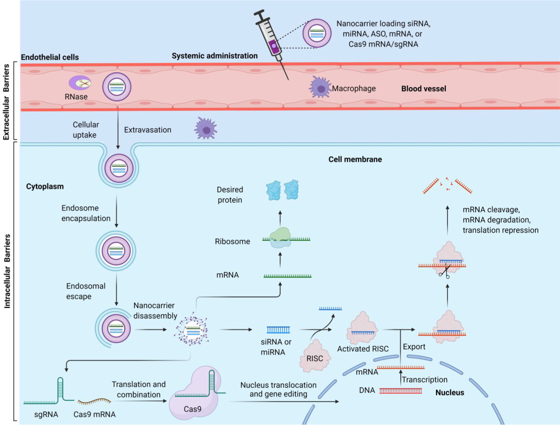
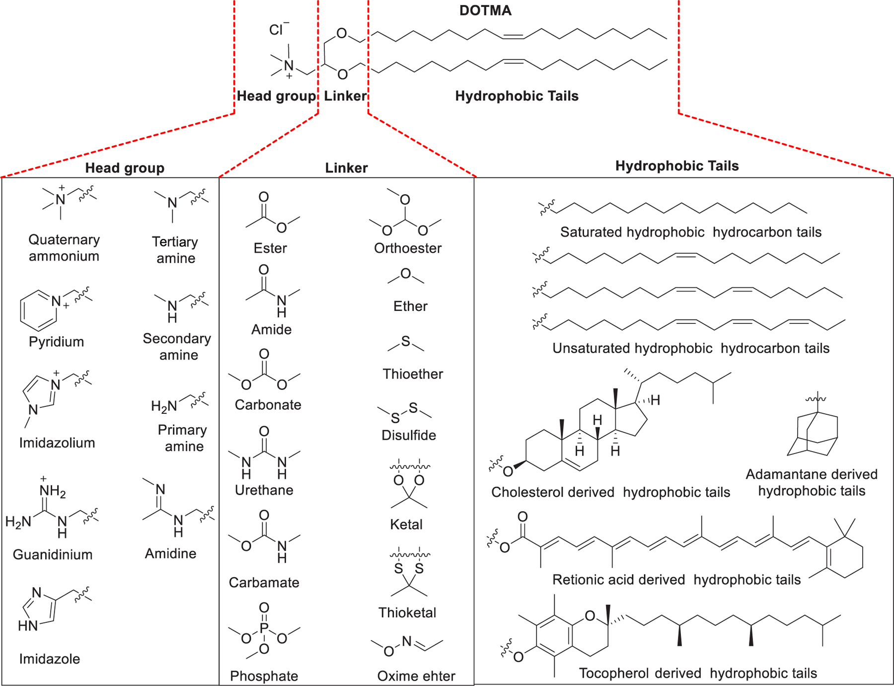
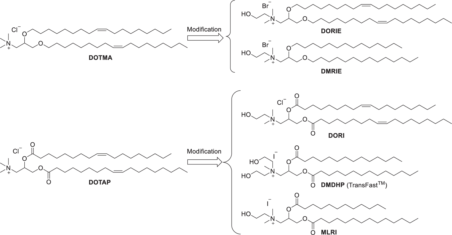
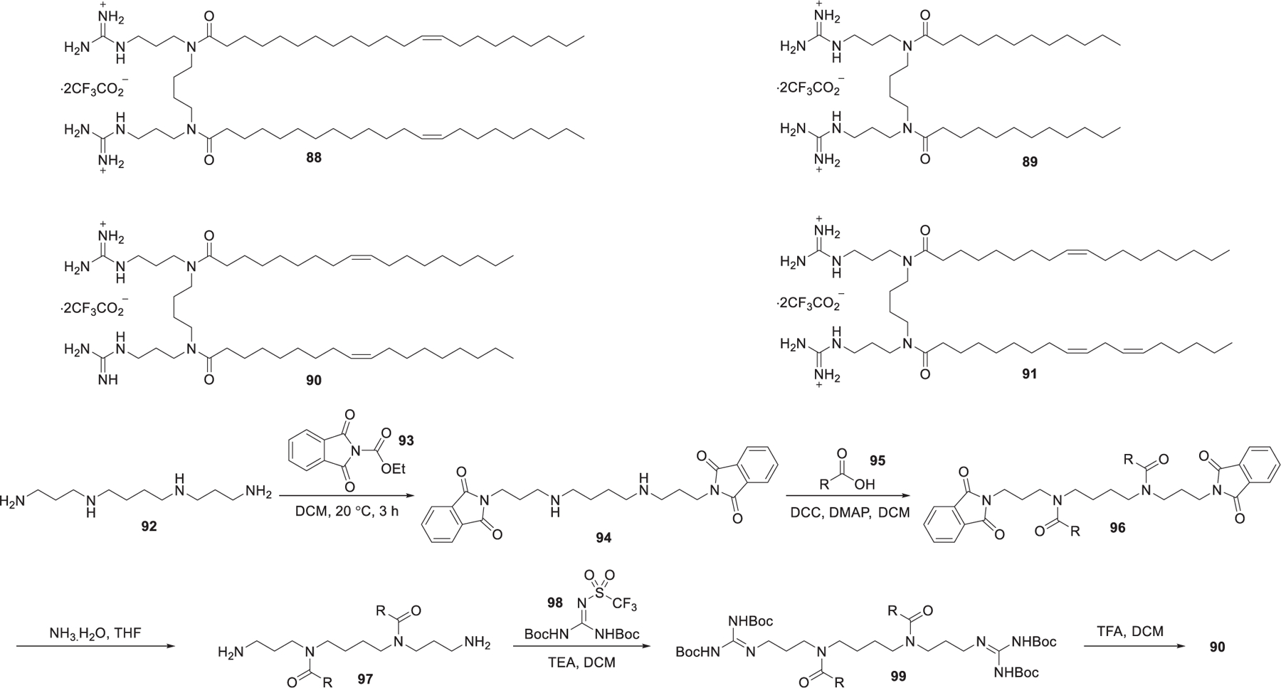
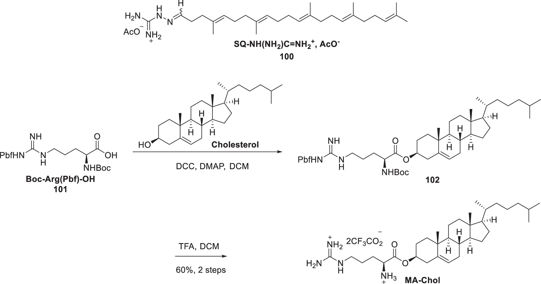
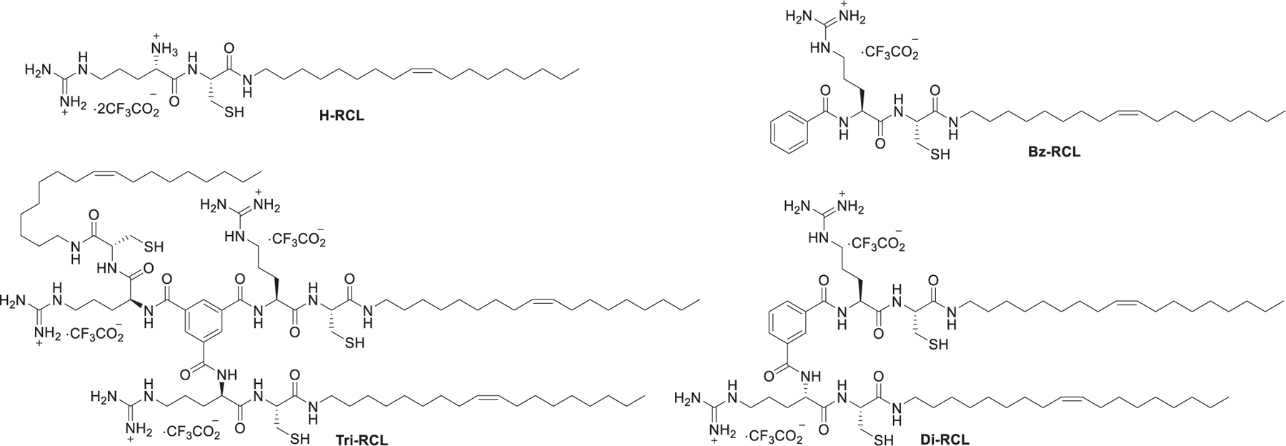
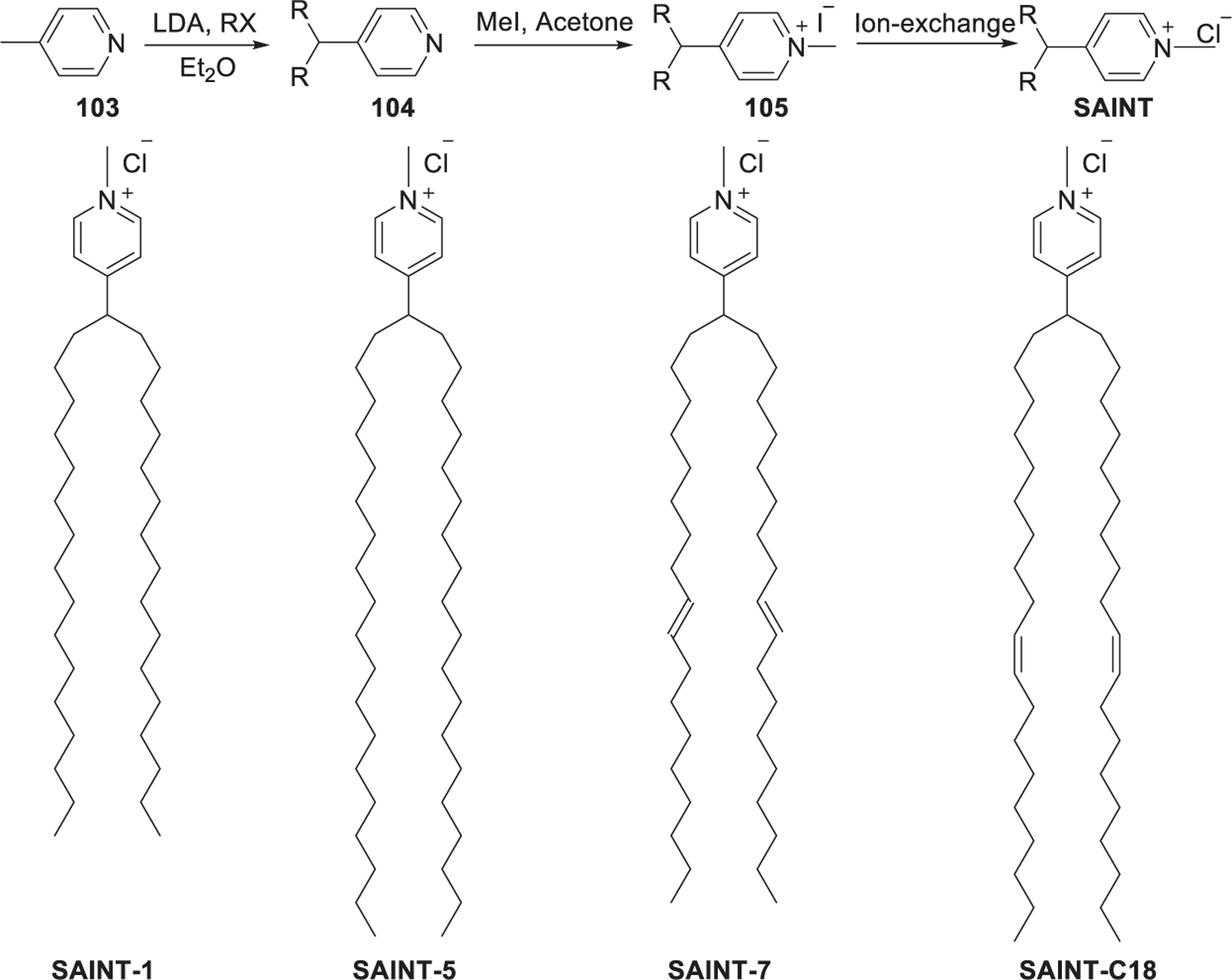
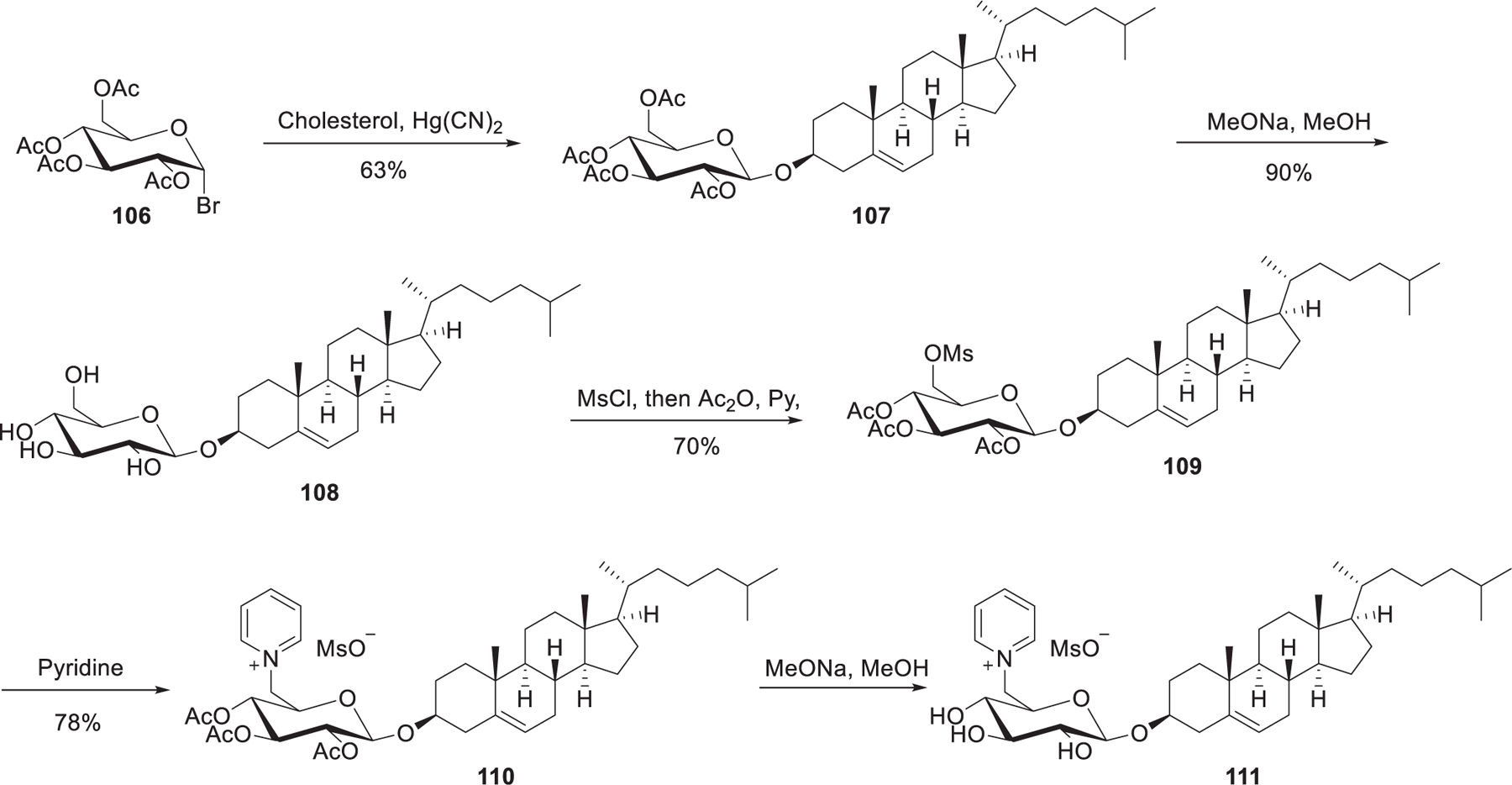
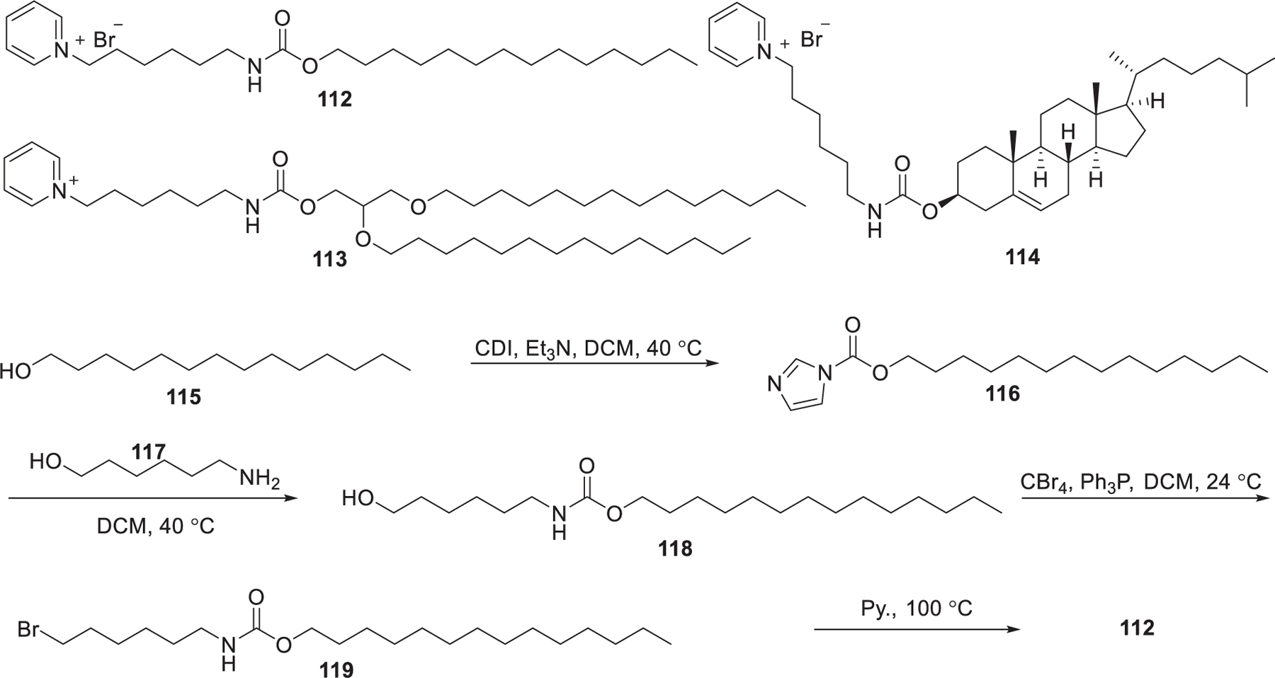
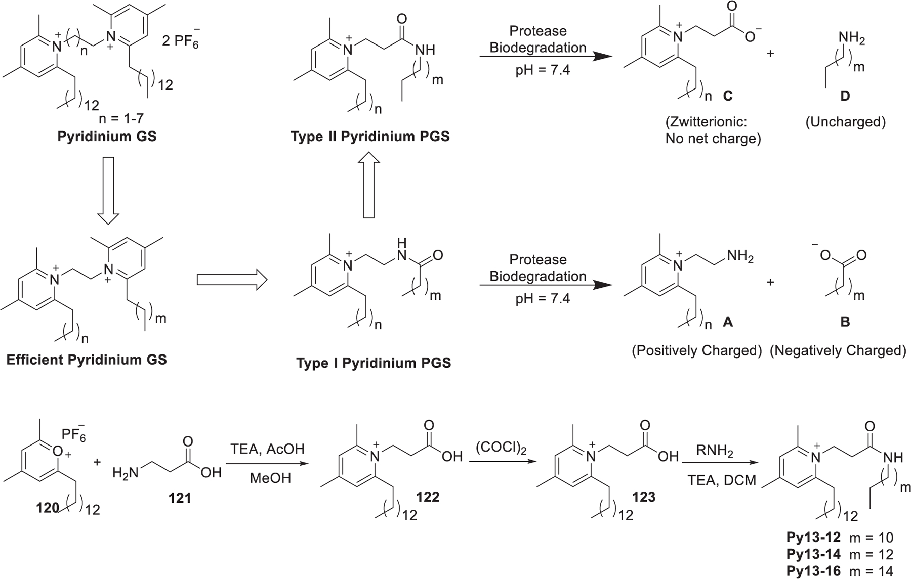
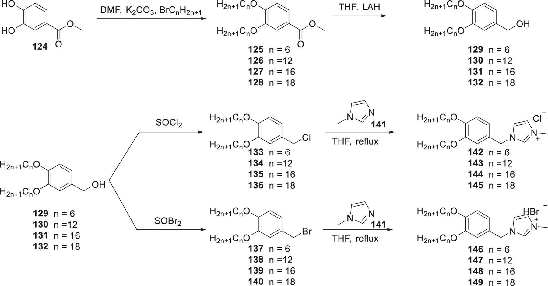
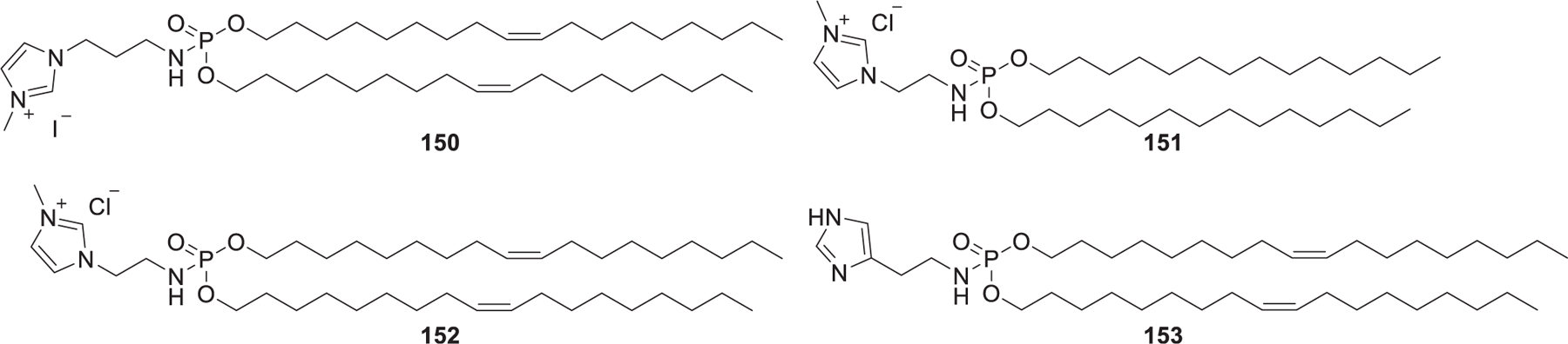
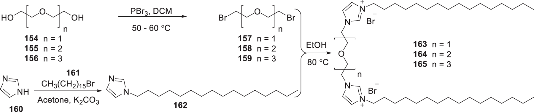
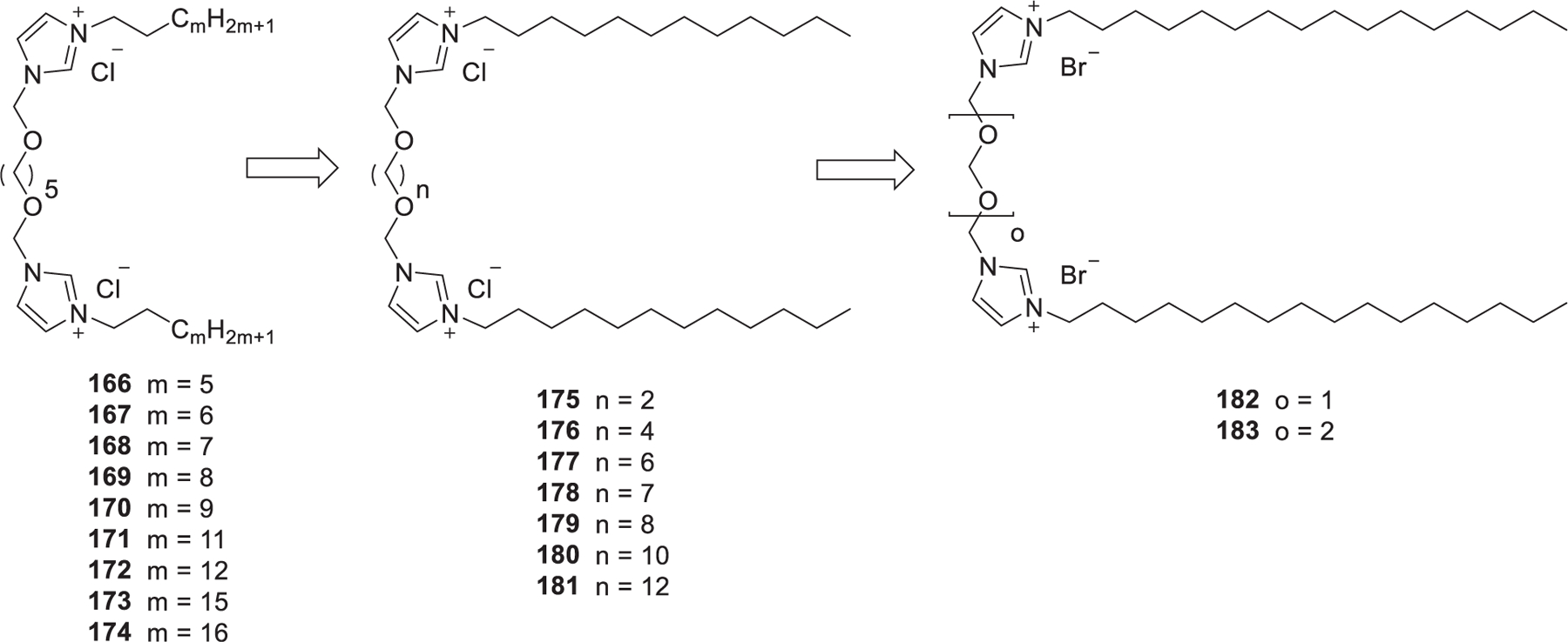
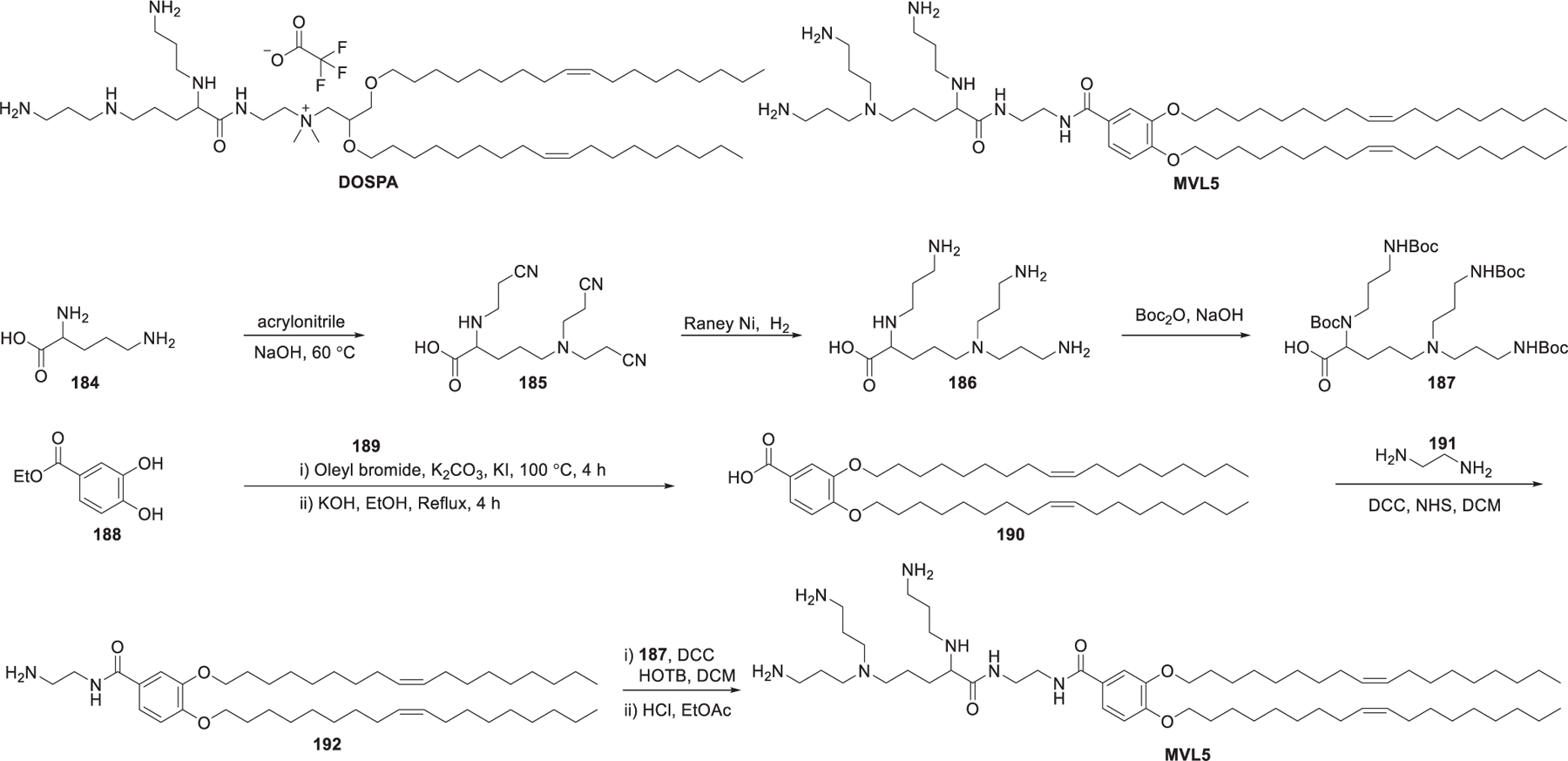
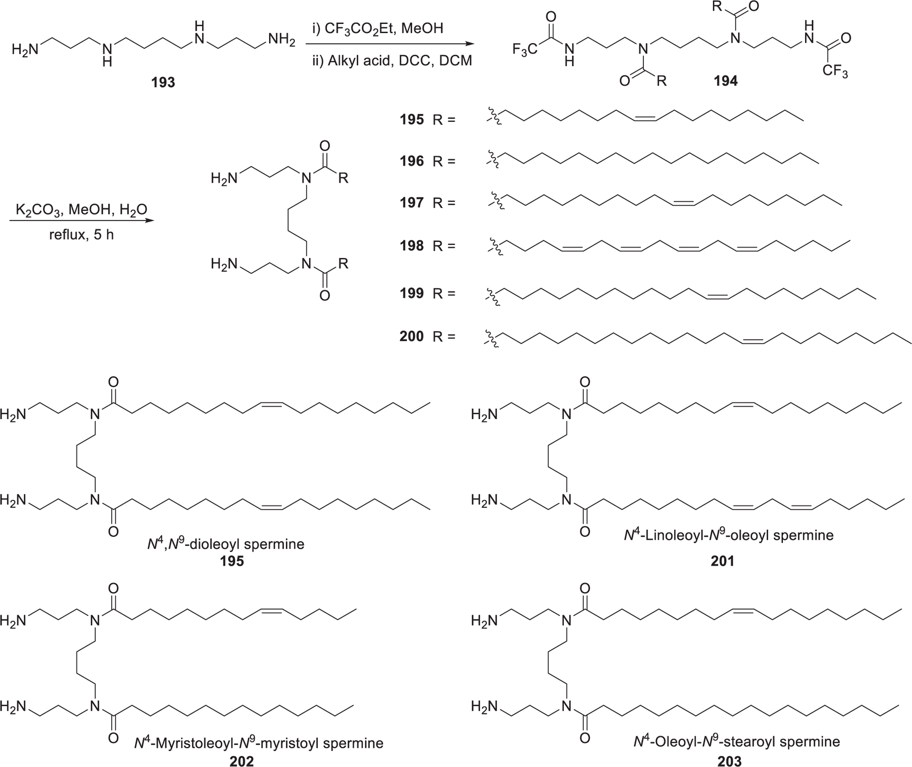
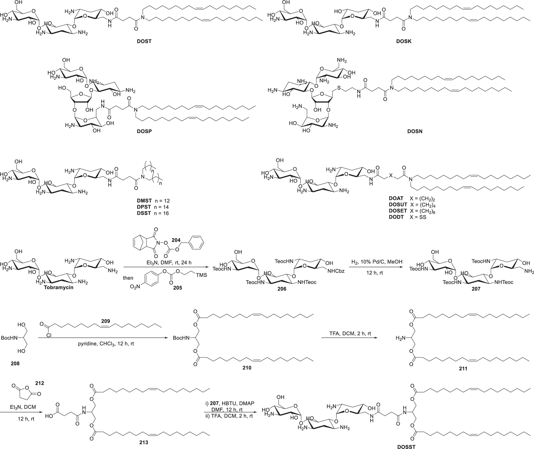
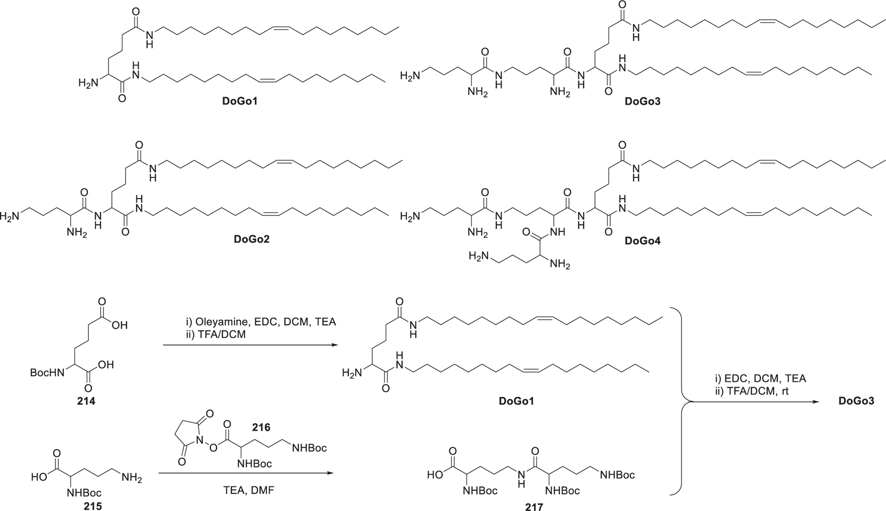
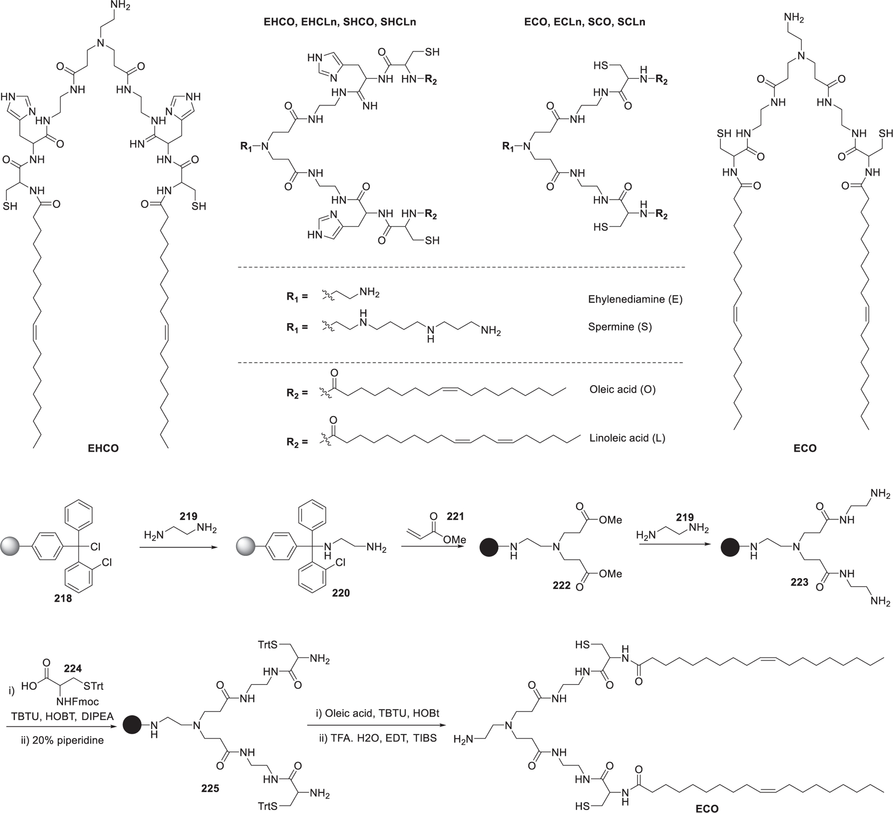
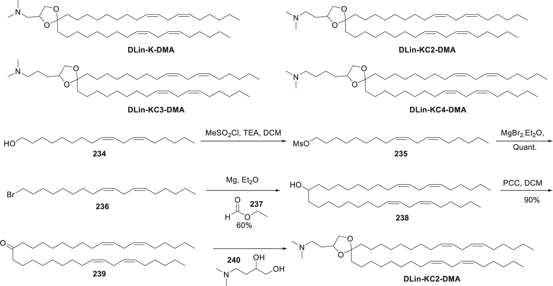
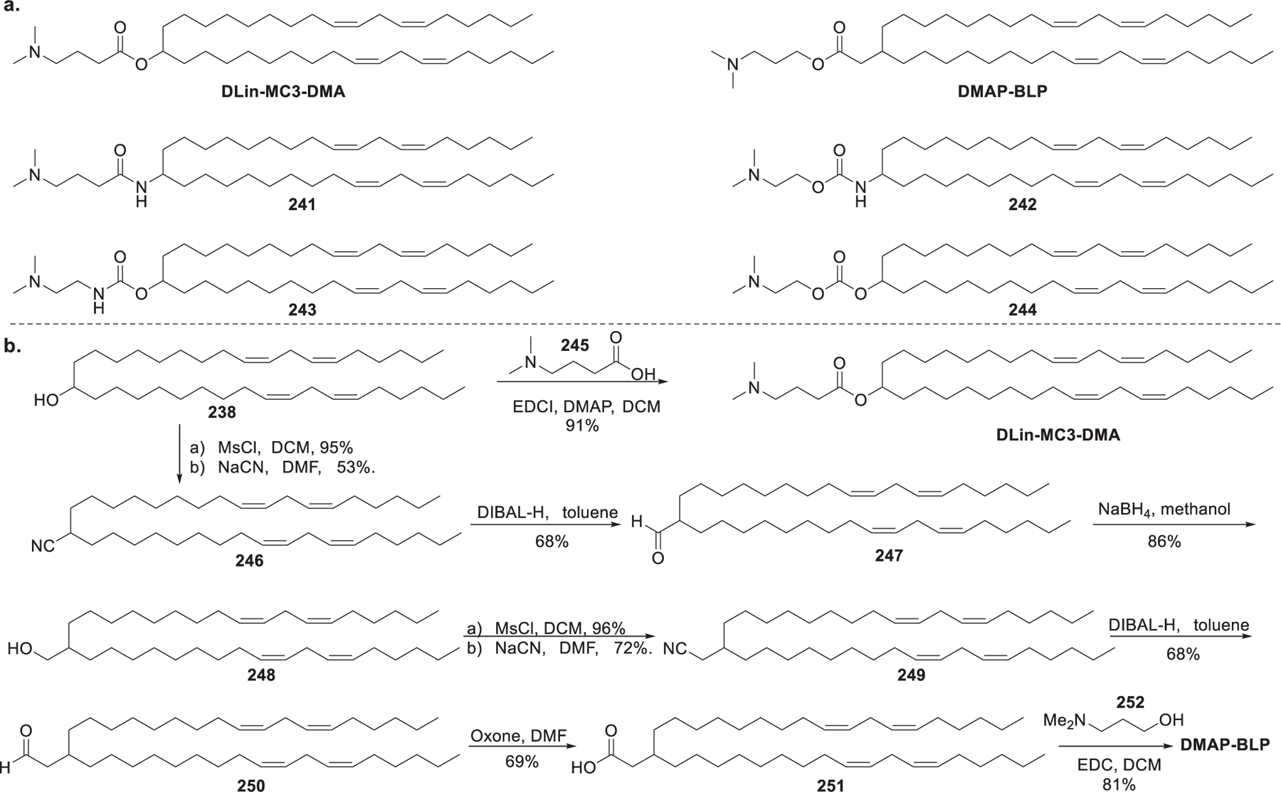
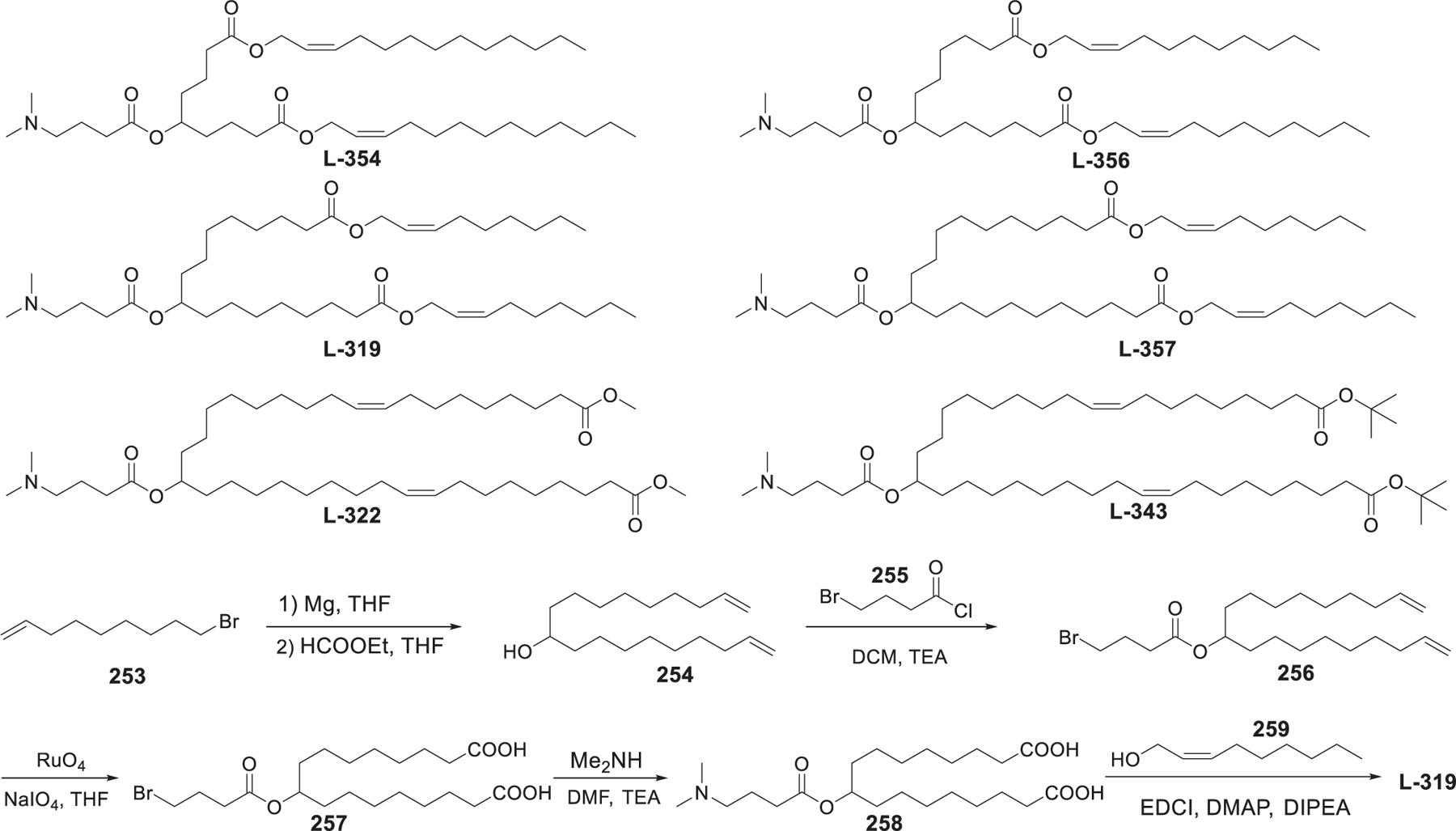
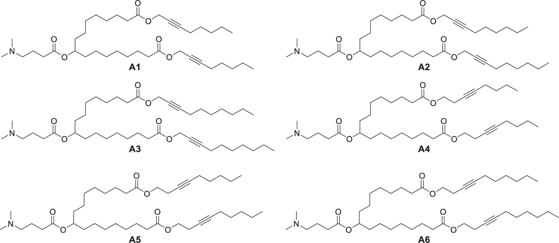
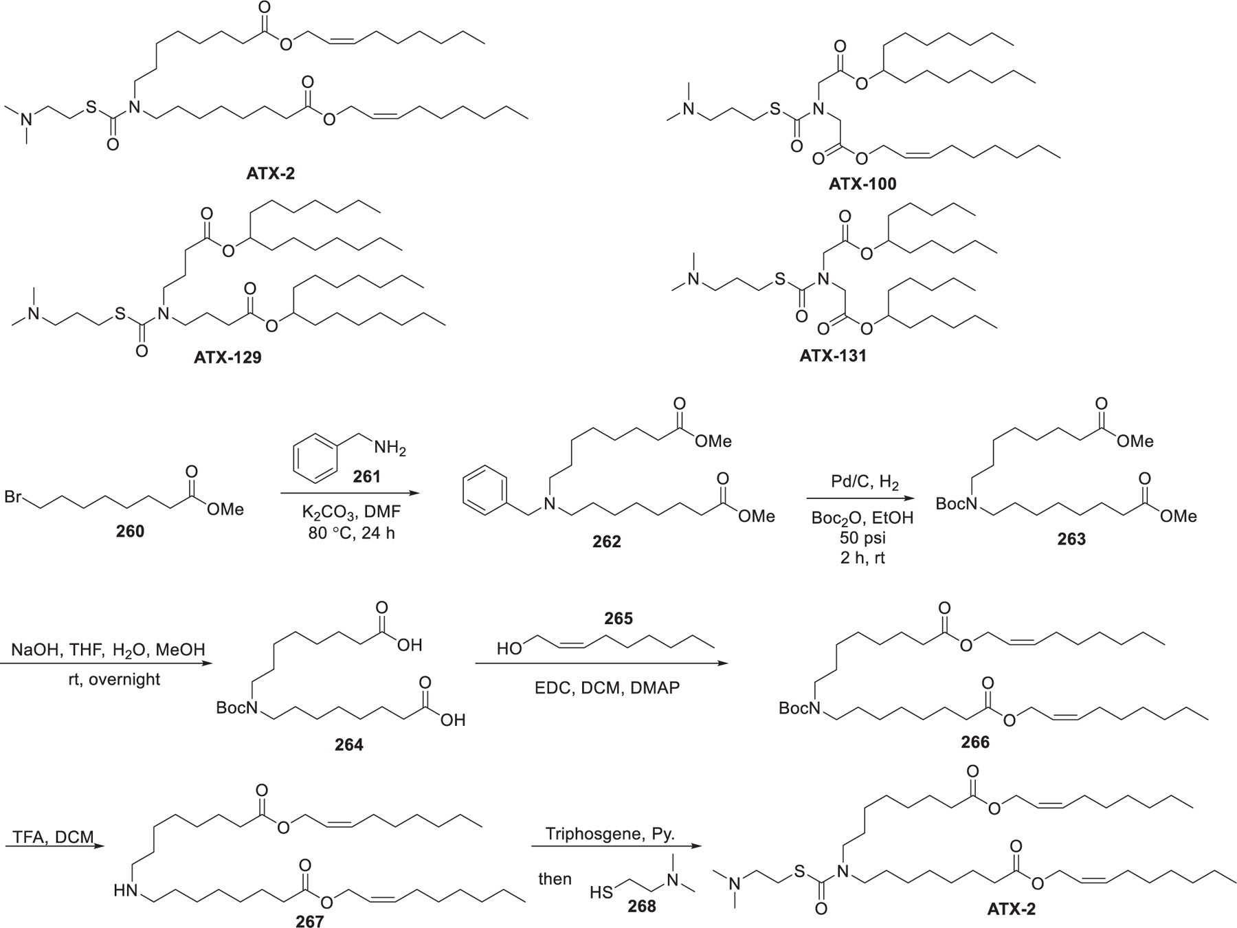
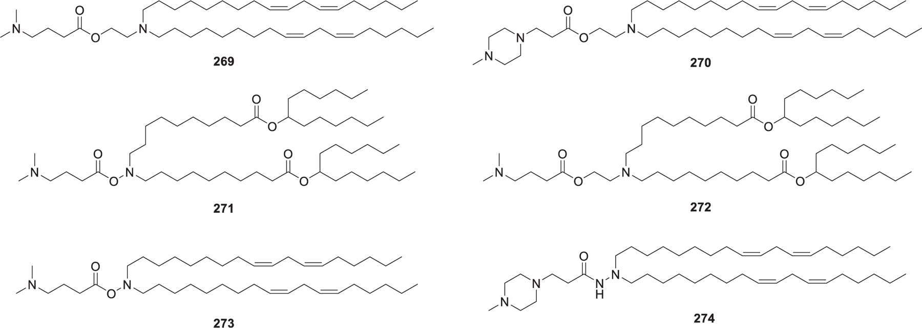
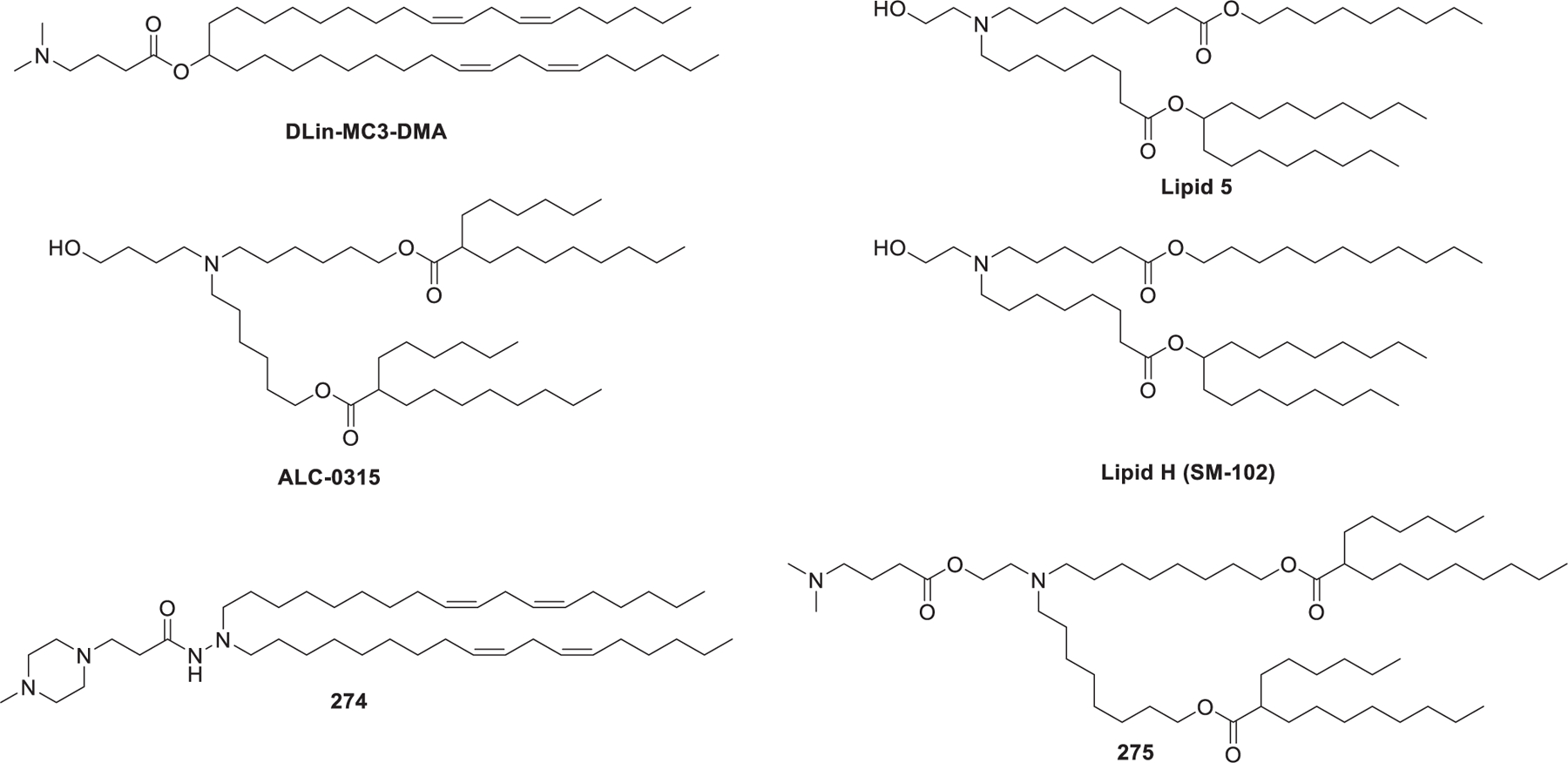
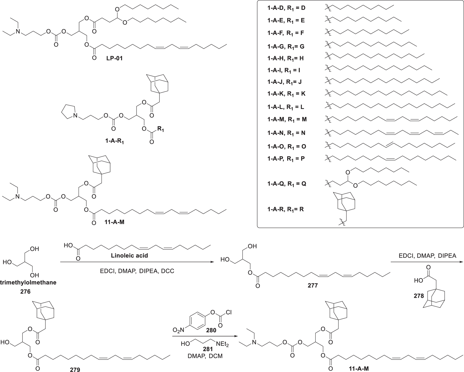
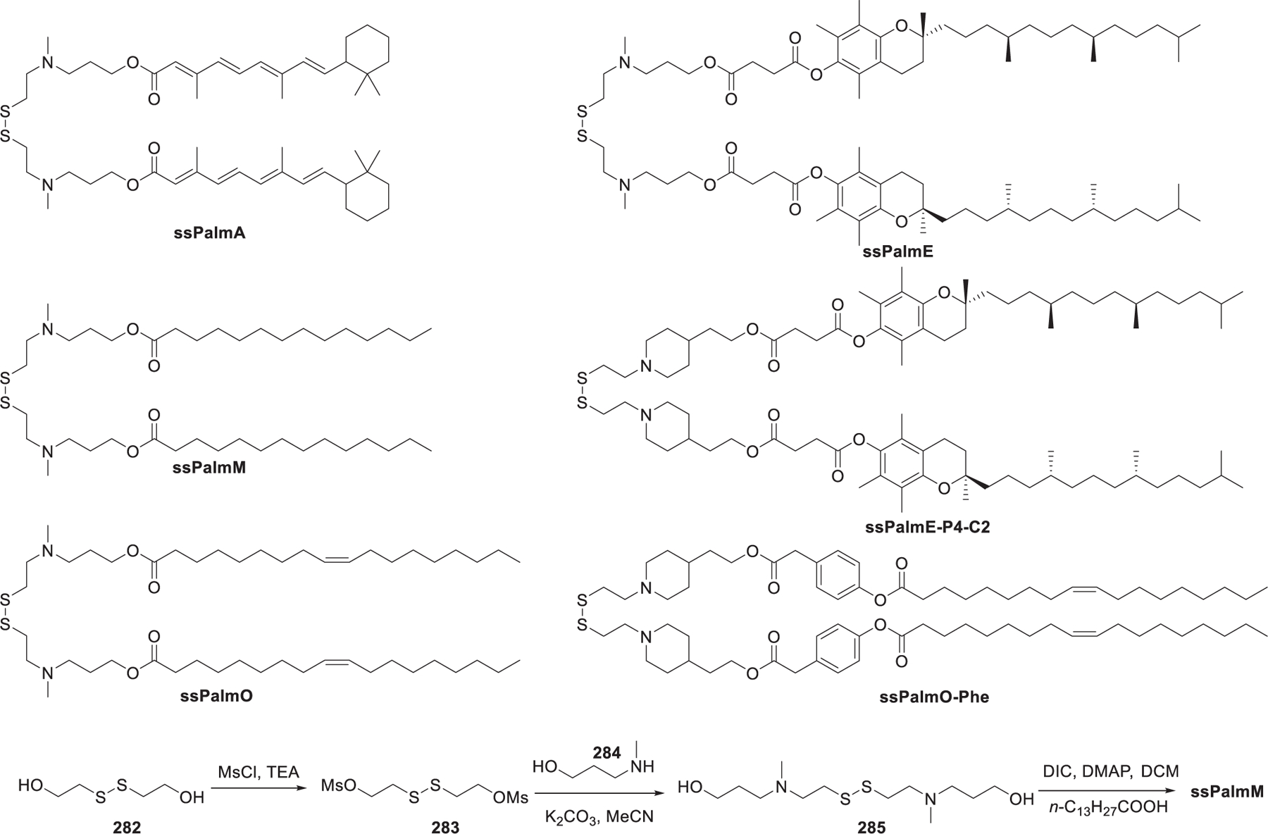
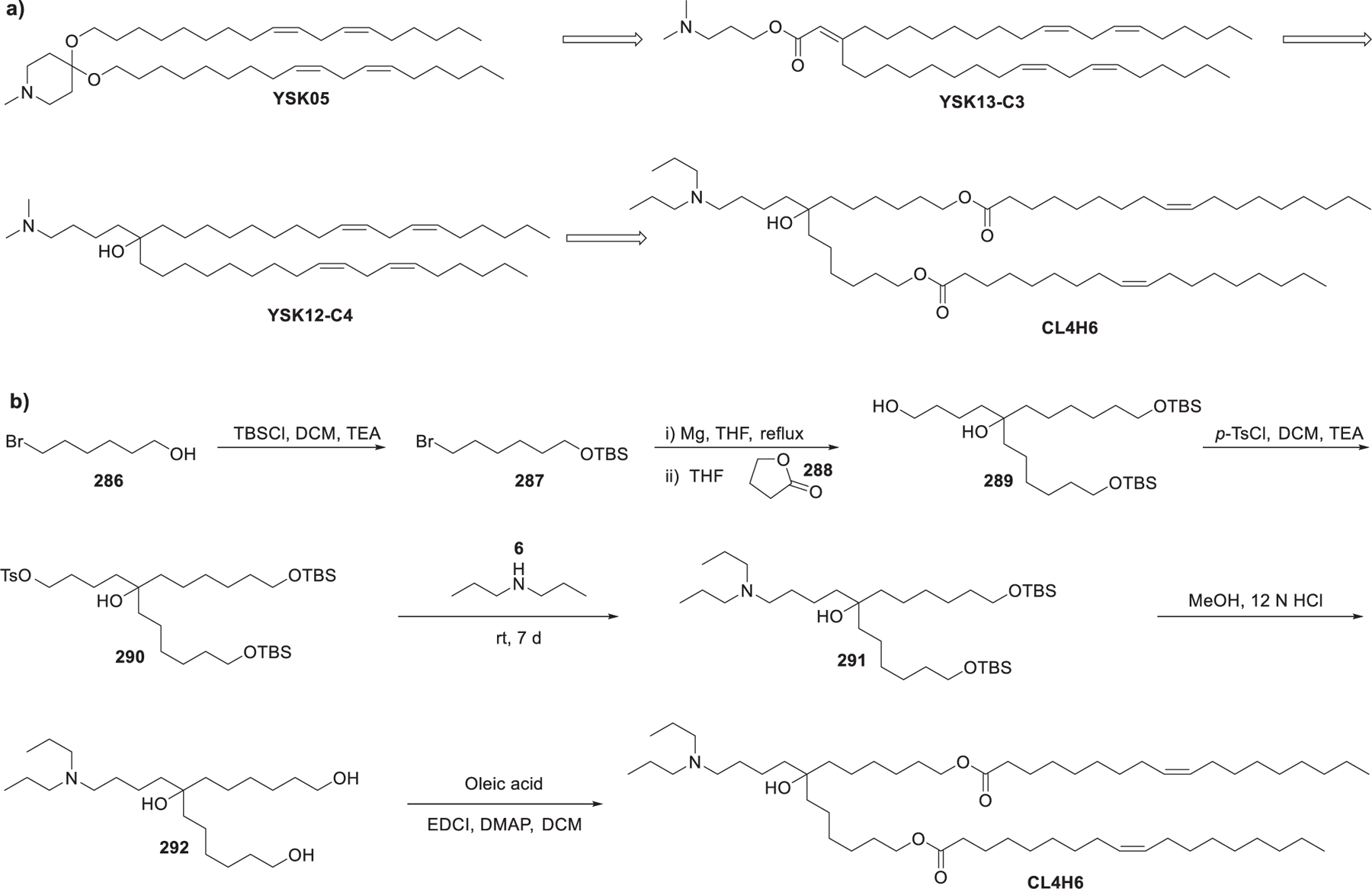
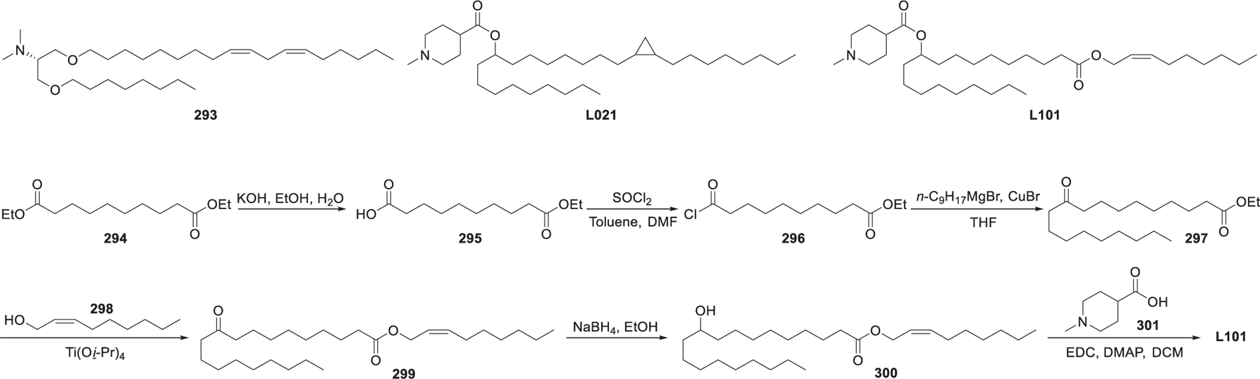
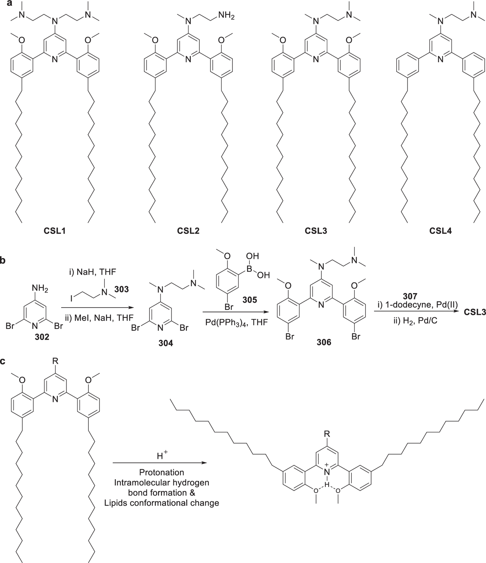
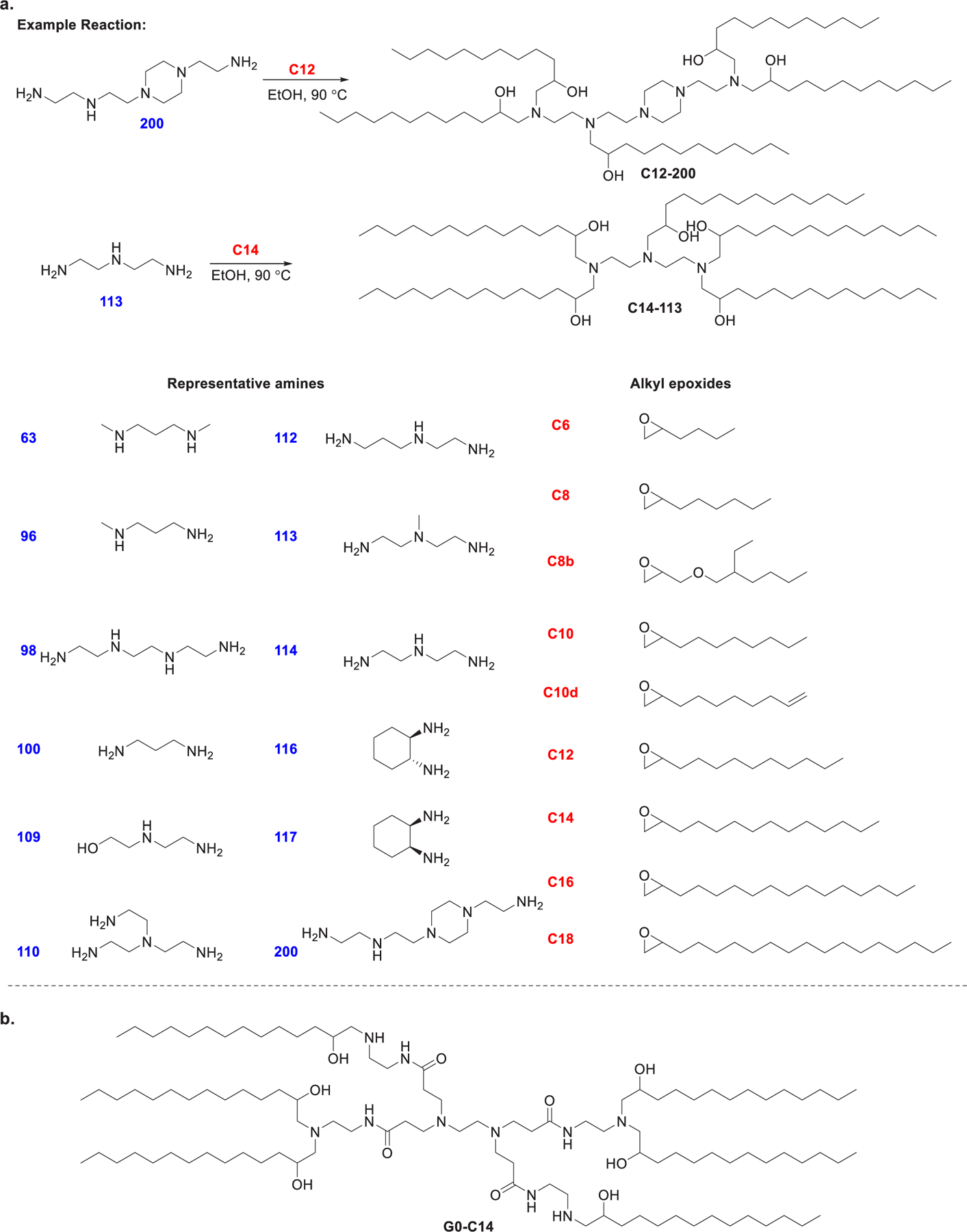
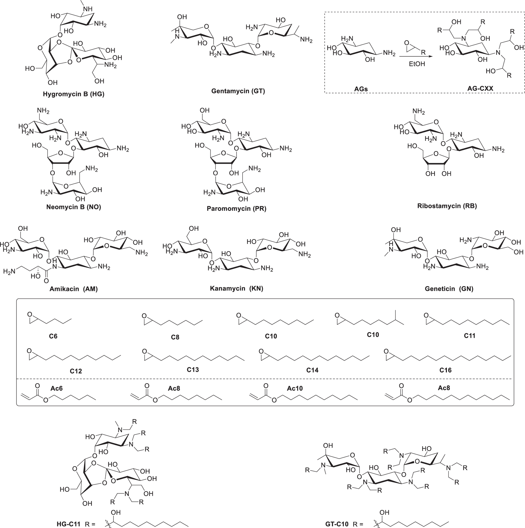
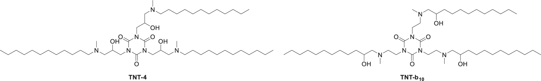
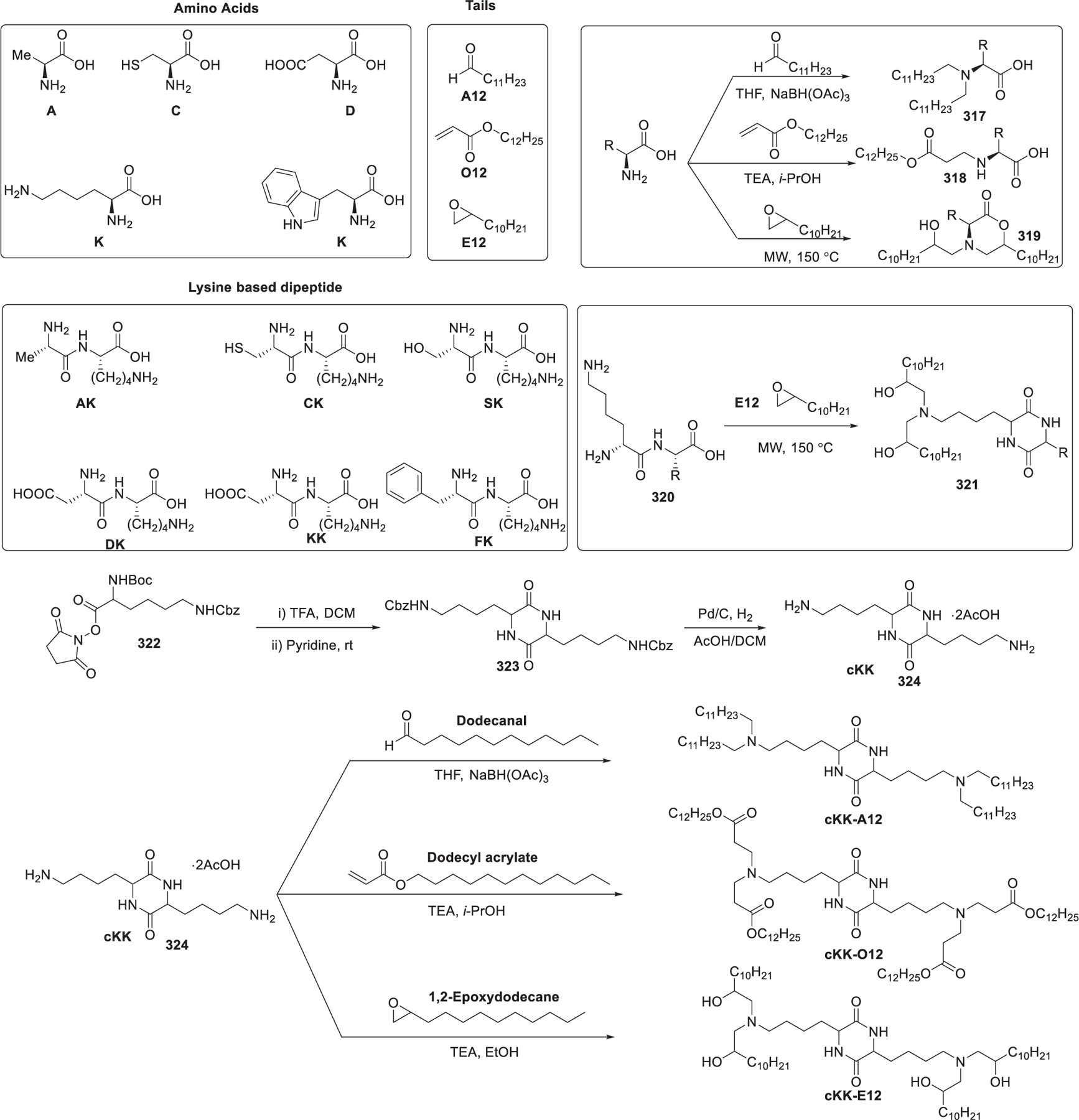
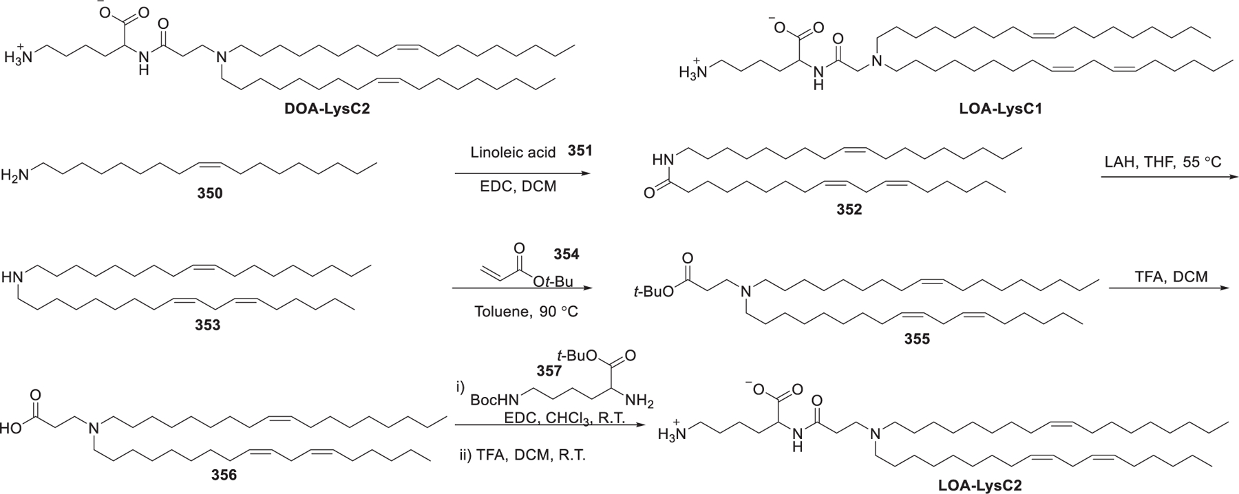
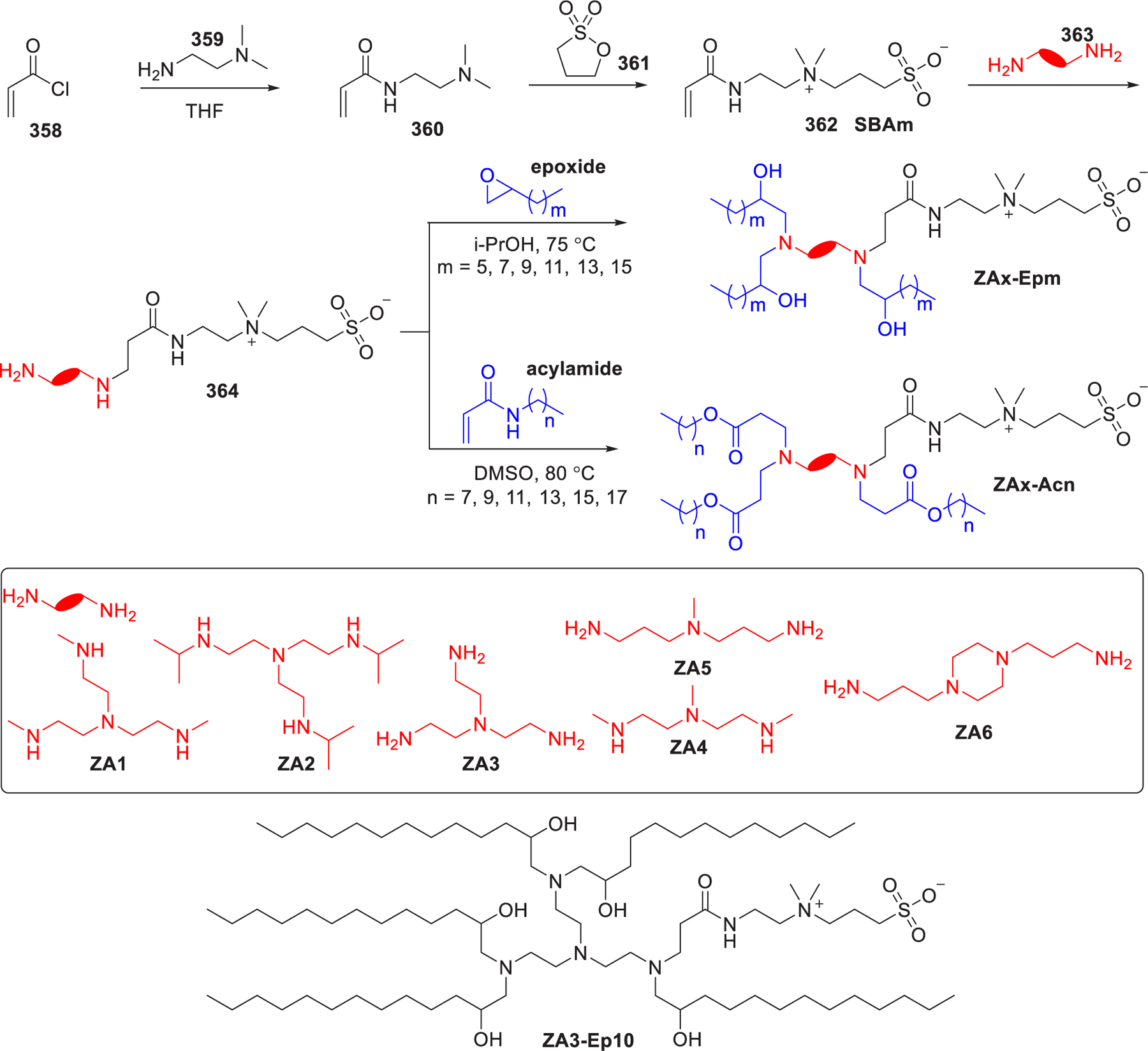
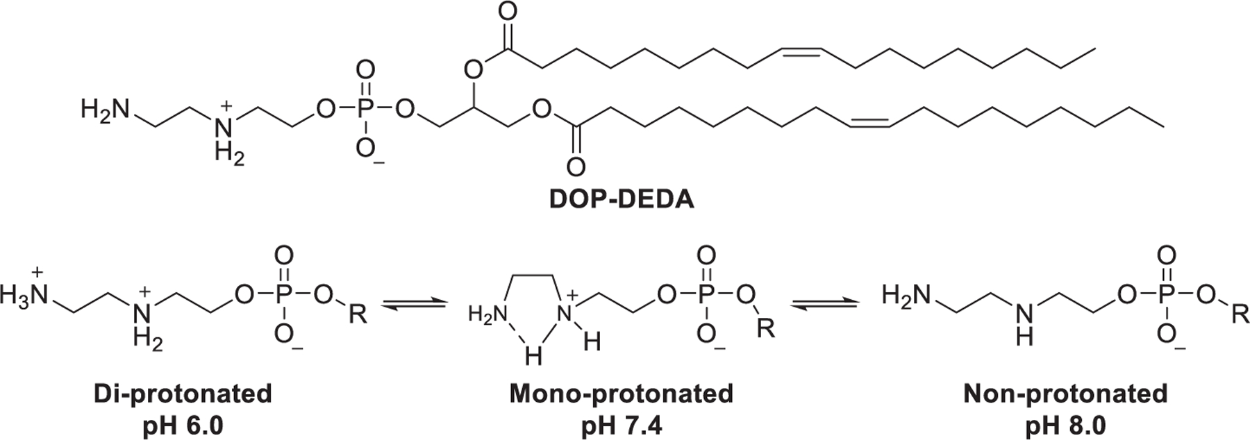
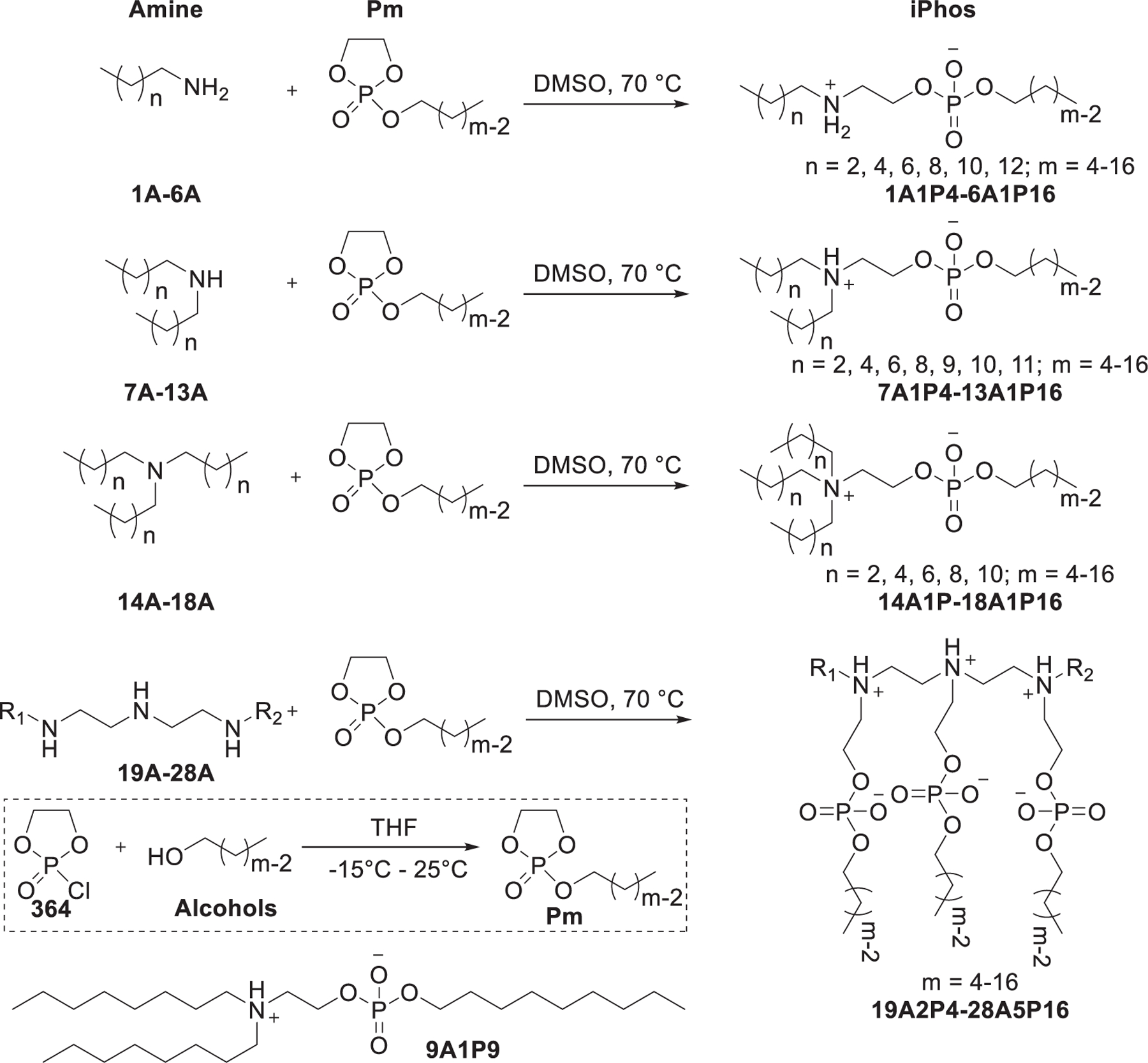
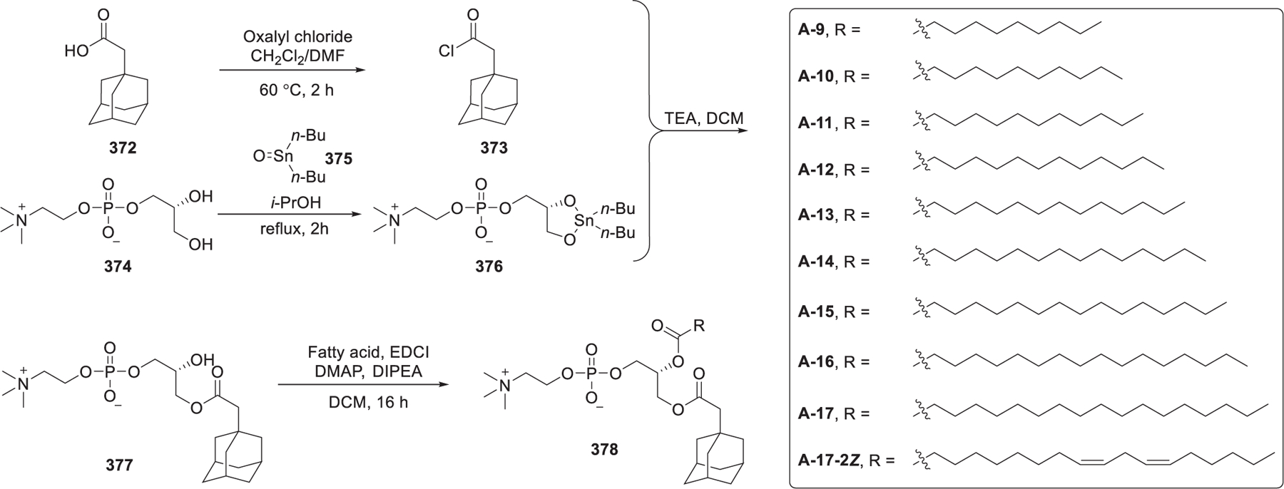
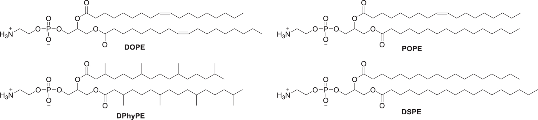
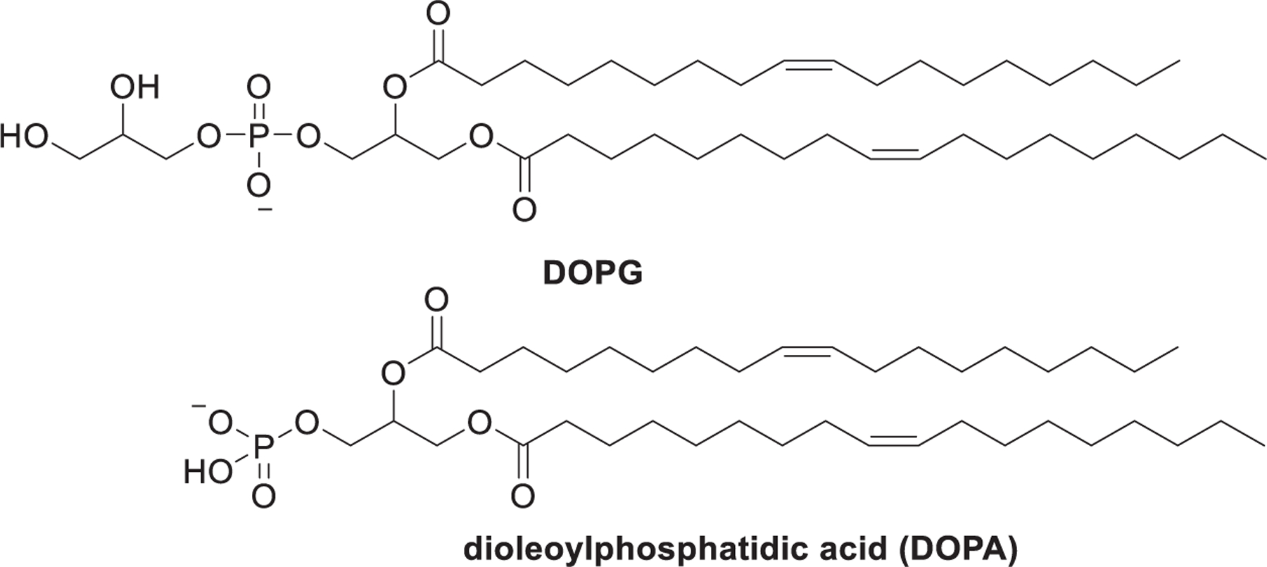
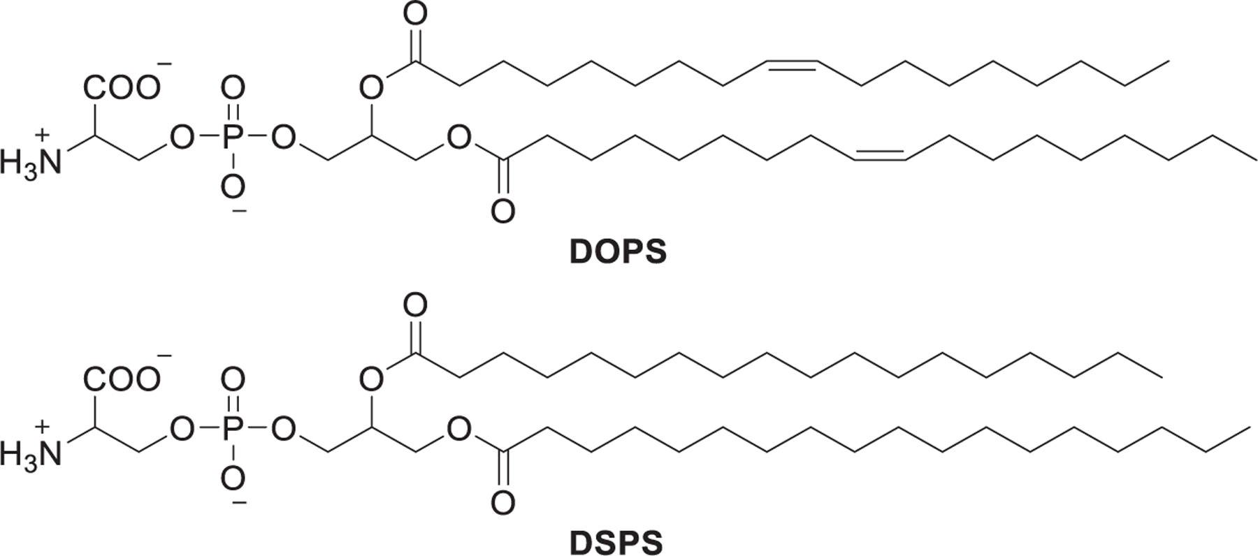
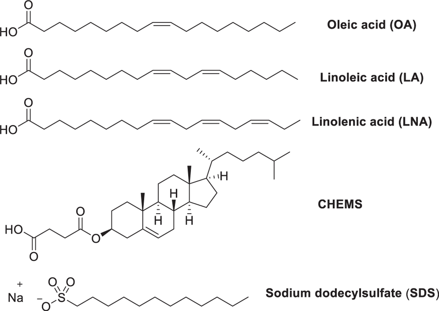
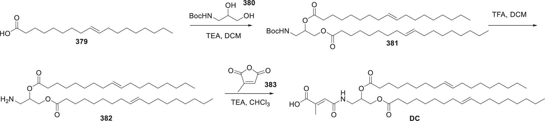
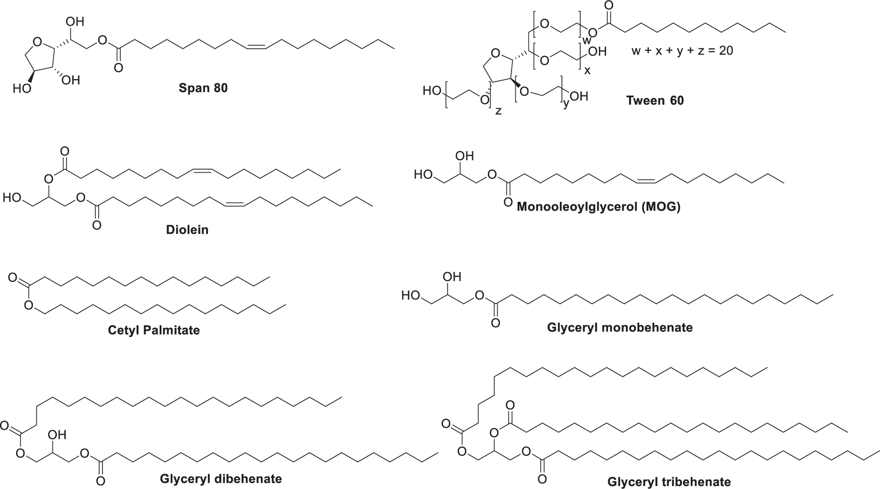
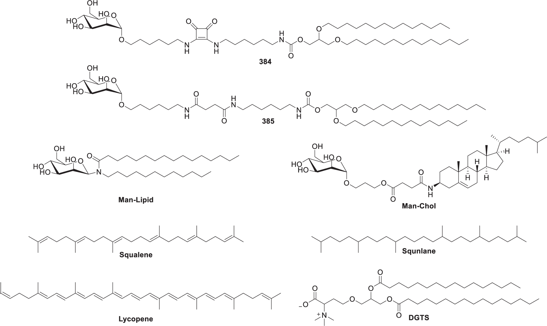
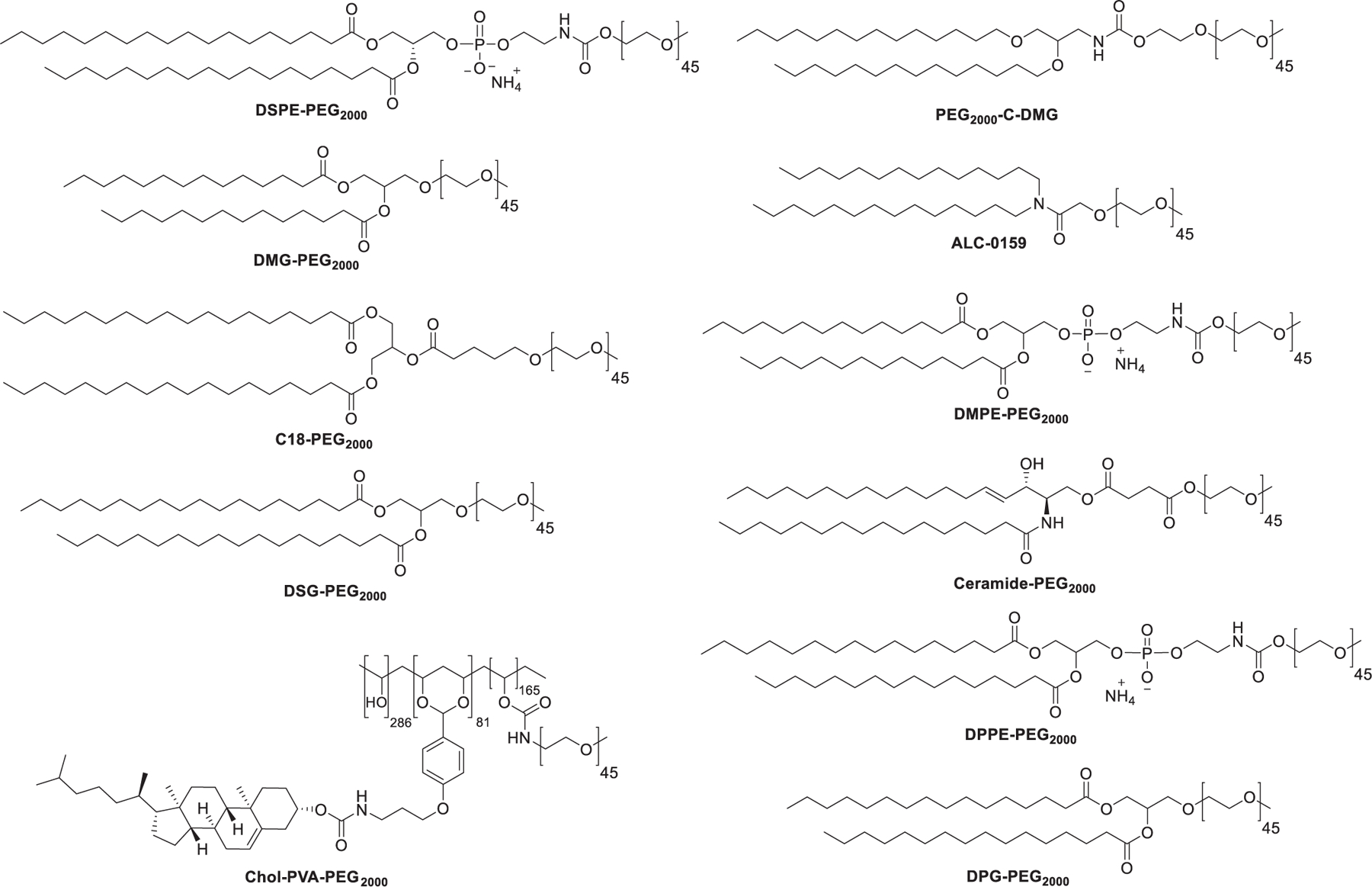
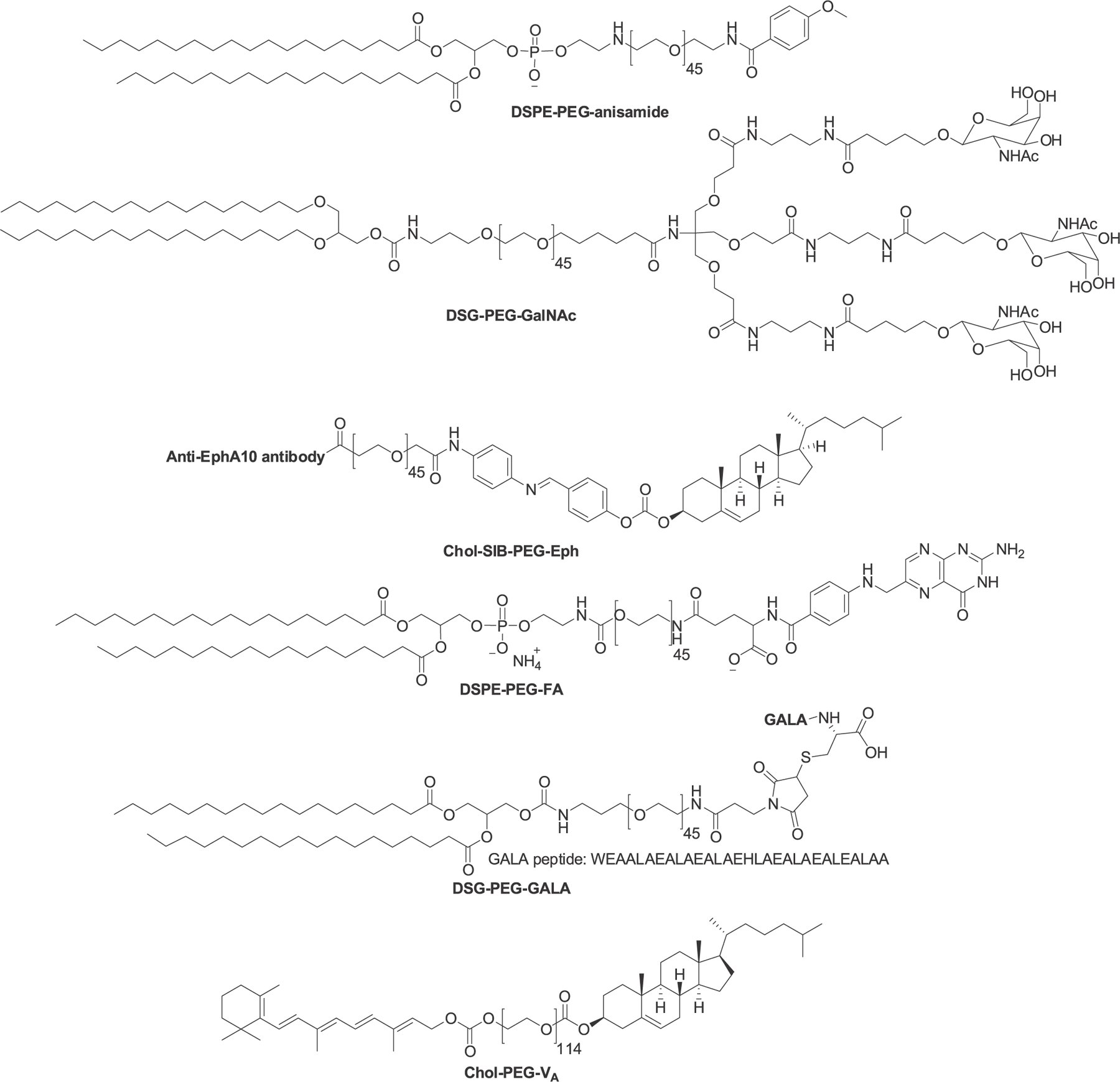
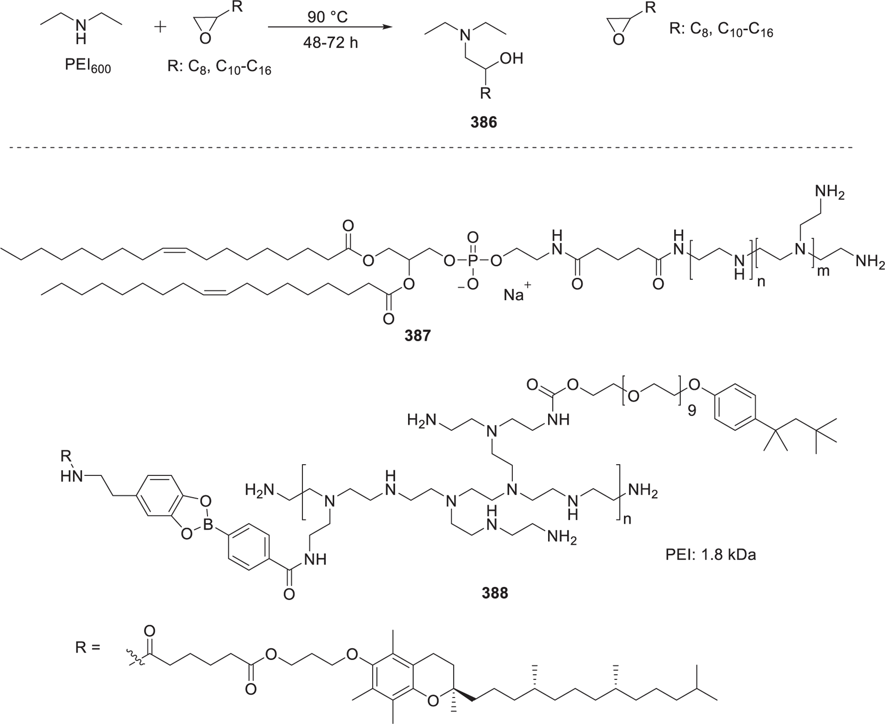
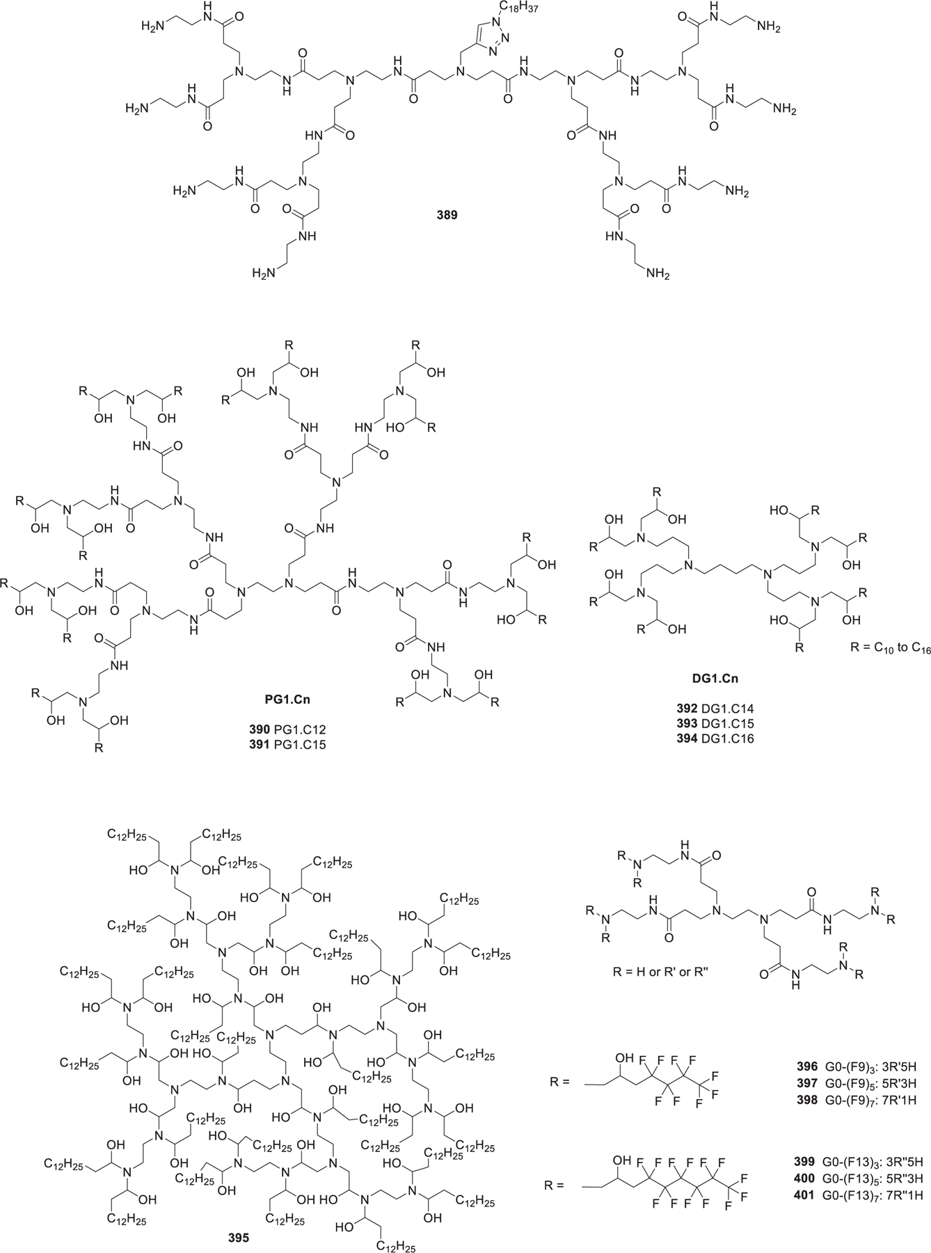
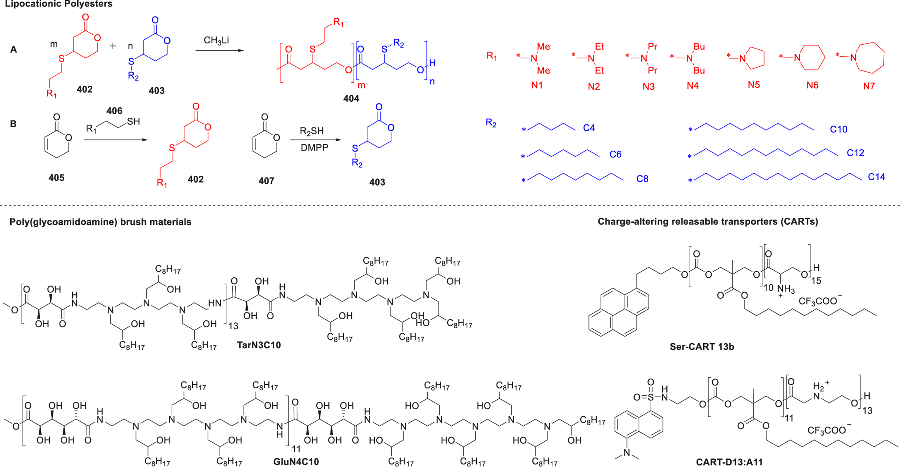
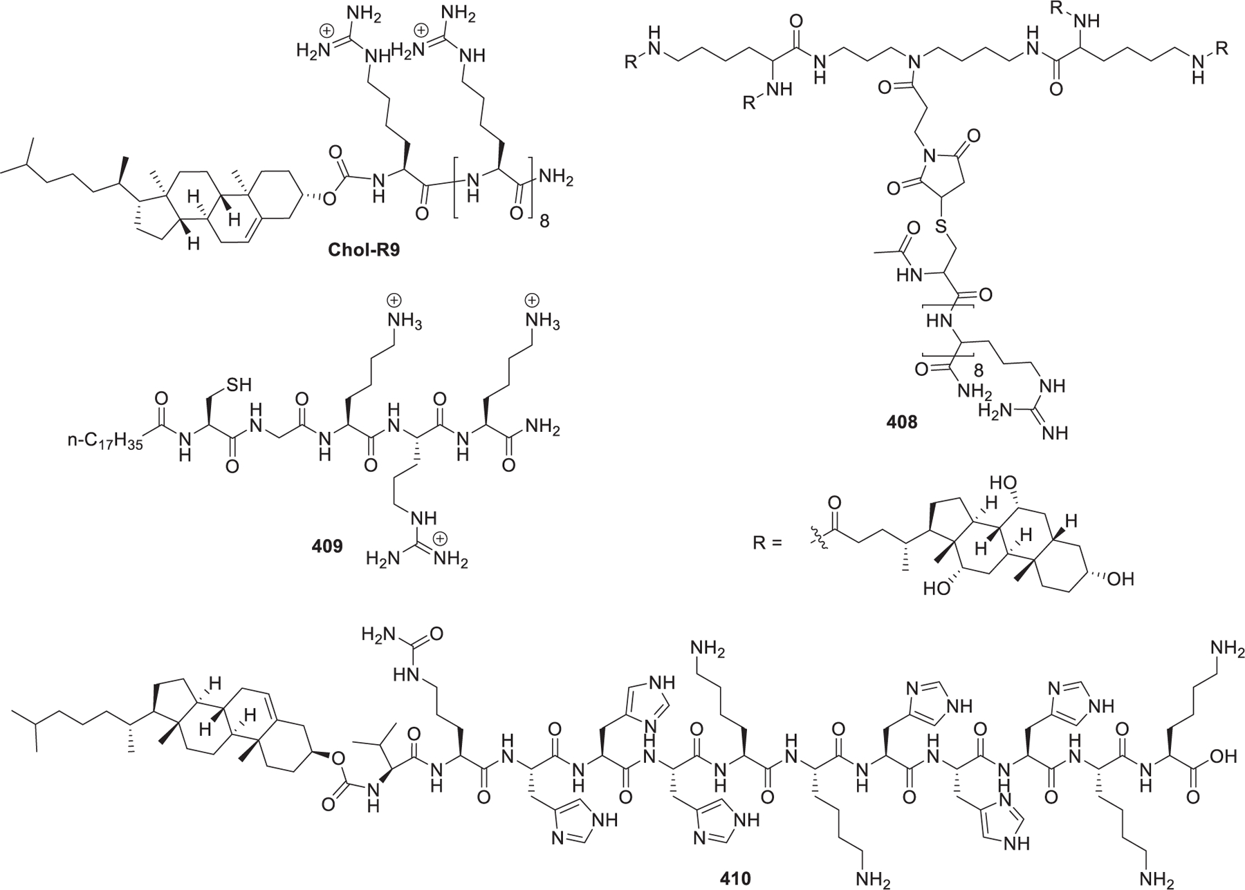
RNA-based therapeutics have shown great promise in treating a broad spectrum of diseases through various mechanisms including knockdown of pathological genes, expression of therapeutic proteins, and programmed gene editing. Due to the inherent instability and negative-charges of RNA molecules, RNA-based therapeutics can make the most use of delivery systems to overcome biological barriers and to release the RNA payload into the cytosol. Among different types of delivery systems, lipid-based RNA delivery systems, particularly lipid nanoparticles (LNPs), have been extensively studied due to their unique properties, such as simple chemical synthesis of lipid components, scalable manufacturing processes of LNPs, and wide packaging capability. LNPs represent the most widely used delivery systems for RNA-based therapeutics, as evidenced by the clinical approvals of three LNP-RNA formulations, patisiran, BNT162b2, and mRNA-1273. This review covers recent advances of lipids, lipid derivatives, and lipid-derived macromolecules used in RNA delivery over the past several decades. We focus mainly on their chemical structures, synthetic routes, characterization, formulation methods, and structure-activity relationships. We also briefly describe the current status of representative preclinical studies and clinical trials and highlight future opportunities and challenges.


Conflict of interest statement

The authors declare the following competing financial interest(s): Y.D. is a scientific advisory board member of Oncorus Inc and serves as a consultant of Rubius Therapeutics.

Figures

Figure 1.
Figure 1.
Schematic illustration of the extracellular and intracellular barriers to effective systemic delivery of RNAs and the mechanism of RNA-based therapeutics. Figure was created with BioRender.com.
Figure 2.
Figure 2.
Chemical structures of the cationic lipid DOTMA and three segments of lipids.
Figure 3.
Figure 3.
Schematic illustration of the shape structure concept of lipids.
Figure 4.
Figure 4.
Chemical structures and synthesis of DOTMA and its analogs.
Figure 5.
Figure 5.
Chemical modifications of DOTMA and DOTAP by introducing hydroxyethyl groups
Figure 6.
Figure 6.
Chemical structure and synthetic route of DC-6-14.
Figure 7.
Figure 7.
Chemical structures and general synthetic route of ethylphosphatidylcholines (ePCs).
Figure 8.
Figure 8.
Representative chemical structures and synthetic routes of DOPC-based lipids.
Figure 9.
Figure 9.
Synthesis of gemini diquaternary ammoniums TODMACS3, TODMACS 6, and CCLA.
Figure 10.
Figure 10.
Chemical structures of piperidinium and morpholinium lipids.
Figure 11.
Figure 11.
Chemical structures and synthetic routes of nucleoside derived lipids.
Figure 12.
Figure 12.
Chemical structures and synthetic route of carotenoid-derived lipids
Figure 13.
Figure 13.
Chemical structure of PBA-BADP.
Figure 14.
Figure 14.
Chemical structure of vectamidine and the delocalization of the positive charge.
Figure 15.
Figure 15.
Chemical structures of AtuFECT01 and DSGLA.
Figure 16.
Figure 16.
Chemical structures of guanidinium lipids and synthetic route of DODAG-9.
Figure 17.
Figure 17.
Chemical structures of guanidinium DiLA2 compounds and C12ANHC18.
Figure 18.
Figure 18.
Chemical structures and synthesis of N1,N12-diamidino-N4,N9-diacylated spermines.
Figure 19.
Figure 19.
Chemical structures of squalene and cholesterol-derived guanidinium lipids.
Figure 20.
Figure 20.
Chemical structures of arginine- and cysteine-derived guanidinium type lipids.
Figure 21.
Figure 21.
Chemical structure and synthesis of SAINT.
Figure 22.
Figure 22.
Synthesis of cholesterol-derived pyridinium lipids.
Figure 23.
Figure 23.
Chemical structures and synthesis of pyridinium lipids
Figure 24.
Figure 24.
Design, synthesis, and proposed biodegradation pattern of pyridinium psudogemini surfactants.
Figure 25.
Figure 25.
Chemical structures and synthesis of imidazolium lipids.
Figure 26.
Figure 26.
Chemical structures of imidazole/imidazolium lipophosphoramidate lipids.
Figure 27.
Figure 27.
Synthesis of gemini imidazolium lipids 163−165.
Figure 28.
Figure 28.
Optimization of imidazolium gemini surfactants.
Figure 29.
Figure 29.
Chemical structures of DOSPA and MVL5 and a synthetic route to lipid MVL5.
Figure 30.
Figure 30.
Synthesis of spermine-derived lipids.
Figure 31.
Figure 31.
Structure of the aminoglycoside-derived ionizable lipids.
Figure 32.
Figure 32.
Chemical structure of lysine-derived lipids.
Figure 33.
Figure 33.
Chemical structures of multifunctional ionizable lipids.
Figure 34.
Figure 34.
Chemical structures of dialkyl phosphate–polyamine conjugates and synthesis of Et-CH2F.
Figure 35.
Figure 35.
Synthesis of MTO-derived ionizable lipids.
Figure 36.
Figure 36.
Chemical structures of the first generation of two-tailed amino lipids.
Figure 37.
Figure 37.
Chemical structures of two-tailed amino lipids with asymmetric tails.
Figure 38.
Figure 38.
Chemical structures of DODAP and DLinDAP.
Figure 39.
Figure 39.
Chemical structures and synthesis of latently biodegradable two-tailed lipids.
Figure 40.
Figure 40.
Representative chemical structures and synthetic routes to biodegradable two-tailed lipids.
Figure 41.
Figure 41.
Synthesis of biodegradable dimethyl amino lipids.
Figure 42.
Figure 42.
Chemical structures of biodegradable alkyne analogs of DLin-MC3-DMA.
Figure 43.
Figure 43.
Chemical structures and synthesis of ATX lipids.
Figure 44.
Figure 44.
Chemical structures of two-tailed lipids with other linkers.
Figure 45.
Figure 45.
Chemical structures of ionizable lipids used in the development of COVID-19 mRNA vaccines.
Figure 46.
Figure 46.
Chemical structures of constrained lipids with small head groups.
Figure 47.
Figure 47.
Chemical structures of representative ssPalm and a synthetic route to ssPalmM.
Figure 48.
Figure 48.
Chemical structures of YSK series lipids and a synthetic route to CL4H6.
Figure 49.
Figure 49.
Chemical structures of lipid 293, L021, and L101 and a synthetic route to L101.
Figure 50.
Figure 50.
(a) Chemical structures of ionizable switchable lipids. (b) Synthetic route to CSL3;. (c) Protonation-induced conformational change of the ionizable switchable lipids.
Figure 51.
Figure 51.
Chemical structures of cholesterol-derived ionizable lipids.
Figure 52.
Figure 52.
Chemical structures of vitamin-derived ionizable lipids.
Figure 53.
Figure 53.
Chemical structures and synthesis of representative phospholipids and glycolipids.
Figure 54.
Figure 54.
Representative reactions for the preparation of lipidoids.
Figure 55.
Figure 55.
Synthesis of lipidoids via Michael addition of amines with acrylamides and acryl ester.
Figure 56.
Figure 56.
Lipidoids synthesized via Michael addition of alkyl-amines (blue) to alkyl-acrylate or methacrylate tails (red).
Figure 57.
Figure 57.
Chemical synthesis of bioreducible lipids.
Figure 58.
Figure 58.
Modular strategy for the synthesis of dendrimer-like lipids and chemical structure of 5A2-SC8.
Figure 59.
Figure 59.
Synthesis of lipids from ring-opening reaction between amines and epoxides.
Figure 60.
Figure 60.
Chemical structures of aminoglycosides, epoxides, and acrylic esters and a representative schematic reaction between aminoglycoside and epoxide.
Figure 61.
Figure 61.
Chemical structures of TNT-4 and TNT-b10.
Figure 62.
Figure 62.
Synthesis of amino acid-derived lipidoids.
Figure 63.
Figure 63.
Chemical structures and synthesis of OF-XX and OF-Deg-Lin.
Figure 64.
Figure 64.
Synthesis of lipidoids via epoxide ring-opening of alkyl epoxides with polyamines.
Figure 65.
Figure 65.
Synthesis of TT and FTT series of ionizable lipids.
Figure 66.
Figure 66.
Synthetic routes to lipids via thiol–yne click chemistry.
Figure 67.
Figure 67.
Synthesis of lipidoids via thiolactones ring-opening reaction followed by the thiol–disulfide exchange reaction. (a) Structures of amines. (b) Structures of pyridyl disulfide derivatives. (c) Structures of thiolactone derivatives. (d) Representative reaction scheme.
Figure 68.
Figure 68.
Isocyanide-mediated three-component reactions for the synthesis of lipidoids.
Figure 69.
Figure 69.
Chemical structure of zwitterionic lipid GDOPE.
Figure 70.
Figure 70.
Chemical structures of zwitterionic lipids.
Figure 71.
Figure 71.
Chemical structures of lysine-derived zwitterionic lipids.
Figure 72.
Figure 72.
Chemical structures and the synthesis of zwitterionic lipid ZA3-Ep10.
Figure 73.
Figure 73.
Chemical structure of DOP-DEDA and schematic illustration of the pH-responsive ability of DOP-DEDA.
Figure 74.
Figure 74.
Chemical structures and synthesis of representative iPhos series lipids.
Figure 75.
Figure 75.
Schematic illustration of the interactions between Zn/DPA, sulfonic acid 365, and phosphate.
Figure 76.
Figure 76.
Chemical structures of representative coordinative amphiphiles (CAs).
Figure 77.
Figure 77.
General chemical structure of phospholipids.
Figure 78.
Figure 78.
Chemical structures of representative phosphatidylcholines.
Figure 79.
Figure 79.
Synthesis of phosphatidylcholines incorporating adamantyl groups.
Figure 80.
Figure 80.
Chemical structures of representative phosphatidylethanolamines.
Figure 81.
Figure 81.
Chemical structures of representative phosphatidylglycerol.
Figure 82.
Figure 82.
Chemical structures of representative phosphatidylserine.
Figure 83.
Figure 83.
Chemical structures of cholesterol variants formulated in LNPs.
Figure 84.
Figure 84.
Chemical structures of sterol variants modified from cholesterol.
Figure 85.
Figure 85.
Chemical structures of three groups of cholesterol analogs.
Figure 86.
Figure 86.
Structural features of C-24 alkyl derivatives of cholesterol.
Figure 87.
Figure 87.
Chemical structures of anionic lipids.
Figure 88.
Figure 88.
Synthesis procedures of DC.
Figure 89.
Figure 89.
Chemical structures of fatty acid esters for RNA delivery.
Figure 90.
Figure 90.
Chemical structures of other helper lipids for RNA delivery.
Figure 91.
Figure 91.
Chemical structures of PEG-lipids.
Figure 92.
Figure 92.
Chemical structures of functionalized PEG-lipids.
Figure 93.
Figure 93.
Chemical structures of PEI-lipids.
Figure 94.
Figure 94.
Chemical structures of dendrimer-lipids.
Figure 95.
Figure 95.
Chemical structures of other types of lipopolymers.
Figure 96.
Figure 96.
Chemical structures of lipopeptides.

References

    1. DeWitt DE; Hirsch IB Outpatient Insulin Therapy in Type 1 and Type 2 Diabetes Mellitus. JAMA 2003, 289, 2254–2264. - PubMed
    1. Akhtar MJ; Ahamed M; Kumar S; Siddiqui H; Patil G; Ashquin M; Ahmad I Nanotoxicity of Pure Silica Mediated Through Oxidant Generation Rather Than Glutathione Depletion in Human Lung Epithelial Cells. Toxicology 2010, 276, 95–102. - PubMed
    1. Lin Y-X; Wang Y; Blake S; Yu M; Mei L; Wang H; Shi J RNA Nanotechnology-Mediated Cancer Immunotherapy. Theranostics 2020, 10, 281–299. - PMC - PubMed
    1. Naldini L Gene Therapy Returns to Centre Stage. Nature 2015, 526, 351–360. - PubMed
    1. Weissman D; Karikó K mRNA: Fulfilling the Promise of Gene Therapy. Mol. Ther 2015, 23, 1416–1417. - PMC - PubMed

Publication types