

Titanium Functionalized with Polylysine Homopolymers: In Vitro Enhancement of Cells Growth
- PMID: 34279306
- PMCID: PMC8269806
- DOI: 10.3390/ma14133735
Titanium Functionalized with Polylysine Homopolymers: In Vitro Enhancement of Cells Growth
Abstract
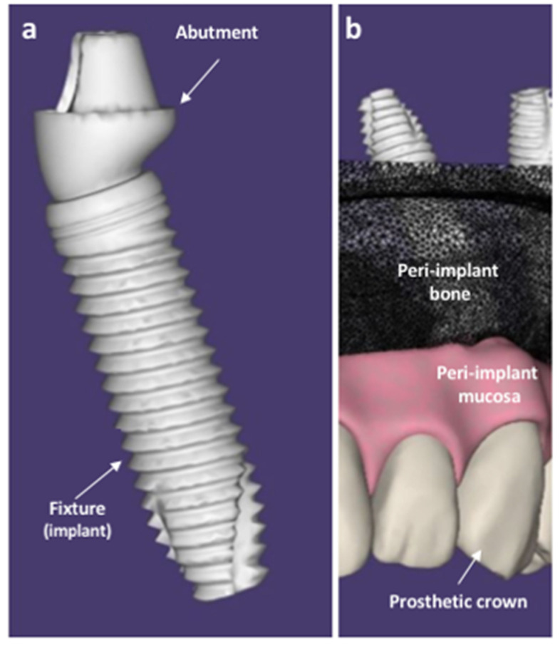
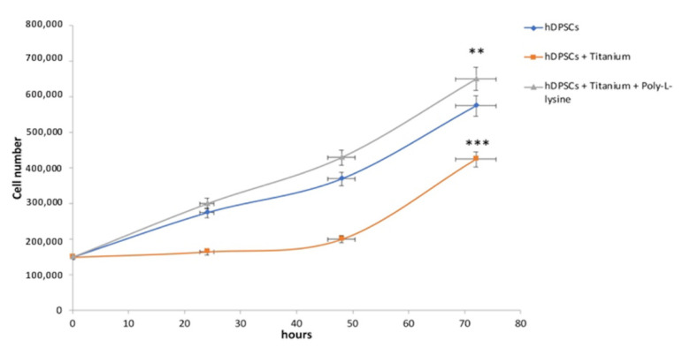
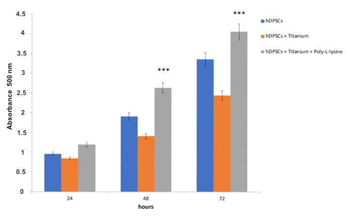
In oral implantology, the success and persistence of dental implants over time are guaranteed by the bone formation around the implant fixture and by the integrity of the peri-implant mucosa seal, which adheres to the abutment and becomes a barrier that hinders bacterial penetration and colonization close to the outer parts of the implant. Research is constantly engaged in looking for substances to coat the titanium surface that guarantees the formation and persistence of the peri-implant bone, as well as the integrity of the mucous perimeter surrounding the implant crown. The present study aimed to evaluate in vitro the effects of a titanium surface coated with polylysine homopolymers on the cell growth of dental pulp stem cells and keratinocytes to establish the potential clinical application. The results reported an increase in cell growth for both cellular types cultured with polylysine-coated titanium compared to cultures without titanium and those without coating. These preliminary data suggest the usefulness of polylysine coating not only for enhancing osteoinduction but also to speed the post-surgery mucosal healings, guarantee appropriate peri-implant epithelial seals, and protect the fixture against bacterial penetration, which is responsible for compromising the implant survival.
Keywords: biomaterials; cell growth; dental implants; epithelial growth; implantology; polylysine; titanium.
Conflict of interest statement
The authors declare no conflict of interest.
Figures
Similar articles
-
An In-Vitro Analysis of Peri-Implant Mucosal Seal Following Photofunctionalization of Zirconia Abutment Materials.Biomedicines. 2021 Jan 15;9(1):78. doi: 10.3390/biomedicines9010078. Biomedicines. 2021. PMID: 33467486 Free PMC article.
-
Effect of immobilized bone morphogenic protein 2 coating of titanium implants on peri-implant bone formation.Clin Oral Implants Res. 2005 Oct;16(5):563-9. doi: 10.1111/j.1600-0501.2005.01143.x. Clin Oral Implants Res. 2005. PMID: 16164462
-
Epithelial and Connective Tissue Sealing around Titanium Implants with Various Typical Surface Finishes.ACS Biomater Sci Eng. 2019 Oct 14;5(10):4976-4984. doi: 10.1021/acsbiomaterials.9b00499. Epub 2019 Sep 13. ACS Biomater Sci Eng. 2019. PMID: 33455245
-
Race to invade: Understanding soft tissue integration at the transmucosal region of titanium dental implants.Dent Mater. 2021 May;37(5):816-831. doi: 10.1016/j.dental.2021.02.005. Epub 2021 Mar 4. Dent Mater. 2021. PMID: 33676764 Review.
-
Biofilm on dental implants: a review of the literature.Int J Oral Maxillofac Implants. 2009 Jul-Aug;24(4):616-26. Int J Oral Maxillofac Implants. 2009. PMID: 19885401 Review.
Cited by
-
Peptide Conjugate on Multilayer Graphene Oxide Film for the Osteogenic Differentiation of Human Wharton's Jelly-Derived Mesenchymal Stem Cells.Polymers (Basel). 2021 Sep 26;13(19):3290. doi: 10.3390/polym13193290. Polymers (Basel). 2021. PMID: 34641106 Free PMC article.
-
Biomimetic Implant Surfaces and Their Role in Biological Integration-A Concise Review.Biomimetics (Basel). 2022 Jun 6;7(2):74. doi: 10.3390/biomimetics7020074. Biomimetics (Basel). 2022. PMID: 35735590 Free PMC article. Review.
-
Bacterial and Cellular Response to Yellow-Shaded Surface Modifications for Dental Implant Abutments.Biomolecules. 2022 Nov 20;12(11):1718. doi: 10.3390/biom12111718. Biomolecules. 2022. PMID: 36421732 Free PMC article.
-
Stability, biomechanics and biocompatibility analysis following different preparation strategies of hierarchical zeolite coatings on titanium alloy surfaces.Front Bioeng Biotechnol. 2023 Dec 22;11:1337709. doi: 10.3389/fbioe.2023.1337709. eCollection 2023. Front Bioeng Biotechnol. 2023. PMID: 38188487 Free PMC article.
-
Design of Materials for Bone Tissue Scaffolds.Materials (Basel). 2021 Oct 12;14(20):5985. doi: 10.3390/ma14205985. Materials (Basel). 2021. PMID: 34683577 Free PMC article.
References
-
- Inchingolo A.D., Inchingolo A.M., Bordea I.R., Xhajanka E., Romeo D.M., Romeo M., Zappone C.M.F., Malcangi G., Scarano A., Lorusso F., et al. The Effectiveness of Osseodensification Drilling Protocol for Implant Site Osteotomy: A Systematic Review of the Literature and Meta-Analysis. Materials. 2021;14:1147. doi: 10.3390/ma14051147. - DOI - PMC - PubMed
-
- Fanali S., Tumedei M., Pignatelli P., Inchingolo F., Pennacchietti P., Pace G., Piattelli A. Implant primary stability with an osteocondensation drilling protocol in different density polyurethane blocks. Comput. Methods Biomech. Biomed. Engin. 2021;24:14–20. doi: 10.1080/10255842.2020.1806251. - DOI - PubMed
-
- Inchingolo F., Paracchini L., DE Angelis F., Cielo A., Orefici A., Spitaleri D., Santacroce L., Gheno E., Palermo A. Biomechanical behaviour of a jawbone loaded with a prosthetic system supported by monophasic and biphasic implants. Oral Implantol. 2017;9:65–70. doi: 10.11138/orl/2016.9.1S.065. - DOI - PMC - PubMed
Grants and funding
LinkOut - more resources
Full Text Sources
