. 2021 Jul 3;26(13):4069.
doi: 10.3390/molecules26134069.

Assessment of the Efficacy of Olive Leaf (Olea europaea L.) Extracts in the Treatment of Colorectal Cancer and Prostate Cancer Using In Vitro Cell Models

Affiliations

Assessment of the Efficacy of Olive Leaf (Olea europaea L.) Extracts in the Treatment of Colorectal Cancer and Prostate Cancer Using In Vitro Cell Models

Sarah Albogami et al. Molecules. .

Abstract

Cancer is one of the most serious public health issues worldwide, ranking second only to cardiovascular diseases as a cause of death. Numerous plant extracts have extraordinary health benefits and have been used for centuries to treat a variety of ailments with few side effects. Olive leaves have a long history of medicinal and therapeutic use. In this study, the anti-cancer properties of an olive leaf extract were investigated in vitro using colorectal and prostate cancer cell lines (HT29 and PC3, respectively). A high-performance liquid chromatography analysis showed that the olive leaf extract contained a high chlorogenic acid content. Accordingly, chlorogenic acid may be related to the observed effects of the aqueous extract on cancer cells, including increased inhibition of cancer cell growth, migration, DNA fragmentation, cell cycle arrest at the S phase, reactive oxygen species (ROS) production, and altered gene expression. The effects of the extracts were greater in HT29 than in PC3 cells. These results suggest that chlorogenic acid, the main constituent in the olive extract, is a promising new anti-cancer agent. Further analyses should focus on its in vivo effects on colorectal tumor models, both alone and in combination with established agents.

Keywords: chlorogenic acid; colorectal cancer; olive leaf extract; prostate cancer.


Conflict of interest statement

The authors declare no conflict of interest.

Figures

Figure 1
Figure 1
HPLC chromatogram of (a) standard polyphenols compounds and (b) the different polyphenolic components of aqueous olive leaf (AOL) extract. Conditions: At a flow rate of 1 mL/min, the mobile phase consisted of (A) water and (B) 0.02% trifluoroacetic acid in acetonitrile. Phase (A) was adjusted stepwise as follows: 0 min (80%), 0–5 min (80%), 5–8 min (40%), 8–12 min (50%), and 12–14 min (80%), followed by monitoring at 280 nm. Approximately 10 µL of each sample solution was injected, and the column temperature was maintained at 35 °C. Retention times are indicated above each peak.
Figure 2
Figure 2
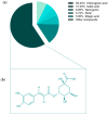
(a) Percentage of total polyphenols, and (b) Chemical structures of chlorogenic acid.
Figure 3
Figure 3
Cellular cytotoxicity of aqueous olive leaf (AOL) extracts on HT29 and PC3 cells. Dose–response curves for HT29 (a) and PC3 cells (b). (c) Multiple comparison of IC50 values between cells. The plotted values represent the means ± SDs of three independent experiments. ns: p > 0.05, ∗ p < 0.05, ∗∗ p < 0.01, ∗∗∗ p < 0.001, ∗∗∗∗ p < 0.0001.
Figure 4
Figure 4
Effect of aqueous olive leaf (AOL) extract on the cell cycle phases in HT29 and PC3 cell lines. Cell cycle phase histogram for (a) untreated HT29 cells, (b) HT29 cells treated with AOL extract, (c) untreated PC3 cells, and (d) PC3 cells treated with AOL extract. (eg) Multiple comparisons between the cell cycle phase (%) values are presented as the means ± SDs of three independent replicates, which were analyzed by two-way ANOVA: ns: p > 0.05, ∗ p < 0.05, ∗∗∗ p < 0.001.
Figure 5
Figure 5
Wound-healing assays demonstrating that treatment with aqueous olive leaf (AOL) extract inhibited cell motility in HT29 and PC3 cell lines. (ac) untreated control HT29 cells, (df) treated HT29 cells, (gi) control PC3 cells, (jl) treated PC3 cells.
Figure 6
Figure 6
Summary of results for the wound-healing assay. Values are presented as the means ± SDs of three independent replicates and were analyzed by two-way ANOVA and multiple comparison tests. (a) HT29 cells treated with aqueous olive leaf (AOL) extracts vs. control (untreated cells), (b) PC3 cells treated with AOL extracts vs. control (untreated cells), (c) HT29 cells treated with AOL extracts vs. PC3 cells treated with AOL extracts. ns: p > 0.05, ∗∗∗ p < 0.001, ∗∗∗∗ p < 0.0001.
Figure 7
Figure 7
Morphological changes in HT29 and PC3 cells treated with aqueous olive leaf (AOL) extract. Cells were treated with AOL extract at their optimum IC50 for 48 h. (a,b) HT29, (c,d) PC3 cells. Apoptotic hallmark represented as: (B) Blebs, (MS) microtubule spikes, (NF) nuclear fragmentation, and (AB) apoptotic bodies.
Figure 8
Figure 8
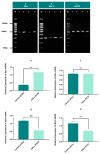
Effects of aqueous olive leaf (AOL) extract treatment on DNA fragmentation in HT29 and PC3 cells after 48 h. (ae) DNA profiles determined by agarose gel electrophoresis. (a) DNA ladder, (b) control (untreated HT29), (c) treated HT29 with AOL extracts, (d) control (untreated PC3), (e) treated PC3 with AOL extracts. (fh) DNA fragmentation percentages after 48 h (means ± SDs of three independent experiments) were analyzed by paired two-tailed t-tests. (f) HT29 cells treated with AOL extracts vs. control (untreated cells), (g) PC3 cells treated with AOL extracts vs. control (untreated cells), (h) HT29 cells treated with AOL extracts vs. PC3 cells treated with AOL extracts. ∗ p < 0.05, ∗∗ p < 0.01.
Figure 9
Figure 9
Effects of aqueous olive leaf (AOL) extract treatment on the expression of Bax and Bcl2 in HT29 and PC3 cells after 48 h. (ac) Bax, Bcl2, and ACTB expression profiles in both the cell lines determined by agarose gel electrophoresis. (a) Bax expression, (b) Bcl2 expression, (c) ACTB expression. (M) DNA ladder, (1) control (untreated HT29), (2) treated HT29 with AOL extracts, (3) control (untreated PC3), (4) PC3 treated with AOL extracts. (eh) Relative expression of Bax and Bcl2 after 48 h (mean values ± SD of three independent experiments) was analyzed by paired two-tailed t-tests. (e) Bax expression in HT29 cells treated with AOL extracts vs. control (untreated cells), (f) Bax expression in PC3 cells treated with AOL extracts vs. control (untreated cells), (g) Bcl2 expression in HT29 cells treated with AOL extracts vs. control (untreated cells), (h) Bcl2 expression in PC3 cells treated with AOL extracts vs. control (untreated cells), ns: p > 0.05, ∗ p < 0.05, ∗∗ p < 0.01.
Figure 10
Figure 10
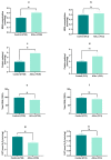
Effect of aqueous olive leaf (AOL) extract on oxidative stress and antioxidant parameters. (a,b) malondialdehyde (MDA) content, (c,d) protein carbonyl content, (e,f) glutathione content, (g,h) catalase activity. Values (expressed as ± SD of three independent experiments) were analyzed by paired two-tailed t-tests. ns: p > 0.05, ∗ p < 0.05.
Figure 11
Figure 11
Molecular mechanism suggested for aqueous olive leaf (AOL) extract in triggering apoptosis in (a) HT29 and (b) PC3 cancer cells.

References

    1. Huang W.-Y., Cai Y.-Z., Zhang Y. Natural phenolic compounds from medicinal herbs and dietary plants: Potential use for cancer prevention. Nutr. Cancer. 2009;62:1–20. doi: 10.1080/01635580903191585. - DOI - PubMed
    1. Sharma A., Kaur M., Katnoria J.K., Nagpal A.K. Polyphenols in food: Cancer prevention and apoptosis induction. Curr. Med. Chem. 2018;25:4740–4757. doi: 10.2174/0929867324666171006144208. - DOI - PubMed
    1. Corrie P.G. Cytotoxic chemotherapy: Clinical aspects. Medicine. 2008;36:24–28. doi: 10.1016/j.mpmed.2007.10.012. - DOI
    1. Allan J.M., Travis L.B. Mechanisms of therapy-related carcinogenesis. Nat. Rev. Cancer. 2005;5:943–955. doi: 10.1038/nrc1749. - DOI - PubMed
    1. Trueb R. Chemotherapy-induced hair loss. Skin Ther. Lett. 2010;15:5–7. - PubMed

MeSH terms