. 2021 Sep:276:121014.
doi: 10.1016/j.biomaterials.2021.121014. Epub 2021 Jul 6.

Injectable catalyst-free "click" organic-inorganic nanohybrid (click-ON) cement for minimally invasive in vivo bone repair

Affiliations

Injectable catalyst-free "click" organic-inorganic nanohybrid (click-ON) cement for minimally invasive in vivo bone repair

Xifeng Liu et al. Biomaterials. 2021 Sep.

Abstract

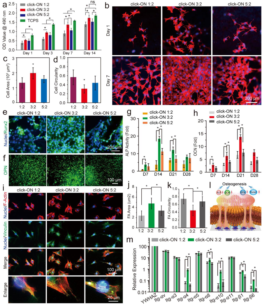
Injectable polymers have attracted intensive attention in tissue engineering and drug delivery applications. Current injectable polymer systems often require free-radical or heavy-metal initiators and catalysts for the crosslinking process, which may be extremely toxic to the human body. Here, we report a novel polyhedral oligomeric silsesquioxane (POSS) based strain-promoted alkyne-azide cycloaddition (SPAAC) "click" organic-inorganic nanohybrids (click-ON) system that can be click-crosslinked without any toxic initiators or catalysts. The click-ON scaffolds supported excellent adhesion, proliferation, and osteogenesis of stem cells. In vivo evaluation using a rat cranial defect model showed outstanding bone formation with minimum cytotoxicity. Essential osteogenic alkaline phosphatase (ALP) and vascular CD31 marker expression were detected on the defect site, indicating excellent support of in vivo osteogenesis and vascularization. Using salt leaching techniques, an injectable porous click-ON cement was developed to create porous structures and support better in vivo bone regeneration. Beyond defect filling, the click-ON cement also showed promising application for spinal fusion using rabbits as a model. Compared to the current clinically used poly (methyl methacrylate) (PMMA) cement, this click-ON cement showed great advantages of low heat generation, better biocompatibility and biodegradability, and thus has great potential for bone and related tissue engineering applications.

Keywords: Bone; Click chemistry; Injectable polymers; Stem cell; Tissue engineering.


Conflict of interest statement

Conflicts of interest

The authors declare no competing financial interest.

Figures

Fig. 1
Fig. 1
Schematic demonstration. a) The two main components of the click-ON bone cement and b) the crosslinking of the two components through catalyst-free strain-promoted alkyne-azide SPAAC “click” reaction. c) the possible molecular biological pathway that stimulate the osteogenesis of cells and promote bone formation. d) Photograph of the click-ON bone cement system and e) the specimens obtained after crosslink.
Fig. 2
Fig. 2
Synthesis and crosslinking. The detailed chemical synthesis of the a) dendritic POSS-PCL-N3 polymer and b) PPF-BCN polymer. c) 1H NMR spectra and corresponding peaks of synthesized hyPCL32-N3 dendrimer and d) PPF-BCN polymers. The variance of two components ratio on the e) gelation time, f) swelling ratio, and g) gel fraction of the click-ON bone scaffolds after crosslinking.
Fig. 3
Fig. 3
Materials properties and in vitro cell studies. The a) schematic demonstration and b) AFM images showing the surface morphology of the click-ON bone scaffolds obtained with varied components ratios. c) Surface roughness calculated from the obtained AFM images. *: Statistically different (p < 0.05). The variance of two components ratio on the d) water contact angle and e) serum protein adsorption on the click-ON scaffolds. The f) strain-stress curve, g) compressive modulus and failure load, h) DSC curve, and i) TGA of the click-ON scaffolds with varied ratios.
Fig. 4
Fig. 4
In vitro cell proliferation, osteogenesis, and cell-material interactions. The a) proliferation and b) morphology of rBMSC stem cells on the click-ON scaffolds. The average c) cell area and d) cell circularity of rBMSCs on the click-ON scaffolds (n=20). *: Statistically different (p < 0.05). Immunofluorescence staining of e) Runx2 and f) OPN in rBMSC stem cells growing on the click-ON scaffolds. The changes of g) ALP activity and h) OCN content in stem cells growing on these substrates. i) Focal adhesion morphology in rBMSC stem cells growing on the click-ON scaffolds and j) focal adhesion area and k) focal adhesion circularity quantified in stem cells growing on these substrates. l) Schematic demonstration of proposed mechanism of osteogenesis induced by signaling from focal adhesion during interactions with extracellular matrix. m) Integrin expression in rBMSC stem cells growing on the click-ON scaffolds. *: Statistically different (p < 0.05); ns: not significant.
Fig. 5
Fig. 5
a) Micro-CT reconstruction of new bone formation (left: top view; right: interior view) in the rat cranial defect with or without click-ON cement injection. b) The quantitative analysis of bone volume / tissue volume ratio. c) Histological H & E stating of varied organs in rats with or without click-ON injection. The d) H & E and e) Masson’s trichrome immunohistochemistry (IHC) staining of tissue samples from rat cranial defects with or without click-ON injection. NB: New Bone; OB: Original Bone; P: Polymer; T: Tissue. *: Statistically different (p < 0.05).
Fig. 6
Fig. 6
a) Immunohistochemistry (IHC) staining of nuclei (blue), ALP (green) and CD31 (red) in tissue samples from rat cranial defects. Quantification of immunofluorescence intensity of b) ALP protein and c) CD31 protein. *: Statistically different (p < 0.05).
Fig. 7
Fig. 7
Injectable click-ON porous scaffolds for bone regeneration. The a) SEM, b) cell F-actin staining, c) OPN marker staining, and d) Runx2 marker staining of rBMSC stem cells growing on the click-ON porous scaffolds. e) The micro-CT reconstruction images of rat cranial defect sites (left: top view; right: interior view) with or without click-ON porous scaffolds. The quantitative analysis of bone area and bone volume to tissue volume ratio (BV/TV). The H & E staining, Masson’s trichrome staining, and Toluidine blue staining of tissue samples from rat cranial defects h) without i) with click-ON injection. j) Histological H & E stating of varied organs in rats with or without click-ON porous scaffolds injection. NB: New Bone; OB: Original Bone; P: Polymer; T: Tissue. *: Statistically different (p < 0.05).
Fig. 8
Fig. 8
The in-situ injection of click-ON cement and X-ray imaging on a) the rat posterolateral spine site and b) the vertebral body subtotal defect (top image: before injection; bottom image: after injection). The micro-CT reconstruction images of rabbit spine filled with moldable click-ON cement viewed from c) back, d) front, e) side and f) top angle. g) The micro-CT slices of the rabbit spinal vertebral bodies with moldable click-ON cement. The rabbit spine micro-CT reconstruction after injection with click-ON cement viewed from c) back, d) front, e) side and f) top angle. l) The micro-CT slices of the rabbit spinal vertebral bodies with injectable click-ON cement. Red arrows point to the new bone formation initiated by the click-ON cement.
Fig. 9
Fig. 9
Comparation with clinically used PMMA bone cement. a) Temperature change during crosslinking of the clinically used PMMA bone cement and click-ON cement. b) Live/dead staining of rBMSC stem cells on the crosslinked click-ON scaffold and PMMA control. The c) flow cytometry and h) MTS analysis of cell apoptosis of rBMSC stem cells on the crosslinked click-ON scaffold and PMMA control. e) Long-term degradation of click-ON scaffold and PMMA control in 1mM NaOH and PBS over a period of 24 months. f) Biodegradation mechanisms for the click-ON cement by hydrolysis of ester bonds within the polymer chains. *: Statistically different (p < 0.05).

References

    1. Salgado CL, Sanchez EM, Zavaglia CA, Almeida AB, Granja PL, Injectable biodegradable polycaprolactone-sebacic acid gels for bone tissue engineering, Tissue Eng Part A 18(1–2) (2012) 137–46. - PubMed
    1. Lasprilla AJ, Martinez GA, Lunelli BH, Jardini AL, Maciel Filho R, Poly-lactic acid synthesis for application in biomedical devices—A review, Biotechnol Adv 30(1) (2012) 321–328. - PubMed
    1. Grayson ACR, Cima MJ, Langer R, Size and temperature effects on poly (lactic-co-glycolic acid) degradation and microreservoir device performance, Biomaterials 26(14) (2005) 2137–2145. - PubMed
    1. Thibault RA, Scott Baggett L, Mikos AG, Kasper FK, Osteogenic differentiation of mesenchymal stem cells on pregenerated extracellular matrix scaffolds in the absence of osteogenic cell culture supplements, Tissue Engineering Part A 16(2) (2009) 431–440. - PMC - PubMed
    1. Henry MG, Cai L, Liu XF, Zhang L, Dong JY, Chen L, Wang ZQ, Wang SF, Roles of Hydroxyapatite Allocation and Microgroove Dimension in Promoting Preosteoblastic Cell Functions on Photocured Polymer Nanocomposites through Nuclear Distribution and Alignment, Langmuir 31(9) (2015) 2851–2860. - PubMed

Publication types