. 2021 Sep;597(7874):103-108.
doi: 10.1038/s41586-021-03817-4. Epub 2021 Jul 19.

Broad sarbecovirus neutralization by a human monoclonal antibody

Affiliations

Broad sarbecovirus neutralization by a human monoclonal antibody

M Alejandra Tortorici et al. Nature. 2021 Sep.

Abstract

The recent emergence of SARS-CoV-2 variants of concern1-10 and the recurrent spillovers of coronaviruses11,12 into the human population highlight the need for broadly neutralizing antibodies that are not affected by the ongoing antigenic drift and that can prevent or treat future zoonotic infections. Here we describe a human monoclonal antibody designated S2X259, which recognizes a highly conserved cryptic epitope of the receptor-binding domain and cross-reacts with spikes from all clades of sarbecovirus. S2X259 broadly neutralizes spike-mediated cell entry of SARS-CoV-2, including variants of concern (B.1.1.7, B.1.351, P.1, and B.1.427/B.1.429), as well as a wide spectrum of human and potentially zoonotic sarbecoviruses through inhibition of angiotensin-converting enzyme 2 (ACE2) binding to the receptor-binding domain. Furthermore, deep-mutational scanning and in vitro escape selection experiments demonstrate that S2X259 possesses an escape profile that is limited to a single substitution, G504D. We show that prophylactic and therapeutic administration of S2X259 protects Syrian hamsters (Mesocricetus auratus) against challenge with the prototypic SARS-CoV-2 and the B.1.351 variant of concern, which suggests that this monoclonal antibody is a promising candidate for the prevention and treatment of emergent variants and zoonotic infections. Our data reveal a key antigenic site that is targeted by broadly neutralizing antibodies and will guide the design of vaccines that are effective against all sarbecoviruses.


Figures

Extended Data Fig. 1.
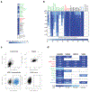
Extended Data Fig. 1.. Breadth of S2X259 mAb across sarbecovirus subgenus.
a, Flow cytometry analysis of S2X259 cross-reactivity with a panel of 29 S glycoproteins representative of sarbecovirus clades 1a, 1b, 2 and 3. The colours represent the lowest concentration of mAb at which binding was observed. b, FACS binding of S2X259 to a panel of sarbecovirus S glycoproteins transiently expressed in ExpiCHO cells. Results represent the binding frequency normalized using the ratio between MFIs for S2X259 and an expression control mAb. c, FACS gating strategy used to assess binding of S2X259 or of the expression control mAb. d, ELISA binding of site I-targeting S2E12, site II-targeting S2X259 and S2X35, and site IV-targeting S309 mAbs to RBDs spanning the different clades of the sarbecovirus subgenus. n=2 independent experiments.
Extended Data Fig. 2.
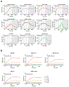
Extended Data Fig. 2.. S2X259 Fab binding to recombinant sarbecovirus RBDs, prefusion SARS-CoV-2 S ectodomain trimer and RBD variants.
a, S or RBD antigens were captured on the sensor chip surface and binding to S2X259 Fab at 11, 33, 100, and 300 nM was monitored successively, in single-cycle kinetics format, by surface plasmon resonance. All data have been fit to a 1:1 binding model (black dashed line) and the equilibrium dissociation constant (KD) is reported. For the S-binding data, we report an apparent KD (KD,app) since kinetics are affected by conformational dynamics between open and closed RBD states. The colouring scheme matches the phylogenetic tree in Figure 1a. b, Biolayer interferometry binding analysis of the S2X259 Fab to wildtype or VOC SARS-CoV-2 biotinylated RBDs immobilized at the surface of SA biosensors. The data are coloured according to the key and fits to a 1:1 binding model is shown as black lines. Equilibrium dissociation constants (KD) are reported above each plot.
Extended Data Fig. 3.
Extended Data Fig. 3.. S2X259 neutralizing activity is not affected by mutations present in circulating SARS-CoV-2 VOCs and recent lineages.
a-b, Neutralization by S2X259 of SARS-CoV-2 S VSV pseudotyped circulating variants shown as IC50 (a) and IC50 fold change as compared to Wuhan-1-D614G (b). Schematic of the SARS-CoV-2 S and the mutation landscape in each variant is shown. Del, deletion. * Tested as single RBD mutant in panels a-b.
Extended Data Fig. 4.
Extended Data Fig. 4.. CryoEM data processing and validation of S2X259-bound SARS-CoV-2 S.
a-b, Representative electron micrographs (a) and class averages (b) of SARS-CoV-2 S in complex with the S2X259 Fab. Scale bar of the micrograph: 500 Å. Scale bar of the class averages: 100 Å. c, Gold-standard Fourier shell correlation curves for the S trimer bound to three S2X259 Fabs (solid black line) and the locally refined reconstruction of the RBD/S2X259 variable domains (dashed black line). The 0.143 cut-off is indicated by a horizontal dashed grey line. d-e, Local resolution map for the open S trimer bound to three S2X259 Fabs (d) and the locally refined reconstruction of the RBD/S2X259 variable domains (e). f, CryoEM data processing flow-chart.
Extended Data Fig. 5.
Extended Data Fig. 5.. The S2X259 angle of approach for binding to the SARS-CoV-2 RBD allows to circumvent the SARS-CoV N357 glycan present in all sarbecovirus RBDs except SARS-CoV-2.
Ribbon diagram showing a superimposition of the S2X259-bound and S2A4-bound (PDB 7JVA) SARS-CoV-2 RBD. The SARS-CoV glycan at position N357 was modelled based on the S230-bound SARS-CoV S structure (PDB 6NB6) and is predicted to sterically hinder S2A4 binding (red star) but not S2X259. The mAb light and heavy chains are coloured magenta and purple (S2X259) or light and dark green (S2A4). N-linked glycans are rendered as blue spheres.
Extended Data Fig. 6.
Extended Data Fig. 6.. S2X259 can bind to the SARS-CoV-2 RBD in presence of site I and IV-targeting mAbs.
a, View of site I-targeting S2E12 (pink), site II-targeting S2X259 (magenta), and site IV-targeting S309 (purple) mAb bound to the SARS-CoV-2 RBD (light blue). b, Competition binding assays for S2X259 vs site I-targeting S2E12 and site IV-targeting S309 mAbs on SARS-CoV-2 RBD as measured by biolayer interferometry. One independent experiment out of two is shown. c, Competition ELISA (blockade-of-binding) between site I-targeting S2H14 or site-II targeting S2X259 and sera or plasma from COVID- 19 convalescent (n=10 biological samples) and vaccinated subjects (n=9 biological samples). Each plot shows the magnitude of inhibition of binding to immobilized RBD in the presence of each mAb, expressed as reciprocal sera or plasma dilution blocking 80% of the maximum binding response. Convalescent donor from whom S2X259 mAb was isolated is shown as a square. Statistical analysis was performed using two-tailed Mann-Whitney test.
Extended Data Fig. 7.
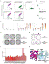
Extended Data Fig. 7.. S2X259 has a high barrier for the emergence of resistance mutants.
a, FACS gates used in DMS to select escape variants. Top row: yeast controls expressing unmutated SARS-CoV-2 RBD labelled at relevant S2X259 concentrations for setting of selection gates. Bottom row: fraction of cells in SARS-CoV-2 mutant libraries falling into the antibody-escape bin. b, Correlation in site-level (top, sum of escape fractions for mutations at a site) and mutation-level (bottom) escape between independently generated and assayed RBD mutant libraries. c, Line plot of escape mutants along all positions in the SARS-CoV-2 RBD (left). Pink lines indicate sites that escape S2X259 binding illustrated at the mutation-level in logoplots (right). In logoplots, the height of a letter scales with its escape fraction. Letters are coloured according to their deleterious consequences for ACE2 binding (middle) or RBD expression (right) as determined in prior DMS experiments. d, Plaque assay using VSV-SARS-CoV-2 chimeric virus on Vero cells with no mAb (−) or S2X259 (+) in the overlay to isolate escape mutants (red arrow). Data are representative of three independent experiments. e, Plaque assays performed to validate the VSV-SARS-CoV-2 G504D mutant in Vero cells in the presence (+) or absence (−) of S2X259 in the overlay. Representative image of two independent experiments is shown. f, S2X259 in vitro neutralizing activity against SARS-CoV-2 S VSV pseudotyped mutants. For each mutant the fold change of the IC50 geometric mean vs SARS-CoV-2 S D614G is reported. *Q506K displayed a 10-fold reduction in viral entry in comparison to the other mutants. Results from two independent experiments are reported. g, Zoomed-in view of the S2X259/RBD interface showing that the G504D substitution would disrupt mAb binding due to steric hindrance (indicated with a red star).
Extended Data Fig. 8.
Extended Data Fig. 8.. S2X259 epitope conservation across sarbecovirus clades.
Protein sequence alignment of representative sarbecovirus RBDs with strictly conserved residues shown as dots. Overall conservation is represented as a bar plot at the bottom of the figure. Residue positions are based on SARS-CoV-2. Residues determined to be most important for S2X259 binding are denoted in dark green. Substitutions at positions D405 and G504 are indicated in pink and blue/orange, respectively. Additional residues representing extended epitope, are denotated grey. Different clades within the sarbecovirus subgenus are overlayed in grey (clade 1a), red (clade 1b), green (clade 2), and light blue (clade 3).
Extended Data Fig. 9.
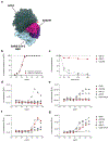
Extended Data Fig. 9.. Inhibition of ACE2 engagement, S1 subunit shedding and activation of FcγRIIa and FcγRIIIa in vitro.
a, S2X259 (purple/pink) and ACE2 (dark green) bind partially overlapping binding sites on the SARS-CoV-2 RBD (blue). b, Pre-incubation of serial dilutions of S2X259 with SARS-CoV-2 (red) or the SARS-CoV (black) RBDs prevents binding to immobilized human ACE2 (hACE2) ectodomain in ELISA. c, mAb-mediated S1 subunit shedding from cell-surface expressed SARS-CoV-2 S as determined by flow-cytometry. S2E12 was included as positive control whereas S2M11 was included as negative control. d-e, NFAT-driven luciferase signal induced in Jurkat cells stably expressing FcγRIIa H131 (a) variant or FcγRIIIa V158 (b) variant by S2X259 binding to full-length wild-type SARS-CoV-2 S on ExpiCHO target cells. f-g, NFAT-driven luciferase signal induced in Jurkat cells stably expressing FcγRIIa H131 (c) or FcγRIIIa V158 (d) variants by S2X259 binding to uncleavable full-length pre-fusion stabilized SARS-CoV-2 S (unable to release the S1 subunit) transiently expressed in ExpiCHO cells. SE12, S2M11, S309, S309-GRLR mAbs are included as controls.
Extended Data Fig. 10.
Extended Data Fig. 10.. Correlation between mAbs concentration and infectious virus in vivo and in vitro neutralizing activity of S2X259/S309 antibody cocktail.
a-c, Infectious virus titers in the lungs at 4 days post-infection plotted as a function of serum mAb concentrations before infection (day 0) with prototypic SARS-CoV-2 (a) and SARS-CoV-2 B.1.351 (c) in prophylactic setting or at 4 days post-infection with prototypic SARS-CoV-2 (b) in therapeutic setting. d-e, SARS-CoV-2 B1.351 VSV-based pseudotypes neutralization (d) and synergy score (e) measured combining S2X259 and S309 mAbs. Synergy was calculated using MacSynergyII (Synergy score: 0.98, Antagonism score: 0). Results represent four replicates of one independent experiment.
Fig. 1.
Fig. 1.. Identification of a broadly neutralizing sarbecovirus mAb.
a, Phylogenetic tree of sarbecovirus RBDs constructed via maximum likelihood analysis of amino acid sequences retrieved from GISAID and GenBank. Cross-reactivity within the sarbecovirus subgenus for S2E12, S309, and ADG-2 is included for comparison. b, S2X259 binding to RBDs representative of the different sarbecovirus clades and SARS-CoV-2 variants as measured by ELISA. c, S2X259-mediated neutralization of SARS-CoV-2-Nluc authentic virus and SARS-CoV-2 S MLV-pseudotyped virus (MLV-pp). d-e, S2X259-mediated neutralization of VSV pseudotypes harbouring SARS-CoV-2 S from isolates representing the B.1.1.7, B.1.351, P.1 and B.1.429 VOC (d) as well as single RBD mutants (e). f-g, S2X259-mediated neutralization of VSV pseudotypes harbouring SARS-CoV-related (clade 1a, f) or SARS-CoV-2-related (clade 1b, g) S glycoproteins. n = 2 independent experiments. Error bars indicate standard deviation of duplicates or triplicates.
Fig. 2.
Fig. 2.. The S2X259 broadly neutralizing sarbecovirus mAb recognizes RBD antigenic site II.
a-b, CryoEM structure of the prefusion SARS-CoV-2 S ectodomain trimer with three S2X259 Fab fragments bound to three open RBDs viewed along two orthogonal orientations (PDB 7RA8 and PDB 7RAL). c, The S2X259 binding mode involving contacts with multiple RBD regions. Residues corresponding to prevalent RBD mutations are shown as red spheres (PDB 7M7W). d-e, Close-up views showing selected interactions formed between S2X259 and the SARS-CoV-2 RBD. In panels a-e, each SARS-CoV-2 S protomer is coloured distinctly (cyan, pink and gold) whereas the S2X259 light and heavy chain variable domains are coloured magenta and purple, respectively. N-linked glycans are rendered as blue spheres in panels a-c.
Fig. 3.
Fig. 3.. S2X259 is resilient to a broad spectrum of escape mutations.
a, Complete mapping of mutations reducing S2X259 binding using yeast-displayed RBD and deep mutational scanning (DMS). Mean mutation effect on ACE2 affinity, RBD folding, and contribution to S2X259 binding of substitutions at each position in the S2X259 epitope is reported. * mutations introducing a N-linked glycosylation sites that may not be tolerated in full spike. b, Frequency of mutants within the S2X259 epitope based on SARS-CoV-2 genome sequences available on GISAID as of May 2021. S2X259 neutralizing activity against selected mutations is reported.
Fig. 4.
Fig. 4.. S2X259 protects hamsters challenged with prototypic and B.1.351 SARS-CoV-2.
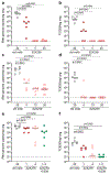
a-b, Viral RNA loads (a) and replicating virus titres (TCID50) (b) in the lungs of Syrian hamsters at 4 days post-intranasal infection with prototypic SARS-CoV-2 following prophylactic administration of S2X259 mAb at 1 (n=6 animals), 5 (n=5 animals), and 25 (n=5 animals) mg/kg. c-d, Viral RNA loads (c) and replicating virus titres (TCID50) (d) in the lungs of Syrian hamsters after therapeutic administration of S2X259 at 5 (n=5 animals), 10 (n=6 animals), and 20 (n=6 animals) mg/kg 24h after infection with Wuhan-related SARS-CoV-2. e-f, Quantification of viral RNA loads (e) and replicating virus titres (TCID50) (f) in the lungs of Syrian hamsters 4 days post intranasal challenge with B.1.351 SARS-CoV-2 VOC following prophylactic administration of S2X259 at 1 mg/kg (n=6 animals), 4 mg/kg (n=6 animals), and in combination with S309 (1+1 mg/kg, n=6 animals). Data from one independent experiment are presented. Two-tailed Mann–Whitney test was used for statistical analysis of significance.

Update of

  • Structural basis for broad sarbecovirus neutralization by a human monoclonal antibody.
    Tortorici MA, Czudnochowski N, Starr TN, Marzi R, Walls AC, Zatta F, Bowen JE, Jaconi S, di Iulio J, Wang Z, De Marco A, Zepeda SK, Pinto D, Liu Z, Beltramello M, Bartha I, Housley MP, Lempp FA, Rosen LE, Dellota E Jr, Kaiser H, Montiel-Ruiz M, Zhou J, Addetia A, Guarino B, Culap K, Sprugasci N, Saliba C, Vetti E, Giacchetto-Sasselli I, Silacci Fregni C, Abdelnabi R, Caroline Foo SY, Havenar-Daughton C, Schmid MA, Benigni F, Cameroni E, Neyts J, Telenti A, Snell G, Virgin HW, Whelan SPJ, Bloom JD, Corti D, Veesler D, Pizzuto MS. Tortorici MA, et al. bioRxiv [Preprint]. 2021 Apr 8:2021.04.07.438818. doi: 10.1101/2021.04.07.438818. bioRxiv. 2021. Update in: Nature. 2021 Sep;597(7874):103-108. doi: 10.1038/s41586-021-03817-4. PMID: 33851169 Free PMC article. Updated. Preprint.

References

    1. Deng X et al. Transmission, infectivity, and neutralization of a Spike L452R SARS-CoV-2 variant. Cell 184, 1–12 (2021). - PMC - PubMed
    1. Challen R et al. Risk of mortality in patients infected with SARS-CoV-2 variant of concern 202012/1: matched cohort study. BMJ 372, n579 (2021). - PMC - PubMed
    1. Davies NG et al. Estimated transmissibility and impact of SARS-CoV-2 lineage B.1.1.7 in England. Science 372, eabg3055 (2021). - PMC - PubMed
    1. Tegally H et al. Detection of a SARS-CoV-2 variant of concern in South Africa. Nature 592, 438–443 (2021). - PubMed
    1. McCallum M et al. SARS-CoV-2 immune evasion by variant B.1.427/B.1.429. bioRxiv 10.1101/2021.03.31.437925 (2021). - DOI

Publication types

MeSH terms

Substances