. 2021 Jul 1;22(13):7108.
doi: 10.3390/ijms22137108.

Overlap Arrhythmia Syndromes Resulting from Multiple Genetic Variations Studied in Human Induced Pluripotent Stem Cell-Derived Cardiomyocytes

Affiliations

Overlap Arrhythmia Syndromes Resulting from Multiple Genetic Variations Studied in Human Induced Pluripotent Stem Cell-Derived Cardiomyocytes

Jacqueline A Treat et al. Int J Mol Sci. .

Abstract

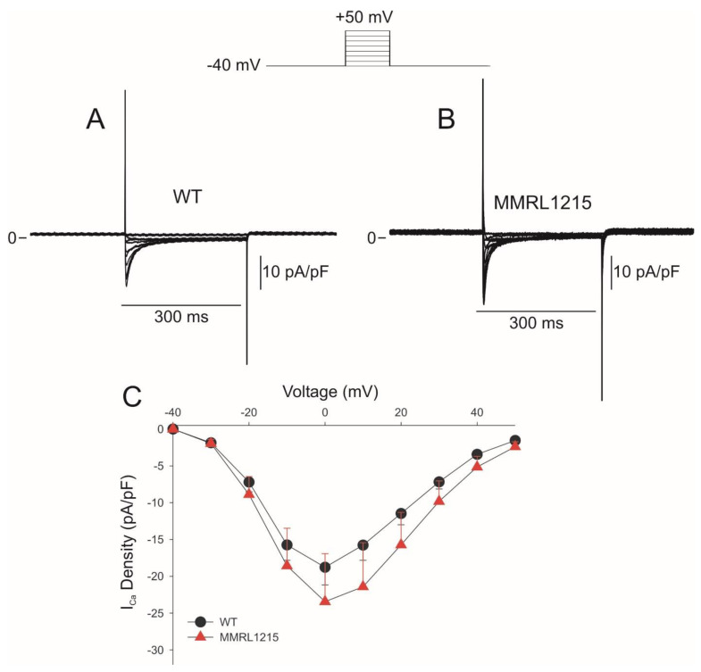
Human induced pluripotent stem cell-derived cardiomyocytes (hiPSC-CMs) are used for genetic models of cardiac diseases. We report an arrhythmia syndrome consisting of Early Repolarization Syndrome (ERS) and Short QT Syndrome (SQTS). The index patient (MMRL1215) developed arrhythmia-mediated syncope after electrocution and was found to carry six mutations. Functional alterations resulting from these mutations were examined in patient-derived hiPSC-CMs. Electrophysiological recordings were made in hiPSC-CMs from MMRL1215 and healthy controls. ECG analysis of the index patient showed slurring of the QRS complex and QTc = 326 ms. Action potential (AP) recordings from MMRL1215 myocytes showed slower spontaneous activity and AP duration was shorter. Field potential recordings from MMRL1215 hiPSC-CMs lack a "pseudo" QRS complex suggesting reduced inward current(s). Voltage clamp analysis of ICa showed no difference in the magnitude of current. Measurements of INa reveal a 60% reduction in INa density in MMRL1215 hiPSC-CMs. Steady inactivation and recovery of INa was unaffected. mRNA analysis revealed ANK2 and SCN5A are significantly reduced in hiPSC-CM derived from MMRL1215, consistent with electrophysiological recordings. The polygenic cause of ERS/SQTS phenotype is likely due to a loss of INa due to a mutation in PKP2 coupled with and a gain of function in IK,ATP due to a mutation in ABCC9.

Keywords: action potentials; depolarization; electrophysiology; sodium current; stem cells.


Conflict of interest statement

The authors report no relationships that can be construed as a conflict of interest.

Figures

Figure 1
Figure 1
Representative ECG of MMRL1215 illustrating Early Repolarization Syndrome phenotype. A slurring of the QRS complex is apparent in multiple leads (denoted by the arrows). In addition, a QTc = 326 ms was noted.
Figure 2
Figure 2
(A): Extracellular field potential (EFP) signals recorded in WT (left side) and MMRL1215 hiPSC-CMs (right side). Compared to WT hiPSC, myocytes derived from MMRL1215 showed a complete lack of a pseudo QRS complex, suggesting a major reduction in inward current(s). Panel (B): Action potential (AP) recordings obtained from WT and MMRL1215 monolayers. MMRL1215 hiPSC-CMs had a shorter action potential duration compared to WT.
Figure 3
Figure 3
Line scans recorded from WT and MMRL1215 hiPSC-CMs showing spontaneous Ca2+ transients. Fluorescence intensity was much lower in MMRL 1215, and in some myocytes the spontaneous activity was episodic in nature.
Figure 4
Figure 4
Representative traces showing ICa recorded from WT (n = 11) and MMRL1215 (n = 9) (A,B). Ca2+ currents were recorded during a 300 ms step depolarization from −40 to +50mV. (C) Current-voltage relationship for ICa.
Figure 5
Figure 5
Representative whole cell INa recordings from WT and MMRL1215 hiPSC-CMs (A,B). Current recordings were obtained at test potentials between −80 and 30 mV (protocol at top of figure). (C): I–V relation for WT (n = 18) and MMRL1215 (n = 25) myocytes. (D): Steady-state activation curves for WT and MMRL1215 myocytes. Data were normalized and plotted against their test potential. * Statistically significant from WT, p < 0.05.
Figure 6
Figure 6
Representative steady-state inactivation recordings from WT and MMRL1215 myocytes (A,B) in response to the voltage clamp protocol (top of the figure). The steady state-inactivation relation for hiPSC-CMs (C) yielded similar mid-inactivation potential for WT (n = 25) and MMRL1215 (n = 29) cells.
Figure 7
Figure 7
Representative recovery from inactivation traces recorded from WT and MMRL1215 myocytes (A,B). Recovery was measured using two identical voltage clamp steps to −20 mV from a holding potential of either −120 mV or −100 mV separated by selected time intervals. (C): Mean data showing recovery time-course of INa recorded at −120 mV for WT (n = 24) and MMRL1215 (n = 26). (D): Recovery time-course of INa recorded at −100 mV for WT (n = 16) and MMRL1215 (n = 19).
Figure 8
Figure 8
Expression profile of genes PKP2, ANK2 and SCN5A obtained using real-time PCR.

Similar articles

Cited by

References

    1. Takahashi K., Tanabe K., Ohnuki M., Narita M., Ichisaka T., Tomoda K., Yamanaka S. Induction of Pluripotent Stem Cells from Adult Human Fibroblasts by Defined Factors. Cell. 2007;131:861–872. doi: 10.1016/j.cell.2007.11.019. - DOI - PubMed
    1. Wu J.C., Garg P., Yoshida Y., Yamanaka S., Gepstein L., Hulot J.-S., Knollmann B.C., Schwartz P.J. Towards Precision Medicine with Human iPSCs for Cardiac Channelopathies. Circ. Res. 2019;125:653–658. doi: 10.1161/CIRCRESAHA.119.315209. - DOI - PMC - PubMed
    1. Terrenoire C., Wang K., Tung K.W.C., Chung W.K., Pass R.H., Lu J.T., Jean J.-C., Omari A., Sampson K.J., Kotton D., et al. Induced pluripotent stem cells used to reveal drug actions in a long QT syndrome family with complex genetics. J. Gen. Physiol. 2012;141:61–72. doi: 10.1085/jgp.201210899. - DOI - PMC - PubMed
    1. El-Battrawy I., Lan H., Cyganek L., Zhao Z., Li X., Buljubasic F., Lang S., Yücel G., Sattler K., Zimmermann W., et al. Modeling Short QT Syndrome Using Human-Induced Pluripotent Stem Cell–Derived Cardiomyocytes. J. Am. Heart Assoc. 2018;7 doi: 10.1161/JAHA.117.007394. - DOI - PMC - PubMed
    1. Sendfeld F., Selga E., Scornik F.S., Pérez G.J., Mills N.L., Brugada R. Experimental Models of Brugada syndrome. Int. J. Mol. Sci. 2019;20:2123. doi: 10.3390/ijms20092123. - DOI - PMC - PubMed

MeSH terms

Supplementary concepts