Review
. 2021 Jul 1;22(13):7112.
doi: 10.3390/ijms22137112.

Chronic Orofacial Pain: Models, Mechanisms, and Genetic and Related Environmental Influences

Affiliations
Review

Chronic Orofacial Pain: Models, Mechanisms, and Genetic and Related Environmental Influences

Barry J Sessle. Int J Mol Sci. .

Abstract

Chronic orofacial pain conditions can be particularly difficult to diagnose and treat because of their complexity and limited understanding of the mechanisms underlying their aetiology and pathogenesis. Furthermore, there is considerable variability between individuals in their susceptibility to risk factors predisposing them to the development and maintenance of chronic pain as well as in their expression of chronic pain features such as allodynia, hyperalgesia and extraterritorial sensory spread. The variability suggests that genetic as well as environmental factors may contribute to the development and maintenance of chronic orofacial pain. This article reviews these features of chronic orofacial pain, and outlines findings from studies in animal models of the behavioural characteristics and underlying mechanisms related to the development and maintenance of chronic orofacial pain and trigeminal neuropathic pain in particular. The review also considers the role of environmental and especially genetic factors in these models, focussing on findings of differences between animal strains in the features and underlying mechanisms of chronic pain. These findings are not only relevant to understanding underlying mechanisms and the variability between patients in the development, expression and maintenance of chronic orofacial pain, but also underscore the importance for considering the strain of the animal to model and explore chronic orofacial pain processes.

Keywords: animal models; environmental factors; genetic factors; orofacial pain; strain differences; trigeminal.


Conflict of interest statement

The author declares no conflict of interest.

Figures

Figure 1
Figure 1
Mechanistic outline of processes by which genetic and environmental risk factors may influence orofacial pain. These include influences on intermediate pain phenotypes and the transition from an acute pain state to a chronic pain state that may further exacerbate these risk factors. From Meloto et al. [15].
Figure 2
Figure 2
Mechanisms involved in initiation and transmission of nociceptive afferent information from orofacial tissues. (A) shows that trigeminal nerve injury or orofacial inflammation can induce hyperexcitability of trigeminal primary afferent neurons, satellite glial cell activation and macrophage accumulation. The hyperactivated trigeminal ganglion (TG) neurons as well as the satellite glial cells and macrophages communicate with each other through a variety of mediators, receptor mechanisms and signalling processes, many of which are shown here. Through this communication, further enhancement of the excitability of TG afferent neurons may occur, as manifested in their hyperexcitable input into components of the trigeminal brainstem sensory nuclear complex, in particular the medullary dorsal horn (also known as trigeminal subnucleus caudalis [Vc]), as well as the upper cervical spinal cord (C1/C2). (B) shows input and output features of nociceptive neurons in the medullary dorsal horn and C1/C2 under normal conditions as well as pathological conditions such as trigeminal nerve injury or orofacial inflammation which produces the hyperexcitable nociceptive afferent input shown in A. This input releases mediators which cause the neurons to become hyperactive, resulting in activation of microglia and astrocytes. Neuron–glial cell communication via various molecules, several of which are shown here, is important in fostering the hyperexcitable state of the neurons, i.e., central sensitization. From Iwata and Sessle [25].
Figure 3
Figure 3
Features of a nociceptive neuron recorded in the rat medullary dorsal horn that exhibited extensive mechanoreceptive field expansion as a reflection of trigeminal central sensitization induced by intramuscular injection of the inflammatory irritant mustard oil. The neuron was a wide dynamic range neuron since it could be activated by tactile stimulation of the cutaneous periorbital area as well as by pinch stimulation of the posterior facial skin and by heavy pressure applied to posterior cranial and cervical tissues. The figurines show that in the control condition (pre-injection), the mechanoreceptive field was limited to the ipsilateral side and was stable for several minutes, and also did not change over a 20-min period following injection of vehicle control (mineral oil) into the tongue musculature. However, note that within 5 min after the injection of the inflammatory irritant mustard oil into the tongue, the mechanoreceptive field had expanded to encompass the ipsilateral ear as well as contralateral facial and cervical areas. The expanded mechanoreceptive field was maintained for at least another 10 min before returning towards pre-injection features by 20 min. Based on data from Yu et al. [89].
Figure 4
Figure 4
Infraorbital nerve injury in the rat induces hypersensitivity that spreads extraterritorially and is genetically dependent. In adult male WKY and LEW rats housed under similar environmental conditions, head withdrawal thresholds to mechanical stimulation of the facial vibrissal pads, ears and hindpaws were assessed bilaterally with von Frey monofilaments before (baseline [BL]) and after receiving unilateral transection (IONX) of the medial branch of the infraorbital nerve (that supplies medial aspect of upper lip, vibrissal pad and anterior teeth) or sham operation (n = 10/group). On the left is an example of the time course of the effect of the partial IONX (in comparison to sham) in inducing extraterritorial spread of sensitivity (ETSS) as reflected in a significant decrease in the withdrawal threshold to mechanical stimulation of the contralateral ear (2-way ANOVA, p < 0.05). This reflection of IONX-induced nociceptive behaviour occurred in both strains but was significantly greater and longer lasting in the WKY rats. Additional evidence of ETSS is illustrated on the right where WKY rats showed, at 3–5 weeks post-IONX, significantly (2-way ANOVA, p < 0.05) decreased mechanical withdrawal thresholds not only in the contralateral ear but also in in the lateral part of the partially denervated ipsilateral vibrissal pad, and ear and hindpaw. LEW rats also displayed significant ETSS but only for the vibrissal pad; their levels of mechanical hypersensitivity in the ear and hindpaw were significantly lower (t-test, p < 0.05) than those in WKY rats. They also showed significantly lower levels of heat hypersensitivity in these sites that were tested by applying noxious infrared laser heat stimuli (200 msec, 18–25 Amp, 2 mm in diameter diode) to these sites (not shown). * p < 0.05 comparison to baseline, ** p < 0.05 and # p < 0.05, ## p < 0.05 comparison between groups. Based on data from Wang et al. [120].
Figure 5
Figure 5
Infraorbital nerve injury in the mouse induces facial mechanical hypersensitivity that is genetically dependent. A/J and C57BL/6J male mice aged 8–12 weeks were maintained under similar environmental conditions, and were tested with von Frey monofilaments for facial mechanical sensitivity before and after unilateral transection (IONX) of the medial branch of the infraorbital nerve (that supplies medial aspect of upper lip, vibrissal pad and anterior teeth) or sham operation; naïve mice were also tested (n = 11/group). Baseline values of facial mechanical sensitivity as reflected in head withdrawal threshold were comparable in A/J and C57BL/6J mice, and following IONX both strains showed evidence of ETSS as reflected in hypersensitivity in the ipsilateral lower lip and contralateral upper lip when compared with the sensitivity in naïve and sham animals. However, the time course of the IONX-induced facial hypersensitivity was significantly different (2 way ANOVA, p < 0.05) between the two strains of mice: hypersensitivity in both A/J and C57BL/6J mice started around post-operative day 1, but in the A/J mice reached its peak throughout post-operative days 3–21 and lasted up to day 49, whereas in the C57BL/6J mice, hypersensitivity had a significantly shorter peak duration. Moreover, hypersensitivity lasted up to days 35 (contralateral upper lip) and 49 (ipsilateral lower lip) in A/J mice but only to days 14 and 21, respectively, in C57BL/6J mice. Sham animals (compared to naïve animals) showed a brief hypersensitivity that also was significantly longer in the A/J mice. * p < 0.05. Based on data from Varathan et al. [121] and Cherkas et al. [168].
Figure 6
Figure 6
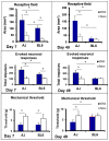
Infraorbital nerve injury in the mouse induces trigeminal central sensitization that is genetically dependent. A/J (AJ, n = 12) and C57BL/6J (BL6, n = 12) male mice aged 8–12 weeks were maintained under similar environmental conditions, and used for electrophysiological recordings made in histologically identified sites in the medullary dorsal horn. A total of 40 nociceptive neurons functionally classified as nociceptive-specific neurons was recorded. At 7 days or 49 days prior to the recording experiment, animals received unilateral transection (IONX) of the medial branch of the infraorbital nerve or no IONX (i.e., naïve rats). IONX-induced trigeminal central sensitization of the neurons was expressed as significant increases (compared to naïve) in neuronal mechanoreceptive field size and responses evoked by graded mechanical stimulation and a significantly decreased mechanical activation threshold (ANOVA, p < 0.05). Note the significant differences between A/J and C57BL6 mice in the magnitude and time course of these three parameters of trigeminal central sensitization following IONX (2-way ANOVA, p < 0.05). Central sensitization was evident at post-operative day 5 in both A/J and C57BL6 mice but at post-operative day 49 it was only present in A/J mice. Note in addition that naïve animals also showed a significant difference between A/J and C57BL6 mice in mechanoreceptive field size. * p < 0.05. Based on data from Varathan et al. [121] and Cherkas et al. [168].

References

    1. Lipton J.A., Ship J.A., Larach-Robinson D. Estimated prevalence and distribution of reported orofacial pain in the United States. J. Am. Dent. Assoc. 1993;124:115–121. doi: 10.14219/jada.archive.1993.0200. - DOI - PubMed
    1. Arendt-Nielsen L., Graven-Nielsen T., Sessle B.J. Mechanisms underlying extraterritorial and widespread sensitization: From animal to chronic pain. In: Graven-Nielsen T., Arendt-Nielsen L., editors. Musculoskeletal Pain: Basic Mechanisms & Implications. Wolters Kluwer Health; Washington, DC, USA: 2015. pp. 417–436.
    1. Macfarlane T.V. Epidemiology of orofacial pain. In: Sessle B.J., editor. Orofacial Pain: Recent Advances in Assessment, Management, and Understanding of Mechanisms. IASP Press; Washington, DC, USA: 2014. pp. 33–52.
    1. Svensson P., Baad-Hansen L., Drangsholt M., Jaaskelainen S. Neurosensory testing for assessment, diagnosis, and prediction of orofacial pain. In: Sessle B.J., editor. Orofacial Pain: Recent Advances in Assessment, Management, and Understanding of Mechanisms. IASP Press; Washington, DC, USA: 2014. pp. 143–164.
    1. Slade G.D., Greenspan J.D., Fillingim R.B., Maixner W., Sharma S., Ohrbach R. Overlap of five chronic pain conditions: Temporomandibular disorders, headache, back pain, irritable bowel syndrome, and fibromyalgia. J. Oral Facial Pain Headache. 2020;34:s15–s28. doi: 10.11607/ofph.2581. - DOI - PMC - PubMed

LinkOut - more resources