Review
. 2021 Jul 1;22(13):7133.
doi: 10.3390/ijms22137133.

Epileptic Mechanisms Shared by Alzheimer's Disease: Viewed via the Unique Lens of Genetic Epilepsy

Affiliations
Review

Epileptic Mechanisms Shared by Alzheimer's Disease: Viewed via the Unique Lens of Genetic Epilepsy

Jing-Qiong Kang. Int J Mol Sci. .

Abstract

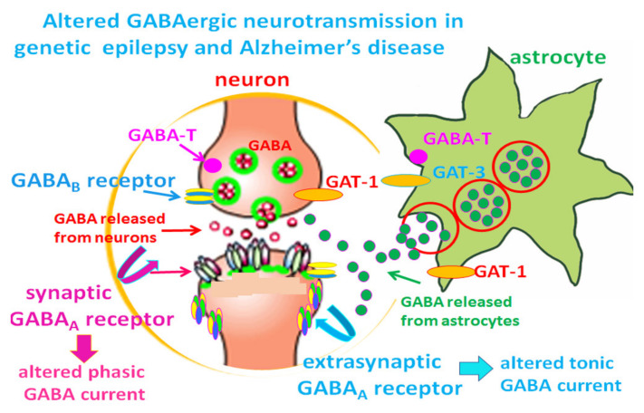
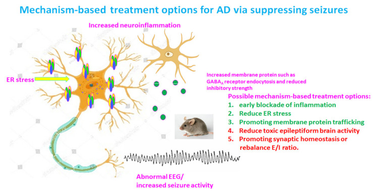
Our recent work on genetic epilepsy (GE) has identified common mechanisms between GE and neurodegenerative diseases including Alzheimer's disease (AD). Although both disorders are seemingly unrelated and occur at opposite ends of the age spectrum, it is likely there are shared mechanisms and studies on GE could provide unique insights into AD pathogenesis. Neurodegenerative diseases are typically late-onset disorders, but the underlying pathology may have already occurred long before the clinical symptoms emerge. Pathophysiology in the early phase of these diseases is understudied but critical for developing mechanism-based treatment. In AD, increased seizure susceptibility and silent epileptiform activity due to disrupted excitatory/inhibitory (E/I) balance has been identified much earlier than cognition deficit. Increased epileptiform activity is likely a main pathology in the early phase that directly contributes to impaired cognition. It is an enormous challenge to model the early phase of pathology with conventional AD mouse models due to the chronic disease course, let alone the complex interplay between subclinical nonconvulsive epileptiform activity, AD pathology, and cognition deficit. We have extensively studied GE, especially with gene mutations that affect the GABA pathway such as mutations in GABAA receptors and GABA transporter 1. We believe that some mouse models developed for studying GE and insights gained from GE could provide unique opportunity to understand AD. These include the pathology in early phase of AD, endoplasmic reticulum (ER) stress, and E/I imbalance as well as the contribution to cognitive deficit. In this review, we will focus on the overlapping mechanisms between GE and AD, the insights from mutations affecting GABAA receptors, and GABA transporter 1. We will detail mechanisms of E/I imbalance and the toxic epileptiform generation in AD, and the complex interplay between ER stress, impaired membrane protein trafficking, and synaptic physiology in both GE and AD.

Keywords: Alzheimer’s disease (AD); GABAergic signaling; amyloid β (Aβ); electroencephlography (EEG); endoplasmic reticulum (ER) stress; excitatory/inhibitory(E/I) imbalance; gamma-aminobutyric acid (GABA); genetic epilepsy (GE); protein trafficking.


Conflict of interest statement

The author declares no conflict of interest for this study.

Figures

Figure 1
Figure 1
Altered GABAergic neurotransmission in both genetic epilepsy and Alzheimer’s disease. The neurotransmitter GABA can be released from both neurons and glia. GABA is synthesized from glutamic acid, the principal excitatory neurotransmitter via glutamic acid decarboxylase (GAD). GABA is catabolized by GABA transaminase (GABA-T), which is a membrane-bound enzyme expressed by neurons and glia. In GABAergic interneurons, GABA is released from vesicles in presynaptic terminals and activates GABA receptors, which include GABAA receptors and GABAB receptors. Under pathological conditions, such as AD, GABA can also be released from reactive astrocytes. GABAA receptors hyperpolarize neurons via Cl influx. The released GABA is taken up by GABA transporters (GAT-1 and GAT-3) back into presynaptic compartments of neurons or into astrocytes. Failure of GABA clearance due to malfunctioning GABA transporters or excessive GABA production by reactive astrocytes will alter GABAergic neurotransmission, leading to a seizure-prone brain state [90].
Figure 2
Figure 2
Potential mechanism-based treatment options for Alzheimer’s disease (AD) via suppressing toxic epileptiform discharges. There are multiple pathologies in a chronic disease state such as AD. In addition to the established amyloid hypothesis and tauopathy, pathological features for AD likely also include increased neuroinflammation, endoplasmic reticulum (ER) stress, impaired protein membrane trafficking, and increased toxic epileptiform discharges due to altered brain excitation and inhibition (E/I) balance. Therefore, any pharmacological or genetic approaches that can block neuroinflammation, reduce ER stress, promote vesicular protein trafficking, or correct E/I imbalance would be beneficial and disease modifying for AD.

Similar articles

Cited by

References

    1. Querfurth H.W., LaFerla F.M. Alzheimer’s disease. N. Engl. J. Med. 2010;362:329–344. doi: 10.1056/NEJMra0909142. - DOI - PubMed
    1. Szaruga M., Munteanu B., Lismont S., Veugelen S., Horré K., Mercken M., Saido T.C., Ryan N.S., De Vos T., Savvides S.N., et al. Alzheimer’s-Causing Mutations Shift Aβ Length by Destabilizing γ-Secretase-Aβn Interactions. Cell. 2017;170:443–456. doi: 10.1016/j.cell.2017.07.004. - DOI - PubMed
    1. Veugelen S., Saito T., Saido T.C., Chavez-Gutierrez L., De Strooper B. Familial Alzheimer’s disease mutations in presenilin generate amyloidogenic Abeta peptide seeds. Neuron. 2016;90:410–416. doi: 10.1016/j.neuron.2016.03.010. - DOI - PubMed
    1. Cacace R., Sleegers K., Van Broeckhoven C. Molecular genetics of early-onset Alzheimer’s disease revisited. Alzheimer’s Dement. 2016;12:733–748. doi: 10.1016/j.jalz.2016.01.012. - DOI - PubMed
    1. Benilova I., Karran E., De Strooper B. The toxic Abeta oligomer and Alzheimer’s disease: An emperor in need of clothes. Nat. Neurosci. 2012;15:349–357. doi: 10.1038/nn.3028. - DOI - PubMed